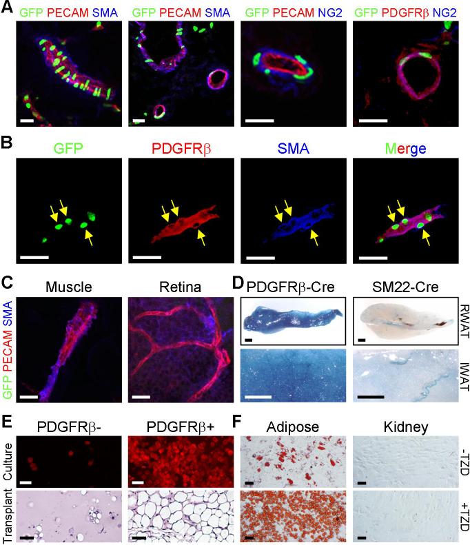
Figure 4. GFP+ cells are present in adipose depot mural cells.
(A) P30 PPARγ-GFP WAT was freshly frozen, cryo-sectioned, and examined with direct fluorescence for GFP and indirect immunofluorescence for the indicated endothelial (PECAM, red) and mural cell (SMA, blue; NG2, blue; PDGFRβ, red) markers.
(B) Cryo-section of a PPARγ-GFP adipose depot showing expression of GFP, PDGFRβ (red) and SMA (blue). Arrows indicate some mural cell nuclei that express GFP. (C) Muscle cryo-sections and retinal whole mount of PPARγ-GFP mice were examined for GFP, PECAM, and SMA as in (A, B). GFP was not expressed in mural cells of these tissues.
(D) RWAT (top panels, 5x) and IWAT (bottom panels, 20x) of P30 PDGFRβ-Cre;R26R and SM22-Cre;R26R mice were stained for β-galactosidase expression (blue).
(E) SV cells were isolated from P30 wild-type mice and sorted with a PDGFRβ antibody. Top panels: confluent PDGFRβ negative and positive cells were cultured in insulin and fat formation assessed with BODIPY (red). Bottom panels: PDGFRβ negative and positive cells were transplanted into nude mice, and the resultant tissues were sectioned and H&E stained.
(F) Adipose SVF and cells dissociated from kidney were sorted with a PDGFRβ antibody, and PDGFRβ positive cells were cultured in the absence (top) or presence of TZD (bottom).
Scale bars: 20μm in confocal images (A-C), 1mm (D), 50μm (E, F).

