Abstract
T helper cells that produce IL-17 (Th17 cells) promote autoimmunity in mice and have been implicated in pathogenesis of human inflammatory diseases. At mucosal surfaces Th17 cells are thought to protect the host from infection while regulatory T (Treg) cells control immune responses and inflammation triggered by the resident microflora1–5. Differentiation of both cell types requires TGF-β, but depends on distinct transcription factors, RORγt for Th17 and Foxp3 for Treg cells6-8. How TGF-β regulates the differentiation of T cells with opposing activities has been perplexing. Here, we demonstrate that together with pro-inflammatory cytokines TGF-β orchestrates Th17 cell differentiation in a concentration-dependent manner. At low concentrations, TGF-β synergizes with IL-6 and IL-21 (ref. 9-11) to promote IL-23R expression, favoring Th17 cell differentiation. High concentrations of TGF-β repress IL-23R expression and favor Foxp3+ Treg cells. RORγt and Foxp3 are co-expressed in naïve CD4+ T cells exposed to TGF-β and in a subset of T cells in the small intestinal lamina propria (LP). In vitro, TGF-β-induced Foxp3 inhibits RORγt function at least in part through their interaction. Accordingly, LP T cells that co-express both transcription factors produce less IL-17 than those that express RORγt alone. IL-6, IL-21 and IL-23 relieve Foxp3-mediated inhibition of RORγt, thereby promoting Th17 cell differentiation. Therefore, the decision of antigen-stimulated cells to differentiate into Th17 or Treg cells depends upon the cytokine-regulated balance of RORγt and Foxp3.
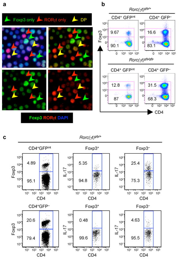
When T lymphocytes are exposed to microbial antigens, they acquire diverse effector functions depending on which cytokines are produced by activated cells of the innate immune system12. Differentiation of pro-inflammatory Th17 cells requires the presence of IL-23, which is produced by activated dendritic cells13-15. In vitro, however, Th17 cell differentiation is independent of IL-23 and is induced by TGF-β plus IL-6 or IL-21 (ref. 6, 9-11). Both in vitro and in vivo differentiation of the Th17 cell lineage require the upregulation of the orphan nuclear receptor RORγt7. TGF-β is also required to restrain inflammatory autoimmune responses16. Among its numerous properties is its ability to induce expression of Foxp3 in naïve antigen-stimulated T cells, endowing the cells with regulatory or suppressor function8. Thus, TGF-β can induce both regulatory and pro-inflammatory T cells, depending on whether pro-inflammatory cytokines such as IL-6 and, potentially, IL-23 are present11,17. Treatment of antigen receptor-stimulated T cells with TGF-β alone induces expression of both Foxp3 and RORγt, but not IL-17 (ref. 7, 11). Following such treatment, a significant proportion of cells co-expressed the two transcription factors (Fig. 1a and supplementary Fig.1a). To determine whether co-expression also occurs in vivo, we examined CD4+ T cells from the small intestinal lamina propria of heterozygous RORγt-GFP knock-in mice, in which IL-17 is produced by TCR+GFPint lymphocytes7. Foxp3 was expressed in about 10% of sorted GFPint (RORγt+) cells (Fig. 1b and Supplementary Fig. 1b). In addition, Foxp3 was expressed in approximately 17-20% of GFP- lamina propria CD4+ T cells, consistent with the relatively large proportion of Treg cells in the intestine.
Figure 1. Co-expression of Foxp3 and RORγt in vitro and in vivo.

a, Naïve CD4+ T cells were stimulated with anti-TCR and 5 ng/ml TGF-β for 48 hours, stained with DAPI (blue nuclear stain), anti-RORγ (red) and anti-Foxp3 (green) mAbs. All panels are of the same section, with Foxp3 and RORγ channels only (bottom), overlay of Foxp3 and RORγ channels (top right), and overlay of all three channels (top left). Foxp3, RORγt, and double expressing (DP) cells are indicated with colored arrows. b, Analysis of Foxp3+RORγt+ cells from the small intestinal lamina propria (LP). CD4+GFPint and CD4+GFP- cells were sorted from LP of RORc(γt)+/gfp and RORc(γt)gfp/gfp mice and Foxp3 expression was examined by intracellular staining. Results are representative of three experiments. c, Expression of IL-17 in Foxp3+RORγt+ and Foxp3-RORγt+ T cells from small intestine. Foxp3 and IL-17 expression was examined by intracellular staining of sorted TCRβ+CD4+GFPint cells from LP of RORc(γt)+/gfp mice.
We next performed a fate-mapping analysis to determine the proportion of IL-17+ small intestinal T cells that had expressed Foxp3 during their ontogeny. Mice expressing Cre recombinase under the regulation of the Foxp3 locus (Rubtsov et al., submitted) were crossed with Rosa26-stop-YFP reporter mice18, and female progeny (Rosa26stop-YFP/+; Foxp3cre/+) were analyzed for expression of YFP. When inactivation of X-linked Foxp3 was taken into account, we found that approximately 15% of IL-17- cells and 25% of IL-17+ cells had expressed Cre at some stage of development (Supplementary Fig. 2). The former represent Foxp3+ Treg cells, while the latter are the minimal proportion of Th17 cells that had expressed Foxp3 at some stage of their differentiation. These data suggest that Foxp3+ T cells can differentiate into Th17 cells in vivo in the presence of pro-inflammatory cytokines.
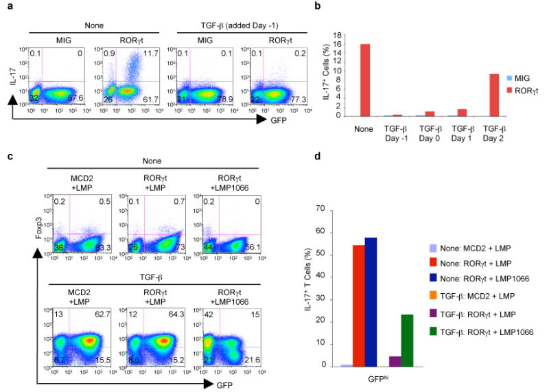
Examination of IL-17 expression in heterozygous RORγt-GFP knock-in mice revealed that RORγt+Foxp3+ lamina propria T cells produced much less IL-17 than RORγt+Foxp3- cells, suggesting that Foxp3 may interfere with the ability of RORγt to induce IL-17 (Fig. 1c). This is consistent with findings showing more than a 1,000-fold increase in IL-17 mRNA, but little change in RORγt, in Treg lineage cells that differentiate in the absence of Foxp3 (ref. 19). To investigate how Foxp3 may influence Th17 cell differentiation, we asked whether its induction would influence the expression of IL-17 in TGF-β-stimulated T cells. In naïve T cells that had been transduced with a retroviral vector encoding RORγt, we found that, whereas IL-6 augmented the proportion of RORγt-IRES-GFP+ cells that expressed IL-17, TGF-β had a profound inhibitory effect even when added one day after transduction (Fig. 2a and 2b). Addition of TGF-β was followed by a sharp increase in expression of Foxp3 in the CD4+ T cells, and both the level of Foxp3 mRNA and proportion of Foxp3+ cells were not affected by the expression of RORγt (Fig. 2c).
Figure 2. TGF-β inhibits RORγt-directed IL-17 production by up-regulating Foxp3.

a, Effect of TGF-β on IL-17 expression following transduction of RORγt. Naïve CD4+ T cells incubated with TGF-β from day -1 were transduced with control vector (MIG) or RORγt-IRES-GFP (RORγt) on day 0 (24 h after TCR activation) and IL-17 intracellular staining was performed on day 5. b, Inhibitory effect of TGF-β when included at different times relative to transduction of RORγt. Percentage of IL-17+ cells among RORγt/GFP+ cells is shown. c, Knockdown of TGFβ-induced Foxp3 expression with an shRNA against Foxp3 (LMP1066). Naïve CD4+ T cells were stimulated as in (a) and co-transduced on days 0 and 1 with retroviral constructs encoding RORγt and the specific shRNA vector (LMP1066) or control vector (LMP). After transduction, the cells were cultured with or without TGF-β, and Foxp3 expression was measured by intracellular staining on day 5. d, Restoration of RORγt-induced IL-17 expression upon knock-down of Foxp3. IL-17 expression was assessed in cells co-transduced as in (c) and gated for the level of GFP expression. Percentage of IL-17+ T cells in GFPhi cell populations is shown. Results with additional shRNA vectors that failed to down-regulate Foxp3 expression were similar to those with the control LMP vector. Representative data from three experiments are shown.
To determine if the inhibitory effect of TGF-β on RORγt is mediated by Foxp3, we knocked down expression of Foxp3 by using a shRNA vector. TGF-β-induced Foxp3 expression was reduced by the Foxp3-specific shRNA vector, but not by control hairpin vectors (Fig. 2c). Accordingly, TGF-β–mediated inhibition of RORγt-directed IL-17 expression was partially reversed by Foxp3 knockdown (Fig. 2d). Consistent with the idea that this inhibition was mediated by Foxp3 upregulation, the most pronounced rescue of IL-17 expression occurred in cells that had lost the most Foxp3 expression (Supplementary Fig. 3). Thus, Foxp3 induced by TGF-β inhibits the function of RORγt.
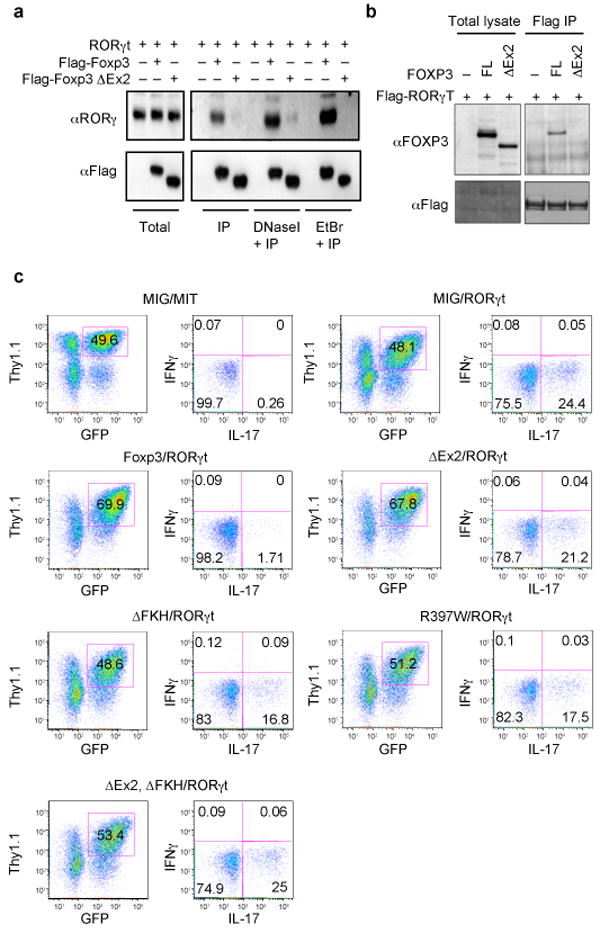
These results prompted us to ask whether Foxp3 interacts with RORγt to inhibit its function. Using a yeast two-hybrid screen, we previously found that human FOXP3 interacts with RORα, and that a splice isoform of FOXP3, lacking exon 2 (ref. 20), was deficient in this interaction21. We therefore examined whether mouse and human Foxp3 could similarly bind to RORγt, and whether such interaction was necessary for inhibition of the RORγt-mediated induction of IL-17. When FLAG epitope-tagged murine Foxp3 was co-expressed with murine RORγt in 293T cells, the two proteins were co-immunoprecipitated (Fig. 3a), even in the presence of DNase I or ethidium bromide, suggesting that the interaction does not involve DNA. A similar interaction was observed between human RORγT and FOXP3 (Fig. 3b). However, both mouse and human Foxp3 lacking the conserved exon 2-encoded sequence (Foxp3ΔEx2) had a substantially reduced association with RORγt (Fig. 3a and 3b). We examined the localization of the two proteins by confocal microscopy of HeLa cells transfected with FLAG-tagged murine Foxp3 constructs with or without murine RORγt. Both Foxp3 and RORγt were localized in the nucleus, but Foxp3 lacking the DNA-binding forkhead domain (Foxp3ΔFKH) remained in the cytoplasm due to deletion of the nuclear localization signal in FKH22 (Supplementary Fig. 4). However, Foxp3ΔFKH translocated to the nucleus when it was co-expressed with RORγt, indicating that the Foxp3-RORγt interaction is independent of FKH. Accordingly, Foxp3ΔFKH co-immunoprecipitated with RORγt in extracts of transfected 293T cells (data not shown). A combined Foxp3DEx2/ΔFKH mutant remained in the cytoplasm even when it was co-expressed with RORγt, further indicating that Foxp3 interacts with RORγt by way of the exon 2-encoded sequence (Supplementary Fig. 4).
Figure 3. Foxp3 interacts with RORγt and inhibits RORγt-directed IL-17 expression.

a, Co-immunoprecipitation of Foxp3 and RORγt from extracts of co-transfected 293T cells with or without DNaseI or ethidium bromide. Cells were transfected with murine RORγt and Flag-tagged wild-type (Flag-Foxp3) or exon 2-deleted Foxp3 (Flag-Foxp3ΔEx2). Anti-Flag immunoprecipitates and total lysates were immunoblotted with anti-RORγt antibody and anti-Flag antibody. b, Cells were transfected with Flag-tagged human RORγt and full-length (FL) or the exon 2-deleted isoform of human FOXP3 (ΔEx2). Anti-Flag immunoprecipitates and total lysates were probed with anti-FOXP3 and anti-Flag antibodies. c, Naïve CD4+ T cells were co-transduced with retroviruses encoding RORγt (MIT vector, Thy1.1 reporter) and various murine Foxp3 constructs (MIG vector, GFP reporter). IL-17 expression was assessed on day 4 in cells gated for expression of both Thy1.1 and GFP. Representative data from at least three experiments are shown in each of the panels.
To investigate the role of the interaction of Foxp3 and RORγt in the repression of RORγt-induced transcription, we co-expressed these transcription factors in naïve CD4+ T cells and examined expression of IL-17. Both murine and human Foxp3 blocked RORγt-directed IL-17 expression, but full suppression required the presence of the exon 2-encoded sequence in Foxp3, suggesting that the interaction between Foxp3 and RORγt is essential (Fig. 3c and supplementary Fig. 5). The ability of both murine and human Foxp3 to repress RORγt-induced IL-17 expression was abrogated by deletion of the FKH domain or a point mutation in this domain (R397W) that impairs FOXP3 DNA binding activity and was identified in X-linked immunodeficiency, polyendocrinopathy, enteropathy (IPEX) syndrome in humans23,24 (Fig. 3c and supplementary Fig. 5). Therefore, Foxp3 can block the activity of RORγt at least in part through an interaction involving a sequence encoded by exon 2, but the requirement for an intact FKH domain suggests that its DNA binding activity also contributes to inhibition of IL-17 expression. Thus, Foxp3 may inhibit RORγt-directed transcription through a mechanism similar to that proposed for its inhibition of IL-2 expression, involving its association with NFAT and Runx1 (ref. 25, 26). However, Foxp3ΔEx2 was as effective as the full-length protein in suppressing expression of IL-2 and IFNγ in primary mouse T cells, indicating that, like the naturally occurring human spliced isoform20, it retains regulatory functions and can, presumably, associate with both NFAT and Runx1 (Supplementary Fig. 6).
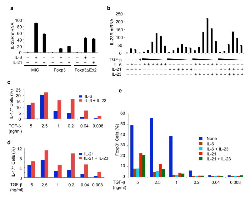
Our results suggest that Foxp3 may inhibit RORγt activity on its target genes during Th17 cell differentiation. To extend our analysis from IL-17 to other potential RORγt transcriptional targets, we examined the effect of TGF-β–induced Foxp3 on IL-23R expression, which also requires the activity of RORγt10,11. Forced expression of wild-type murine Foxp3 inhibited IL-6/IL-21-induced IL-23R expression, whereas Foxp3ΔEx2 had less inhibitory activity (Fig. 4a), consistent with the notion that Foxp3 inhibits the function of RORγt through an interaction involving the sequence encoded by exon 2. Similar results were observed with IL-22 expression in response to IL-6 or IL-21 (data not shown). Expression of IL-22 and of IL-23R in response to either IL-6 or forced expression of RORγt was also inhibited by high concentrations of TGF-β (Ref. 11, 27 and data not shown). However, at low concentrations, TGF-β synergized with IL-6 and IL-21 to enhance expression of IL-23R mRNA (Fig. 4b). As a consequence, addition of IL-23 to cultures containing high concentrations of TGF-β had no effect on IL-17 expression, but significantly increased the number of IL-17+ cells and the level of IL-17 expression per cell when low concentrations of TGF-β were used (Figs. 4c and 4d, and supplementary Fig. 7). In contrast, induction of Treg (Foxp3+) cells was optimal at high concentrations of TGF-β, but there was little induction at TGF-β concentrations at which IL-23 had synergistic effect on expression of IL-17 (Fig. 4e).
Figure 4. TGF-β concentration influences IL-23R expression and levels of IL-17 in response to Th17-inducing cytokines.
a, Foxp3-mediated inhibition of IL-6/IL-21-induced IL-23R expression. Naïve CD4+ T cells were transduced with MIG, full length Foxp3, or Foxp3ΔEx2 viruses and were treated with the indicated cytokines. RNA was isolated from GFP+ cells at day 2. IL-23R expression was measured by real-time RT-PCR and was normalized to the actin level. Error bars represent standard deviations obtained using the standard curve method. b, Induction of IL-23R mRNA in response to cytokines. Naïve CD4+ T cells were stimulated with anti-CD3 and anti-CD28 throughout the culture period in the presence of the indicated combinations of cytokines. TGF-β was titrated into the cultures at the concentrations of 5 ng/ml, 2.5 ng/ml, 1 ng/ml, 200 pg/ml, 40 pg/ml, or 8 pg/ml. IL-23R mRNA expression was measured after 48 h by real-time RT-PCR and was normalized to the actin expression level. c and d, IL-23 enhancement of IL-17 expression at low concentrations of TGF-β. Percent IL-17+ cells at 96 hours of stimulation with the indicated cytokines is shown. Results in histogram format are shown in Supplemental Fig. 7. e, Induction of Foxp3 at different concentrations of TGF-β. Representative data from at least three experiments are shown for each set of panels.
TGF-β-induced Foxp3 expression is inhibited by IL-6 (ref. 17), IL-21 (ref. 10), and IL-23 (Supplementary Fig. 8). However, a substantial number of Foxp3+ cells differentiated in response to TGF-β even in the presence of IL-6, and many of these cells also expressed IL-17 (Supplementary Fig. 9a). Conversely, many of the IL-17+ cells also expressed Foxp3. Thus, RORγt-dependent IL-17 expression can occur in the presence of Foxp3, but the level of Foxp3 may be insufficient to block RORγt function or, alternatively, IL-6 may overcome the inhibitory function of Foxp3. To examine this possibility, we added IL-6 or IL-21 to cultures of cells transduced with both RORγt and Foxp3. Under these conditions, the inhibitory effect of Foxp3 on IL-17 induction was largely circumvented, even though the level of Foxp3 protein was not affected (Supplementary Fig. 9b and data not shown), suggesting that IL-6 and IL-21 may have an additional post-translational effect on either Foxp3 or RORγt.
Our data collectively suggest that T cells receiving a TGF-β signal can acquire the potential to develop into either the Treg or Th17 lineage. Foxp3 induction restrains the differentiation of inflammatory Th17 cells in response to TGF-β in the absence of other pro-inflammatory cytokines by inhibiting the activity of RORγt. In the presence of pro-inflammatory cytokines, the suppression of Foxp3 expression and inhibitory function, together with the concurrent upregulation or stabilization of RORγt expression, leads to full progression towards the Th17 lineage (Supplementary Fig. 10). This process may be especially relevant in the intestinal LP, where TGF-β can promote either Th17 or Treg cell lineage differentiation, depending on its local concentration. In this setting, a fine balance between RORγt and Foxp3 may be critical for immune homeostasis. In line with the observation that more Foxp3+ Tregs were present in the gut of RORγt-deficient mice (Fig. 1b), these mutant mice were also protected from autoimmune disease (ref. 7 and data not shown). Conversely, a decrease of Foxp3 expression and function and an increase of RORγt expression tips the Treg/Th17 balance toward the Th17 cell lineage. This may occur in some autoimmune diseases, as suggested by the finding that an IL-23R polymorphism correlates with protection from Crohn's disease28. These results therefore have important implications for how peripheral tolerance is maintained in the presence of potentially pro-inflammatory cytokines.
Methods Summary
Mice
C57BL/6 mice (Taconic), mice with a GFP reporter cDNA knocked-in at the RORγt translation initiation site29, mice with an IRES-YFP-Cre cDNA knocked into the 3′ UTR of the Foxp3 locus (Rubtsov et al, submitted), and Rosa26stop-YFP (ref. 18) mice were kept in SPF conditions at the animal facility of the Skirball Institute. All animal experiments were performed in accordance with approved protocols for the NYU institutional Animal Care and Usage Committee.
Cell culture
Naïve CD4+ T Cells were purified and cultured as described previously7. Briefly, 1.5×06 naive CD4+ T cells were cultured in wells of 24-well plates (or 0.7×10⁶ cells per well in 48-well-plates) containing plate-bound anti-CD3 (5 μg/ml) and soluble anti-CD28 (1 μg/ml). Cultures were supplemented with 2 μg/ml anti-IL-4 (BD Pharmingen), 2 μg/ml anti-IFN-γ (BD Pharmingen) with or without 80 U/mL human IL-2 (a kind gift from S. Reiner), 20 ng/ml IL-6 (eBioscience), 5 ng/ml TGF-β (PeproTech), 50 ng/ml IL-21(R&D Systems) and 10 ng/ml IL-23 (eBioscience). Viral transduction was performed as described previously, unless indicated otherwise in the text7. T cells were isolated from the small intestinal lamina propria as described previously7.
General
All DNA constructs were generated by PCR-based methodology and confirmed by sequencing. Retroviral production and transduction were as described previously7. Protein-protein interaction was deteced by co-immunoprecipitation and confocal microscopy in 293T cells and HeLa cells. Gene expression analysis was monitored by real-time RT-PCR using gene specific primers and probes. IL-17 and Foxp3 protein expression were examined by intracellular staining performed according to the manufacturer's protocol. Co-expression of RORγt and Foxp3 was examined by immunofluorescence using anti-RORγ30 and anti-Foxp3 antibodies.
Methods
Plasmids and retrovirus production
MIG, MIT, and MCD2 are retrovirus-based vectors containing GFP, Thy1.1, and hCD2, respectively, under regulation of an internal ribosome entry site (IRES). The RORγt cDNA was PCR amplified and cloned into MIG (RORγt-IRES-GFP), MIT (RORγt-IRES-Thy1.1), and MCD2 (RORγt-IRES-hCD2). The wild-type full-length Foxp3 cDNA and various Foxp3 mutant cDNAs were PCR amplified with a 5′ FLAG-tag primer and a 3′ corresponding primer and cloned into MIG (Foxp3-IRES-GFP). IL-23R cDNA was PCR amplified and cloned into MIG (IL-23R-IRES-GFP). Human FOXP3 cDNAs were PCR amplified and cloned into MIG. MSCV-LTRmiR30-PIG (LMP) is a commercial vector from Openbiosystems. A double-stranded DNA oligo that targets the coding region of Foxp3 was cloned into LMP (LMP1066) according to the manufacturer's protocol (the target sequence of Foxp3: 5′-GGCAGAGGACACTCAATGAAAT-3′). Retrovirus production was as described previously7.
Cell culture and retroviral transduction
Naïve CD4+ T Cells were purified and cultured as described previously7. Briefly, 1.5×10⁶ naive CD4+ T cells were cultured in wells of 24-well plates (or 0.7×10⁶ cells per well in 48-well-plates) containing plate-bound anti-CD3 (5 μg/ml) and soluble anti-CD28 (1 μg/ml). Cultures were supplemented with 2 μg/ml anti-IL-4 (BD Pharmingen), 2 μg/ml anti-IFN-γ (BD Pharmingen) with or without 80 U/mL human IL-2 (a kind gift from S. Reiner), 20 ng/ml IL-6 (eBioscience), 5 ng/ml TGF-β (PeproTech), 50 ng/ml IL-21(R&D Systems) and 10 ng/ml IL-23 (eBioscience). Viral transduction was performed as described previously, unless indicated otherwise in the text7. T cells were isolated from the small intestinal lamina propria as described previously7.
Surface and intracellular staining and carboxyfluoroscein succinimidyl ester (CFSE) labeling
For intracellular staining, cells obtained from in vitro culture or isolated from the small intestinal lamina propria were incubated for 4-5 hours with 50 ng/ml PMA (Sigma) and 500 ng/ml Ionomycin (Sigma), plus 2 μg/ml Brefeldin A (Sigma) during the last 2 hours. The cells were kept in a tissue culture incubator at 37 °C. Surface staining was performed for 15-20 min with the corresponding cocktail of fluorescently labeled antibodies. After surface staining, the cells were resuspended in Fixation/Permeabilization solution (BD Cytofix/Cytoperm kit-BD pharmingen), and intracellular cytokine staining was performed according to the manufacturer's protocol. For intracellular staining of Foxp3, the Foxp3 Staining Buffer Set (Fixation/Permeabilization and Permeabilization Buffers) was used (eBioscience) and performed as per the manufacturer's protocol. For CFSE labeling, sorted naïve CD4+ T cells were washed twice with HBSS (Invitrogen), and labeled with 5 μM CFSE (Sigma) in HBSS for 10 min at room temperature. The labeling was then stopped by adding 1/5 vol. of FCS. The labeled cells were washed twice with the T cell culture medium before they were seeded and stimulated as described in the text.
Real-time RT-PCR
cDNA was synthesized and analyzed by real-time quantitative PCR as described previously7. The starting quantity (SQ) of the initial cDNA sample was calculated from primer-specific standard curves by using the iCycler Data Analysis Software. The expression level of each gene was normalized to actin expression level using the Standard Curve Method. The primer sets and probes for real-time PCR were described elsewhere7,11.
Co-immunoprecipitation and western blot
293T cells were transfected with the indicated constructs using Lipofectamine 2000 (Invitrogen). Forty-eight hours after transfection, whole cell extracts were made as in the lysis buffer containing 50mM Tris-HCl pH 8.0, 120mM NaCl, 4mM EDTA, 1% NP-40, 50 mM NaF, 1mM Na3VO4 and protease inhibitors. After the insoluble material was removed by centrifugation, the lysate was immunoprecipated overnight with anti-Flag M2 agarose beads (Sigma). After extensive washes with the lysis buffer, samples were resolved in an SDS-PAGE gel and transferred to nitrocellulous membrane. Western blotting was performed with an anti-FLAG monoclonal antibody (Sigma), an anti-Foxp3 monoclonal antibody (eBioscience), and an anti-RORγt hamster monoclonal antibody30.
Confocal microscopy
HeLa cells were plated on 8 well glass slides (Lab-Tek II Chamber Slide System) before transfection with the indicated constructs using Lipofectamine 2000 (Invitrogen). Forty-eight hours after transfection, cells were washed once in PBS, fixed for 15 mins in 2% paraformaldehyde in Phosphate Buffer (PBS without saline), and then washed twice in PBS. Cells were blocked and permeabilized in PBS-XG (10% goat serum (Sigma) in PBS containing 0.1% Triton X-100) for one hour at room temperature. The cells were then incubated overnight at 4 °C with anti-RORγt hybridoma supernatant30 (1:2 dilution in PBS-XG). After 2 washes in PBS, the cells were incubated for 1 hour at room temperature with Cy3-conjugated goat anti-hamster antibody (Jackson Immuno Research Laboratory) at 1:400 dilution in PBS-XG. The cells were then washed 3 times in PBS and incubated for 1 hour at room temperature with anti-FLAG M2 monoclonal antibody (Sigma) at 1:1000 dilution in PBS-XG. After two washes in PBS, the cells were incubated for 1 hour at room temperature with anti-mouse Alexa 633 (Molecular Probes) at 1:200 dilution in PBS-XG. The cells were then washed twice in PBS and incubated for 5 mins at room temperature with 1 μg/ml 4′, 6-diamidino-2-phenylindole-2HCl (DAPI; Sigma), washed two more times in PBS and mounted with Fluoromount-G (Southern Biotechnology Associates). The cells were examined with a Zeiss ZMD510 microscope with CCD camera and images were processed with Zeiss LSM Image Browser 4.0 and Adobe Photoshop 7.0.
Immunofluorescence
Naive T cells were sorted as described before and stimulated in the presence of the indicated cytokines as described7. Lamina propria lymphocytes from Rorc(γt)gfp/+ small intestines were isolated as described7 and CD4+GFPint and CD4+GFP- cells were sorted on a MoFlo cytometer (DAKO Cytomation). Naive T cells or sorted LP T cells were then cytospinned on glass slides and fixed in 2% paraformaldehyde in phosphate buffer (PBS without saline) for 20 minutes. After blocking, immunofluorescence staining was performed by incubating consecutively with the anti-RORγ antibody30 (hybridoma supernatant 1:4) and biotin anti-mouse/rat Foxp3 monoclonal antibody (eBioscience clone FJK-16 1:200 dilution). The blocking solution contained PBS, 0.1% Triton-X100, and 10% goat serum. Secondary goat anti-Armenian-hamster Cy3 conjugated antibody (Jackson Immunoresearch) and streptavidin-APC (eBioscience), both at 1:400 dilution, were used to detect the RORγ and Foxp3 primary antibodies, respectively.
Supplementary Material
Acknowledgments
We thank Peter Lopez and John Hirst for assistance with cell sorting. We also thank Juan Lafaille and Derya Unutmaz for critical reading of the manuscript and members of the Littman laboratory for their helpful suggestions. L.Z., M.M.W.C. and I.I.I., were supported, respectively, by fellowships from the Irvington Institute for Immunological Research, the Cancer Research Institute and the Crohn's and Colitis Foundation of America. This work was supported by the Howard Hughes Medical Institute (D.R.L., A.Y.R.), the Sandler Program for Asthma Research (D.R.L.), the National Multiple Sclerosis Society (D.R.L.), the Helen and Martin Kimmel Center for Biology and Medicine (D.R.L.), NIH grant AI48779 (S.F.Z.), and the JDRF Collaborative Center for Cell Therapy (S.F.Z.).
Footnotes
Author contributions: L.Z., J.E.S., M.M.W.C, I.I.I., and Y.S. performed the experiments with assistance from R.M. and G.D.V. Y.P.R. and A.Y.R. provided mice for fate-mapping experiments. L.Z., S.F.Z., and D.R.L. designed the experiments, and L.Z. and D.R.L. wrote the manuscript with input from the co-authors.
References
- 1.Weaver CT, Harrington LE, Mangan PR, Gavrieli M, Murphy KM. Th17: an effector CD4 T cell lineage with regulatory T cell ties. Immunity. 2006;24:677–688. doi: 10.1016/j.immuni.2006.06.002. [DOI] [PubMed] [Google Scholar]
- 2.Weaver CT, Hatton RD, Mangan PR, Harrington LE. IL-17 family cytokines and the expanding diversity of effector T cell lineages. Annu Rev Immunol. 2007;25:821–852. doi: 10.1146/annurev.immunol.25.022106.141557. [DOI] [PubMed] [Google Scholar]
- 3.McKenzie BS, Kastelein RA, Cua DJ. Understanding the IL-23-IL-17 immune pathway. Trends Immunol. 2006;27:17–23. doi: 10.1016/j.it.2005.10.003. [DOI] [PubMed] [Google Scholar]
- 4.Lee E, et al. Increased expression of interleukin 23 p19 and p40 in lesional skin of patients with psoriasis vulgaris. J Exp Med. 2004;199:125–130. doi: 10.1084/jem.20030451. [DOI] [PMC free article] [PubMed] [Google Scholar]
- 5.Witowski J, Ksiazek K, Jorres A. Interleukin-17: a mediator of inflammatory responses. Cell Mol Life Sci. 2004;61:567–579. doi: 10.1007/s00018-003-3228-z. [DOI] [PMC free article] [PubMed] [Google Scholar]
- 6.Veldhoen M, Hocking RJ, Atkins CJ, Locksley RM, Stockinger B. TGFβ in the context of an inflammatory cytokine milieu supports de novo differentiation of IL-17-producing T cells. Immunity. 2006;24:179–189. doi: 10.1016/j.immuni.2006.01.001. [DOI] [PubMed] [Google Scholar]
- 7.Ivanov II, et al. The orphan nuclear receptor RORγt directs the differentiation program of proinflammatory IL-17+ T helper cells. Cell. 2006;126:1121–1133. doi: 10.1016/j.cell.2006.07.035. [DOI] [PubMed] [Google Scholar]
- 8.Chen W, et al. Conversion of peripheral CD4+CD25- naive T cells to CD4+CD25+ regulatory T cells by TGF-β induction of transcription factor Foxp3. J Exp Med. 2003;198:1875–1886. doi: 10.1084/jem.20030152. [DOI] [PMC free article] [PubMed] [Google Scholar]
- 9.Korn T, et al. IL-21 initiates an alternative pathway to induce proinflammatory TH17 cells. Nature. 2007;448:484–487. doi: 10.1038/nature05970. [DOI] [PMC free article] [PubMed] [Google Scholar]
- 10.Nurieva R, et al. Essential autocrine regulation by IL-21 in the generation of inflammatory T cells. Nature. 2007;448:480–483. doi: 10.1038/nature05969. [DOI] [PubMed] [Google Scholar]
- 11.Zhou L, et al. IL-6 programs TH-17 cell differentiation by promoting sequential engagement of the IL-21 and IL-23 pathways. Nature Immunol. 2007;8:967–974. doi: 10.1038/ni1488. [DOI] [PubMed] [Google Scholar]
- 12.Murphy KM, Reiner SL. The lineage decisions of helper T cells. Nat Rev Immunol. 2002;2:933–944. doi: 10.1038/nri954. [DOI] [PubMed] [Google Scholar]
- 13.Langrish CL, et al. IL-23 drives a pathogenic T cell population that induces autoimmune inflammation. J Exp Med. 2005;201:233–240. doi: 10.1084/jem.20041257. [DOI] [PMC free article] [PubMed] [Google Scholar]
- 14.Aggarwal S, Ghilardi N, Xie MH, de Sauvage FJ, Gurney AL. Interleukin-23 promotes a distinct CD4 T cell activation state characterized by the production of interleukin-17. J Biol Chem. 2003;278:1910–1914. doi: 10.1074/jbc.M207577200. [DOI] [PubMed] [Google Scholar]
- 15.Cua DJ, et al. Interleukin-23 rather than interleukin-12 is the critical cytokine for autoimmune inflammation of the brain. Nature. 2003;421:744–748. doi: 10.1038/nature01355. [DOI] [PubMed] [Google Scholar]
- 16.Letterio JJ, Roberts AB. Regulation of immune responses by TGF-β. Annu Rev Immunol. 1998;16:137–161. doi: 10.1146/annurev.immunol.16.1.137. [DOI] [PubMed] [Google Scholar]
- 17.Bettelli E, et al. Reciprocal developmental pathways for the generation of pathogenic effector TH17 and regulatory T cells. Nature. 2006;441:235–238. doi: 10.1038/nature04753. [DOI] [PubMed] [Google Scholar]
- 18.Srinivas S, et al. Cre reporter strains produced by targeted insertion of EYFP and ECFP into the ROSA26 locus. BMC Dev Biol. 2001;1:4. doi: 10.1186/1471-213X-1-4. [DOI] [PMC free article] [PubMed] [Google Scholar]
- 19.Gavin MA, et al. Foxp3-dependent programme of regulatory T-cell differentiation. Nature. 2007;445:771–775. doi: 10.1038/nature05543. [DOI] [PubMed] [Google Scholar]
- 20.Allan SE, et al. The role of 2 FOXP3 isoforms in the generation of human CD4+ Tregs. J Clin Invest. 2005;115:3276–3284. doi: 10.1172/JCI24685. [DOI] [PMC free article] [PubMed] [Google Scholar]
- 21.Du J, Huang C, Zhou B, Ziegler SF. Isoform-specfic inhibition of RORα-mediated transcriptional activation by human FOXP3. J Immunol. doi: 10.4049/jimmunol.180.7.4785. in press. [DOI] [PubMed] [Google Scholar]
- 22.Schubert LA, Jeffery E, Zhang Y, Ramsdell F, Ziegler SF. Scurfin (FOXP3) acts as a repressor of transcription and regulates T cell activation. J Biol Chem. 2001;276:37672–37679. doi: 10.1074/jbc.M104521200. [DOI] [PubMed] [Google Scholar]
- 23.Wildin RS, et al. X-linked neonatal diabetes mellitus, enteropathy and endocrinopathy syndrome is the human equivalent of mouse scurfy. Nature Genet. 2001;27:18–20. doi: 10.1038/83707. [DOI] [PubMed] [Google Scholar]
- 24.Lopes JE, et al. Analysis of FOXP3 reveals multiple domains required for its function as a transcriptional repressor. J Immunol. 2006;177:3133–3142. doi: 10.4049/jimmunol.177.5.3133. [DOI] [PubMed] [Google Scholar]
- 25.Ono M, et al. Foxp3 controls regulatory T-cell function by interacting with AML1/Runx1. Nature. 2007;446:685–689. doi: 10.1038/nature05673. [DOI] [PubMed] [Google Scholar]
- 26.Wu Y, et al. FOXP3 controls regulatory T cell function through cooperation with NFAT. Cell. 2006;126:375–387. doi: 10.1016/j.cell.2006.05.042. [DOI] [PubMed] [Google Scholar]
- 27.Zheng Y, et al. Interleukin-22, a TH17 cytokine, mediates IL-23-induced dermal inflammation and acanthosis. Nature. 2007;445:648–651. doi: 10.1038/nature05505. [DOI] [PubMed] [Google Scholar]
- 28.Duerr RH, et al. A genome-wide association study identifies IL23R as an inflammatory bowel disease gene. Science. 2006;314:1461–1463. doi: 10.1126/science.1135245. [DOI] [PMC free article] [PubMed] [Google Scholar]
- 29.Eberl G, et al. An essential function for the nuclear receptor RORγt in the generation of fetal lymphoid tissue inducer cells. Nature Immunol. 2004;5:64–73. doi: 10.1038/ni1022. [DOI] [PubMed] [Google Scholar]
- 30.Sun Z, et al. Requirement for RORγ in thymocyte survival and lymphoid organ development. Science. 2000;288:2369–2373. doi: 10.1126/science.288.5475.2369. [DOI] [PubMed] [Google Scholar]
Associated Data
This section collects any data citations, data availability statements, or supplementary materials included in this article.


