Abstract
Background
Studies have shown that topical administration of exogenous opioid drugs impairs wound healing by inhibiting the peripheral release of neuropeptides, thereby inhibiting neurogenic inflammation. This delay is immediate and peaks during the first days of wound closure. This study examined the effects of topical morphine treatment in a cutaneous wound healing model in the rat.
Methods
Full-thickness 4mm diameter wounds were placed on the periscapular region of rats that subsequently received twice-daily topical applications of IntraSite Gel (Smith+Nephew, Hull, United Kingdom) alone or gel infused with 5 mM morphine sulfate on days 0–3 or 4–10 post-wounding or throughout the time course. Wound tissue was taken on days 1, 3, 5, 8, and 18 post-wounding and immunostained for myofibroblast and macrophage markers or stained with hematoxylin and eosin.
Results
Delays in wound closure observed during morphine application on days 0–3 post-wounding mimicked those seen in wounds treated with morphine throughout the entire healing process. However, no significant delays in closure were seen in wounds treated with morphine beginning on day 4 post-wounding. Treatment of wounds with morphine significantly reduced the number of myofibroblasts and macrophages in the closing wound. Additionally, morphine application resulted in decreases in skin thickness and an increase in residual scar tissue in healed skin.
Conclusions
These findings demonstrate the time-dependent and persistent nature of the detrimental effects of topical morphine on cutaneous wound healing. The data identify specific limitations that could be ameliorated to optimize topical opioid administration as an analgesic therapeutic strategy in the treatment of painful cutaneous wounds.
Introduction
Opioid drugs, such as morphine, remain the standard course of care in providing analgesia to patients with chronic cutaneous wounds. However, pain management with systemic, centrally-acting opioid drugs is restricted by dose-limiting side effects such as respiratory depression, sedation, nausea, and constipation, as well as the development of tolerance. These shortcomings have led to the investigation of peripheral opioid analgesia as an alternative approach to providing analgesia for cutaneous wounds. For years opioid analgesia was believed to originate exclusively via the activation of opioid receptors within the central nervous system. Accumulating evidence over the past decade reveals the analgesic efficacy of peripheral opioids, offering a promising new alternative in the treatment of pain. Analgesia during peripheral opioid administration can be achieved via activation of opioid receptors located on afferent sensory nerve terminals in peripheral tissues 1–3, avoiding the negative effects seen upon stimulation of central opioid receptors. The topical application of opioids has been explored as a strategy for reducing the pain associated with cutaneous wounds 4–6.
Anti-inflammatory effects of opioids have been well documented 7–9. Activation of peripheral opioid receptors on primary afferent neurons reduces the excitability of these neurons and suppresses the antidromic release of pro-inflammatory neuropeptides such as substance P and calcitonin gene-related peptide 3,10. Previous studies demonstrate that sensory neuropeptides play an essential role in wound repair, as healing is enhanced by their exogenous application 11–13 and impaired by their depletion 14–17. Neuropeptides facilitate wound healing by regulating blood flow and modulating the function of many types of cells in the closing wound, including immunocompetent and inflammatory cells, as well as epithelial and endothelial cells. Therefore, inhibition of their action by opioid agonists negatively impacts the rate of wound healing.
Studies within this lab have revealed that topical application of morphine sulfate significantly delays cutaneous wound closure rates in rats. The delay occurs in a concentration-dependent manner (consistent with opioid-receptor mediated effects) and can be reversed by the addition of the tachykinins substance P or neurokinin A 18. Interestingly, the morphine-induced delays in wound closure were immediately apparent, occurring only during the early phase of wound healing. This transient delay was followed by acceleration in wound closure, resulting in morphine-treated wounds closing at times similar to controls. Therefore, this study was designed primarily to assess the temporal effects of topical morphine application on wound closure rates and the impact morphine has on inflammatory and parenchymal cells essential in the healing process. This study also addressed whether endogenous opioids play a direct role in wound closure, since their production by peripheral immune cells has been implicated in cutaneous analgesia 19,20. Finally, although morphine-treated wounds ultimately closed at times similar to controls, disruption of normal closure may produce long-term effects that outlast the duration of topical morphine administration. Accordingly, the structural architecture of healed skin was evaluated after closure of cutaneous wounds treated with morphine.
Materials & Methods
Animals and Experimental Design
A cutaneous wound healing model was utilized to evaluate wound closure rates in rats. A total of 78 male Sprague-Dawley rats (Harlan, Indianapolis, IN) approximately 8 weeks of age (200–220 grams body weight) were randomly assigned to one of five treatment groups. Rats were subsequently anesthetized by intraperitoneal administration of 65 mg/kg ketamine hydrochloride and 5.5 mg/kg xylazine hydrochloride and the mid-periscapular region clipped and shaved. A skin biopsy punch was used to excised a 4mm diameter full-thickness skin flap from the midline below the scapulae to a depth just above the panniculus carnosus muscle 21. All rats were then housed individually to prevent cage mates from grooming or otherwise disturbing the wound. Animal facilities were temperature-and humidity-controlled with a 12-h dark–light cycle and food and water ad libitum. All surgical procedures and animal handling were performed in accordance with National Institutes of Health laboratory care standards and were approved by the University of Kansas Medical Center Animal Care and Use Committee (Kansas City, KS).
Drug Preparation and Gel Administration
Morphine sulfate (25 mg/ml) (Abbott Laboratories, Inc., North Chicago, IL) was infused into IntraSite Gel (amorphous hydrogel; Smith+Nephew, Hull, United Kingdom) at a concentration of 5 mM. Gel and drug were combined in 3 mL syringes by repeated passage through a Luer-lock stopcock. Beginning one hour post-surgery, 150 μL IntraSite Gel (Smith+Nephew) alone or morphine-infused IntraSite Gel (Smith+Nephew) was applied topically to the wound twice daily. Animals were divided into four treatment groups. Control animals received applications of IntraSite Gel (Smith+Nephew) alone throughout the entire time course (n=37). Saline was added to control gel to match the saline content of the morphine-infused gel. A second treatment group received 5 mM morphine sulfate on days 0–3 post-wounding and gel-only treatments for the remaining 7 days (n=5). The third group received gel-only applications on wound days 0–3 and morphine sulfate-infused gel on days 4–10 post-wounding (n=6). A fourth group was treated with 5 mM morphine sulfate throughout the entire time course or until euthanasia (n=25). The final treatment group received the opioid receptor antagonist naltrexone (1 mM) throughout the entire time course (n=5).
Wound Imaging and Analysis
Wound images were captured each morning prior to treatment using a hand-held digital camera. A bar attached to the camera provided a fixed focal distance target for wound imaging. A size standard with known surface area was affixed to the target bar and included in each image. Wound surface area was determined using a computerized planimetric program (Scion Image, Fredrick, MD). The area occupied by the wound was defined by the boundary created by the granulation tissue or scab/intact tissue interface. Wound area data generated by Scion Image were converted from pixels to area units of mm2 by comparison to the known area of the fixed size standard.
Tissue Harvesting
Rats were decapitated and wound tissue including approximately 1.0 cm of surrounding intact skin was harvested. Skin was dissected from rats in each treatment group on post-wound day 0 (n=3), 1 (n=8), 3 (n=6), 5 (n=6), 8 (n=6), and 18 (n=9). Tissue was embedded in tissue freezing medium (Electron Microscopy Sciences, Hatfield, PA), frozen on dry ice and cryosectioned serially throughout the wound site at 14 μm thickness. Sections at ten-section intervals were placed on slides and stored at −80°C until staining.
Histochemistry
Sections from day 18 post-wounding were post-fixed at room temperature with 4% paraformaldehyde for 5 minutes and stained with hematoxylin and eosin. Slides were mounted with Permount (Fisher, St. Louis, MO). Images were captured using light microscopy at a magnification of 20× (Nikon Eclipse 80i microscope, Nikon Digital Sight Fi1 camera, Melville, NY). Epidermal and full-skin thicknesses were quantified in 3 sections equally spaced throughout each wound. In each analyzed section, two regions were selected randomly. All measurements were performed blind with ImageJ (NIH, Bethesda, MD) analysis software.
Immunohistochemistry
Tissue sections from post-wound days 0, 1, 3, 5, and 8 were post-fixed in 4% paraformaldehyde for 5 minutes, blocked in 5% goat serum for 1 hour, and directly immunostained (90 min at room temp.) with antibodies for the macrophage marker macrosialin (rat polyclonal IgG, fluorescein isothiocyanate conjugated, 1:200, clone FA-11, Serotec, Oxford, United Kingdom), and for the myofibroblast marker α-smooth muscle actin (α-SMA, mouse monoclonal IgG 2A, Cy3 conjugated, 1:200, clone 1A4, Sigma, St. Louis, MO). Rat serum was added to the blocking serum of sections stained with macrosialin antisera to negate the impact of non-specific staining. In addition, a rat IgG antibody conjugated to fluorescein isothiocyanate (1:100, Serotec, clone YTH71.3) was utilized as a negative control, and no specific staining was detected with this approach. In pilot studies, ED1 mouse monoclonal IgG (Chemicon, Temecula, CA), CD68 goat polyclonal (Santa Cruz, Santa Cruz, CA), ED1 mouse monoclonal IgG (Serotec), and CD68 mouse monoclonal IgG (Biomeda, Foster City, CA) antibodies were evaluated and they demonstrated similar staining patterns. However, they exhibited reduced sensitivity compared to the antibody selected and were thus not utilized for quantification.
Imaging and Quantification
Slides were mounted with Fluoromount G (Fisher) and viewed using a Nikon Eclipse 80i microscope. Images were captured with a Nikon Digital Sight Fi1 camera and adjusted for brightness and contrast. Immunoreactive (-ir) morphological structures were quantified in 3 sections equally spaced throughout each wound. In each analyzed section, four regions, each equaling an area of 0.35 mm2, were selected randomly; two images from within the wound and two at the edge of the lesion were analyzed.
Numbers of cells macrosialin-ir were averaged and expressed as counts per unit area. The α-SMA-ir area was obtained by threshold discrimination, divided by the total area analyzed and expressed as a percent of field area. α-SMA-ir vasculature was excluded from analysis and only cells demonstrating a spindle-shaped myofibroblast morphology were included. All images were evaluated by a blind observer using ImageJ (NIH) analysis software.
Statistical Analyses
Statistical analyses were performed using SigmaStat (San Jose, CA). Data are reported as mean ± SEM. The effects of drug treatment and time on wound closure, macrophage infiltration and myofibroblast activation were evaluated using separate two-way repeated measures analyses of variance. Differences between treatment groups and within treatment groups over time were identified using Tukey post-hoc tests. The long-term effects of morphine application on skin thickness and residual scar area were analyzed using unpaired t-tests. Differences between means were considered significant when p < 0.05.
Results
Time-dependent effects of topical morphine sulfate administration on cutaneous wound closure rates
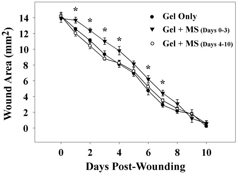
The effects of topical morphine sulfate application at different times throughout healing on cutaneous wounds were assessed using a standardized model of cutaneous wound healing in rats. A previous concentration-response study conducted in this lab demonstrated that 5 mM morphine sulfate causes a significant, but not maximal, delay in wound closure and was, therefore chosen as the concentration to be used in subsequent experiments. Topical morphine treatment on wound days 0–3 significantly delayed wound closure when compared to controls (p = 0.017). Animals treated with 5 mM morphine sulfate on days 0–3 post-wounding had significantly larger wounds on days 1, 2, 3, 4, 6, and 7 when compared to gel-only treated controls (Fig. 1). Total wound area over the complete time course of animals receiving 5 mM morphine sulfate treatments for the first 4 days was 14% larger than both gel-only treated control rats and rats treated with morphine sulfate for the last 7 days. In addition, no significant differences were seen in animals treated with 5 mM morphine sulfate on days 4–10 post-wounding when compared to gel-only treated controls. In rats treated topically with gel infused with 1 mM naltrexone, the area of the wounds treated with the opioid receptor antagonist did not differ significantly from control wounds (data not shown).
Fig. 1. Wound closure time course for rats receiving morphine sulfate-infused gel treatments.
IntraSite Gel (Smith+Nephew, Hull, United Kingdom) alone or containing 5 mM morphine sulfate (MS) was applied to the wound twice daily through wound day 10. Wound size is presented as area (mm2) mean ± SEM and was determined by analysis of digital images. Note that wounds treated with MS on wound days 0–3 were significantly larger compared to gel-only controls, while those treated with MS on days 4–10 post-wounding showed no change (n=5). * p < 0.05 comparison between morphine 0–3 group and gel-only treated controls (two-way ANOVA, Tukey’s post-hoc test).
Impact of topical morphine sulfate treatment on the infiltration of macrophages into the healing wound
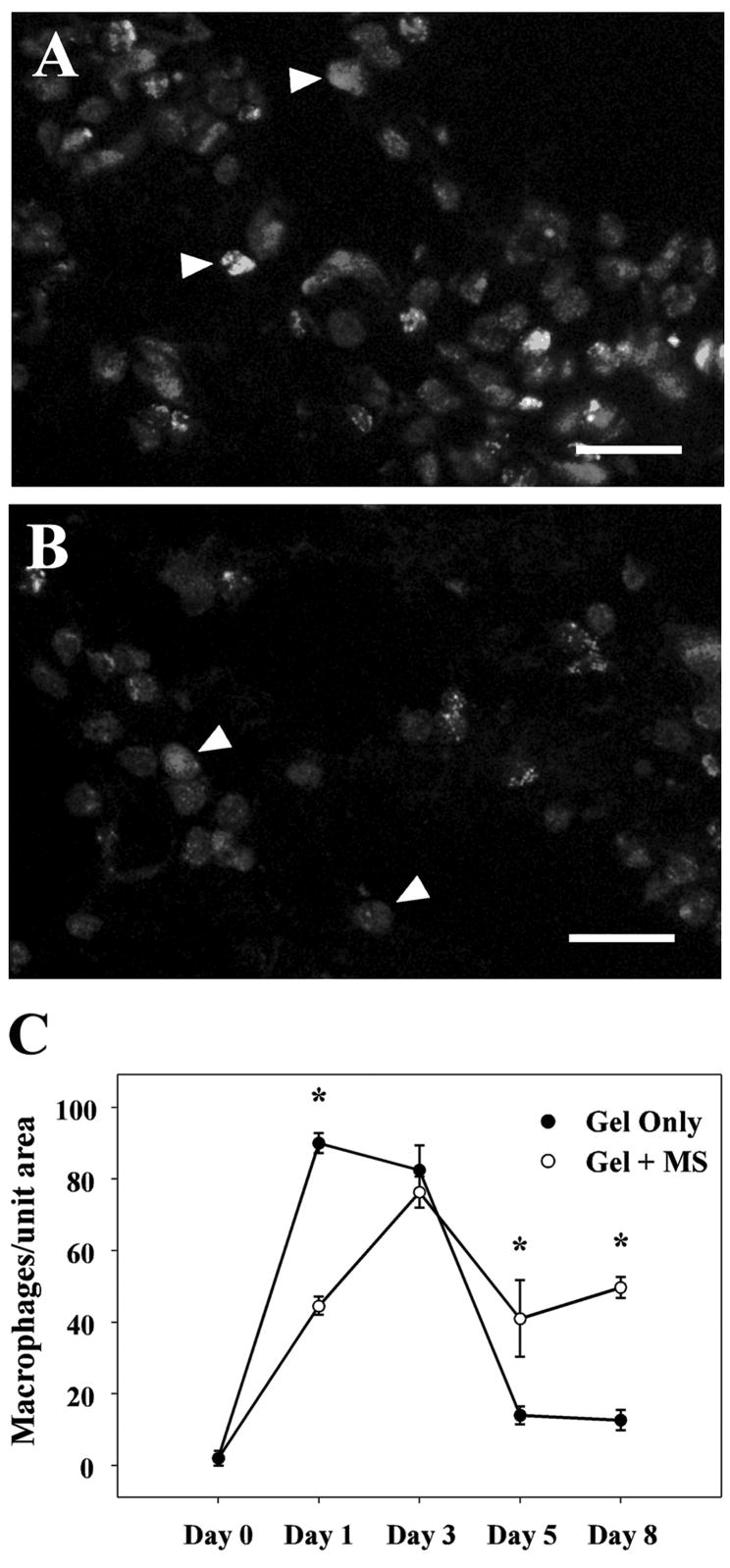
Macrophage infiltration into the closing wound was assessed with immunohistochemistry using the macrophage marker macrosialin. Very few macrosialin-ir cells were observed in control skin adjacent to the peri-wound area. Macrosialin-ir cells in gel-only treated wounds were greatest on day 1 post-wounding (Fig. 2). A slight decrease in the number of macrophages in the wound was seen on wound day 3 followed by a return to near-baseline levels on day 5 and 8 post-wounding. Appearance of macrosialin-ir cells was significantly delayed in morphine-treated wounds (p = 0.01). Macrophage concentrations in wounds treated with morphine sulfate did not peak until wound day 3. A significant decrease in macrosialin-ir cells numbers of 51% in morphine-treated wounds was observed on wound day 1 when compared to gel-only treated controls. While macrosialin-ir cell numbers declined in morphine-treated wounds on day 5 post-wounding, they were significantly higher than control levels and remained elevated through wound day 8 compared to gel-only treated controls.
Fig. 2. Effect of morphine on immunolabeled macrophages in the healing cutaneous wound.
Cutaneous wounds were treated twice daily with IntraSite Gel (Smith+Nephew, Hull, United Kingdom) alone or gel infused with 5 mM morphine sulfate (MS). (A, B) Macrosialin-ir cells (arrows) in granulation tissue of healing wounds. Fluorescence photomicrographs of gel-only (A) and morphine-treated (B) wounds were obtained 1 day post-wounding. Note a significant decrease in the number of macrophages present in morphine-treated wounds as compared to controls. Scale bar = 100 μm. (C) Time course of macrophages present in the periwound area. Data are presented as number of macrosialin-ir cells per unit area (mm2) of histological sections analyzed. Macrophage infiltration is decreased and delayed in morphine-treated wounds (n=3 or 4). * p < 0.05 comparison between morphine and gel-only treated controls (two-way ANOVA, Tukey’s post-hoc test).
Myofibroblast density in control versus morphine-treated wounds
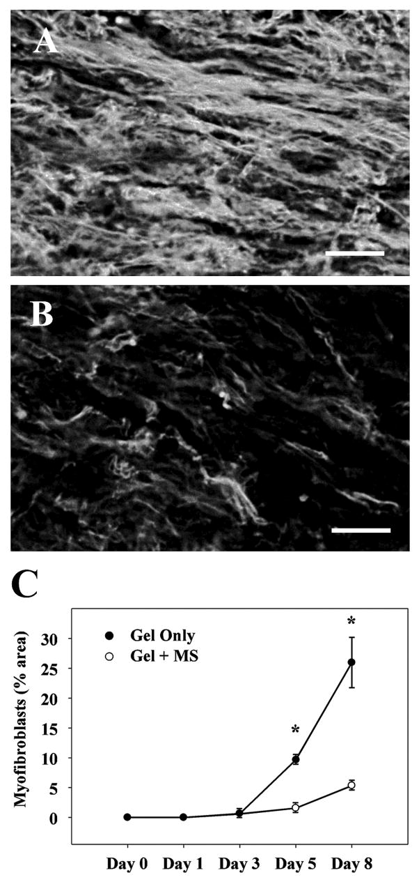
α-SMA-immunoreactivity was utilized to measure myofibroblast density in the healing wounds. α-SMA-ir cells first appear on wound day 3, although at negligible levels. In gel-only treated wounds, α-SMA-ir cell density increased rapidly at day 5 post-wounding and still further at wound day 8 (Fig. 3). However, this increase was markedly reduced in wounds treated with topical morphine sulfate (p < 0.001). α-SMA-ir cell density was significantly lower in morphine-treated wounds on days 5 and 8 post-wounding compared to controls, with an 84% and 79% reduction seen on days 5 and 8, respectively.
Fig. 3. Myofibroblast accumulation in the healing cutaneous wound.
Wounds received twice-daily treatment with IntraSite Gel (Smith+Nephew, Hull, United Kingdom) alone or gel infused with 5 mM morphine sulfate (MS). (A, B) αSMA-ir cells in granulation tissue of healing wounds 8 days following wounding. Fluorescence photomicrographs of gel-only (A) and morphine-treated (B) wounds. Note a significant decrease in myofibroblast density in morphine-treated wounds as compared to controls. Scale bar = 100 μm. (C) Time course of myofibroblasts present in the periwound area. Data are presented as a percent field area occupied by α-SMA immunoreactive cells (mm2). Myofibroblast density in wounds treated with morphine was significantly reduced compared to gel-only treated controls (n=3 or 4). * p < 0.05 comparison between morphine and gel-only treated controls (two-way ANOVA, Tukey’s post-hoc test).
Architectural changes of rat skin following topical application of morphine sulfate during wound healing
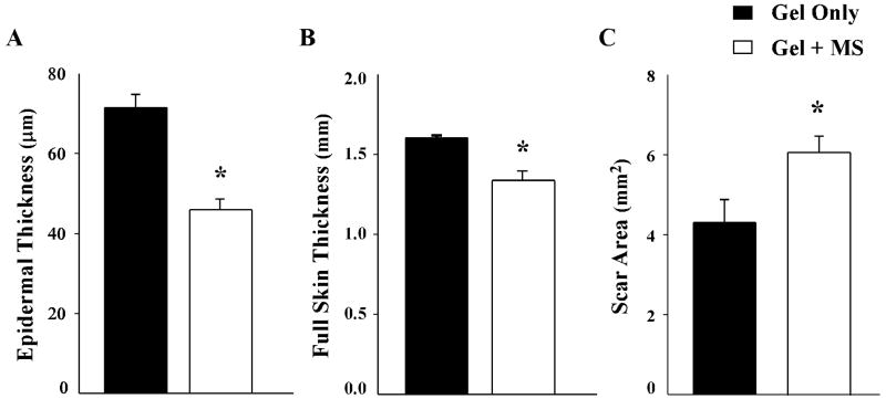
The long-term impact of topical morphine sulfate treatment during cutaneous wound healing on the structural architecture of healed skin was assessed at post-wound day 18 using measures of epidermal and dermal thickness and the extent of scar tissue formation in rats receiving topical 5 mM morphine sulfate applications days 0–18 post-wounding. Control wounds treated with gel alone had an average epidermal thickness of 71.5 μm, while the full thickness of the skin (epidermis + dermis) was 1.6 mm. Closed wounds of rats treated with topical morphine had significantly thinner (35% p = 0.019) epidermis over the wound compared to gel-only treated controls (Fig. 4A). The full-skin thickness was 19% (p = 0.014) less in closed morphine-treated wounds as well (Fig. 4B). In addition, topical morphine treatment resulted in significantly larger (41% p = 0.023, Fig. 4C) scar tissue surface area on day 18 post-wounding.
Fig. 4. Effects of topical morphine on skin architecture of fully-closed cutaneous wounds.
Rats were treated with IntraSite Gel (Smith+Nephew, Hull, United Kingdom) alone or gel infused with 5 mM morphine sulfate (MS) twice daily through wound day 18. (A) Epidermal thickness of new skin in wounds. Note that topical morphine treatment resulted in significantly thinner epidermis than that in control wounds. (B) Full thickness of the new skin covering closed cutaneous wounds following topical morphine treatment. Similarly, the full skin thickness was diminished following topical morphine administration. (C) Area of scar tissue covering closed cutaneous wounds following topical morphine treatment. Scar tissue was defined as the visibly distinct new epithelium covering the original wound site. Scar size is presented as area (mm2) mean ± SEM and was determined by analysis of digital images of closed wounds on day 18 after wounding. Treatment of the wounds with 5 mM morphine sulfate significantly increased the scar area compared to gel-only controls (n=4–7). * p < 0.05 comparison between morphine and gel-only treated controls (unpaired t test).
Discussion
Previous experimental studies in rats have demonstrated that treatment with topical morphine results in a delay in the closure of cutaneous wounds via inhibition of peripheral neuropeptide release from primary afferent neurons 18. The onset of the delay begins within the first 24 hours after wounding and continues for the first few days of healing. After approximately 4 days following wounding, wound closure accelerates with wounds ultimately closing fully at times not significantly different than controls. These results suggest a time-dependent effect of topical morphine treatment on wound closure. Therefore, these studies were designed to begin to characterize the temporal characteristics of morphine’s impact on wound healing. A standardized model of cutaneous wound healing was used that is sensitive enough to detect age-related variations in wound closure as well as the negative impact of partial sensory denervation on wound healing 17,21.
The results of the current study demonstrate that the closure of wounds treated with morphine on days 0–3 post-wounding only is nearly identical to that of wounds treated with morphine throughout the entire healing process 18, with an immediate delay in closure followed by acceleration and full closure at approximately day 10 post-wounding. However, wounds treated with morphine beginning on day 4 post-wounding and continuing through the remainder of the time course were not significantly different from controls treated with gel alone. These results demonstrate that physiological events in wound healing that facilitate morphine-induced delays in closure occur early in the healing process.
Neuropeptides have proinflammatory actions and serve as a link between the nervous and immune systems. For example, substance P promotes chemotaxis and increased function of macrophages 22, as well as cytokine release from human monocytes 23,24. Macrophages are essential for normal wound healing and are the predominant cell type in the wound in the early inflammatory phase of wound healing 25. Macrophages not only clear the wound of bacteria and cellular debris but also secrete a number of growth factors and other cytokines that induce angiogenesis and stimulate keratinocytes and fibroblasts 26–30. Accordingly, the effect of topical morphine treatment on macrophage density within the healing wound was assessed.
Activation of opioid receptors located on primary afferent neurons inhibits the antidromic release of neuropeptides from these terminals. Neuropeptides both attract and activate macrophages in states of inflammation. Therefore, decreased concentrations of neuropeptides within the periwound area may result in reduced macrophage infiltration. Results from this study demonstrate the inhibitory effect of topical morphine on macrophage infiltration into the closing wound. Macrophage density within control wounds peaked 1 day following injury. The number of macrophages in the wound began to decline on day 3 post-wounding and continued through days 5 and 8 after wounding. The number of macrophages in the wound decreased to near-baseline levels by day 5 after wounding, a time consistent with the resolution of the inflammatory phase in normal wound healing. However, macrophage migration was significantly delayed in morphine-treated wounds. In this group, macrophage numbers were significantly less on day 1 post-wounding and a maximum was not reached until wound day 3. Decreased macrophage density would result in reduced induction of the parenchymal cells essential for wound closure 29–31. Hence, the results of this study suggest that topical morphine delays wound closure, at least in part, by inhibiting the recruitment of macrophages into the closing wound. In addition to delaying macrophage infiltration, topical morphine application prolonged the presence of macrophages within the wound. Macrophage density in morphine-treated wounds was significantly higher at later time points (wound days 5 and 8) than in gel-only treated control wounds. This finding suggests that morphine inhibits resolution of the inflammatory phase, which may result in aberrant healing, propagate chronic inflammation, and result in undue tissue damage.
Wound healing is a dynamic process consisting of a complex, overlapping pattern of events that have been artificially divided into three phases: the inflammatory phase, including coagulation and inflammation; the proliferative phase, consisting of angiogenesis, the formation of granulation tissue, and reepithelialization; and the maturation phase, involving matrix formation and tissue remodeling 32. Myofibroblasts are transdifferentiated fibroblasts that express α-SMA, giving them contractile capabilities 33,34. Myofibroblasts are the predominant cell recruited by macrophages to initiate the proliferative phase of wound healing 35,36. Closure of excision wounds occurs primarily through the contractile actions of myofibroblasts. The presence of α-SMA-immunoreactive myofibroblasts appeared within the wound in this study on day 3 post-wounding in control animals. The density of myofibroblasts was significantly increased through day 8. This trend is consistent with the pattern of normal wound healing. However, the presence of myofibroblasts was drastically reduced in morphine-treated wounds, with only a small increase occurring by wound day 8. These data are consistent with the idea that topical morphine application prolongs the inflammatory phase of wound closure and concomitantly delays progression into the proliferation phase of wound healing.
Examining the effects of exogenous opioids on the closure of cutaneous wounds led us to questions whether endogenous opioids play a role in regulating their closure. Previous studies have demonstrated that endogenous opioids produced by resident immune cells produce antinociceptive effects in inflamed tissues 19,37. Accordingly, production of local endogenous opioids could impact wound closure rates. The possibility of delayed wound closure by activation of peripheral opioid receptors by endogenous opioids during normal wound closure was tested in this study when the opioid antagonist naltrexone was applied topically to wounds at a concentration of 1 mM; however, no significant differences in wound closure rates resulted from this treatment (data not shown). This suggests that while local endogenous opioids can contribute to peripheral analgesia, their actions do not significantly impact wound closure and, therefore, do not modulate the effects of topical morphine on wound closure dynamics.
Although morphine-treated wounds ultimately closed at times similar to control wounds, it was reasonable to suspect that morphine could have longer-term effects than that of delaying wound closure. Alterations in the cellular content of the healing wound or modulation of other mechanisms of wound closure by morphine could result in aberrant organization of newly-formed skin despite apparent wound closure. Evaluation of the architecture of healed wound skin on day 18 demonstrated a significant decrease in the thickness of the healed skin over the wound following morphine treatment. Topical morphine applied throughout the entire time course of wound closure also resulted in a significant increase in the residual scar area. Analysis of the myofibroblast density during the healing process revealed a significant decrease in the numbers of activated myofibroblasts in morphine-treated wounds, which supports the likelihood that a larger residual scar is formed due to the lack of contractility of the newly-formed tissue. These results demonstrate that topical morphine has lasting effects beyond the duration of its application to the healing wound and suggest that its use as a topical analgesic agent could produce persistent detrimental effects that would reduce the strength of healed skin.
Taken together, these studies provide evidence that the delays in wound closure seen with topical morphine application can be attributed to alterations in the initiation and duration of essential, early processes during wound healing. Morphine-induced delays in the onset of inflammation produce a shift in the timing of essential subsequent events (such as myofibroblast activation) in the healing process. Alterations in the temporal processes of wound healing not only results in delayed wound closure but also results in long-term architectural deficits, jeopardizing the integrity of the healed skin following topical morphine administration. Topical morphine application is most widely used clinically on wounds of elderly patients with pressure ulcers or burn patients with highly inflamed wounds. It is important to acknowledge that the current studies were conducted on young, healthy animals with aseptic wounds. Further study is required to characterize fully the effects of topical morphine on wounds with co-morbid disease states.
Acknowledgments
The authors would like to thank Michelle Winter, B.S., Research Associate, Department of Pharmacology, Toxicology and Therapeutics, University of Kansas Medical Center (Kansas City, KS, USA) and Donald Warn, Ph.D., Imaging Center Managing Director, University of Kansas Medical Center (Kansas City, KS, USA) for their expert technical assistance and the contributions of the University of Kansas Medical Center Kansas Intellectual and Developmental Disabilities Research Center core facilities.
Funding: This study was supported in part by Research Grant DA12505 from the National Institute on Drug Abuse (Bethesda, MD, USA), a Basic Science Research Pilot Grant from the Lied Foundation/University of Kansas Medical Center Research Institute (Kansas City, KS, USA) and a Predoctoral Training award from the University of Kansas Medical Center Biomedical Research Training Program (Kansas City, KS, USA).
Footnotes
Work is attributed to the Department of Pharmacology, Toxicology and Therapeutics, University of Kansas Medical Center (Kansas City, KS, USA)
Summary Statement: Alterations in the temporal processes of wound healing not only result in delayed wound closure but also produce long-term architectural deficits, jeopardizing the integrity of the healed skin following topical morphine administration.
References
- 1.Janson W, Stein C. Peripheral opioid analgesia. Curr Pharm Biotechnol. 2003;4:270–4. doi: 10.2174/1389201033489766. [DOI] [PubMed] [Google Scholar]
- 2.Oeltjenbruns J, Schafer M. Peripheral opioid analgesia: clinical applications. Curr Pain Headache Rep. 2005;9:36–44. doi: 10.1007/s11916-005-0073-9. [DOI] [PubMed] [Google Scholar]
- 3.Stein C. Peripheral mechanisms of opioid analgesia. Anesth Analg. 1993;76:182–91. doi: 10.1213/00000539-199301000-00031. [DOI] [PubMed] [Google Scholar]
- 4.Long TD, Cathers TA, Twillman R, O’Donnell T, Garrigues N, Jones T. Morphine-Infused silver sulfadiazine (MISS) cream for burn analgesia: a pilot study. J Burn Care Rehabil. 2001;22:118–23. doi: 10.1097/00004630-200103000-00006. [DOI] [PubMed] [Google Scholar]
- 5.Tran QN, Fancher T. Achieving analgesia for painful ulcers using topically applied morphine gel. J Support Oncol. 2007;5:289–93. [PubMed] [Google Scholar]
- 6.Zeppetella G, Paul J, Ribeiro MD. Analgesic efficacy of morphine applied topically to painful ulcers. J Pain Symptom Manage. 2003;25:555–8. doi: 10.1016/s0885-3924(03)00146-5. [DOI] [PubMed] [Google Scholar]
- 7.Jin S, Lei L, Wang Y, Da D, Zhao Z. Endomorphin-1 reduces carrageenan-induced fos expression in the rat spinal dorsal horn. Neuropeptides. 1999;33:281–4. doi: 10.1054/npep.1999.0040. [DOI] [PubMed] [Google Scholar]
- 8.Khalil Z, Sanderson K, Modig M, Nyberg F. Modulation of peripheral inflammation by locally administered endomorphin-1. Inflamm Res. 1999;48:550–6. doi: 10.1007/s000110050502. [DOI] [PubMed] [Google Scholar]
- 9.Dinda A, Gitman M, Singhal PC. Immunomodulatory effect of morphine: therapeutic implications. Expert Opin Drug Saf. 2005;4:669–75. doi: 10.1517/14740338.4.4.669. [DOI] [PubMed] [Google Scholar]
- 10.Schroeder JE, Fischbach PS, Zheng D, McCleskey EW. Activation of mu opioid receptors inhibits transient high- and low-threshold Ca2+ currents, but spares a sustained current. Neuron. 1991;6:13–20. doi: 10.1016/0896-6273(91)90117-i. [DOI] [PubMed] [Google Scholar]
- 11.Delgado AV, McManus AT, Chambers JP. Exogenous administration of Substance P enhances wound healing in a novel skin-injury model. Exp Biol Med (Maywood) 2005;230:271–80. doi: 10.1177/153537020523000407. [DOI] [PubMed] [Google Scholar]
- 12.Engin C. Effects of calcitonin gene-related peptide on wound contraction in denervated and normal rat skin: a preliminary report. Plast Reconstr Surg. 1998;101:1887–90. doi: 10.1097/00006534-199806000-00017. [DOI] [PubMed] [Google Scholar]
- 13.Kjartansson J, Dalsgaard CJ. Calcitonin gene-related peptide increases survival of a musculocutaneous critical flap in the rat. Eur J Pharmacol. 1987;142:355–8. doi: 10.1016/0014-2999(87)90073-2. [DOI] [PubMed] [Google Scholar]
- 14.Khalil Z, Helme R. Sensory peptides as neuromodulators of wound healing in aged rats. J Gerontol A Biol Sci Med Sci. 1996;51:B354–61. doi: 10.1093/gerona/51a.5.b354. [DOI] [PubMed] [Google Scholar]
- 15.Kjartansson J, Dalsgaard CJ, Jonsson CE. Decreased survival of experimental critical flaps in rats after sensory denervation with capsaicin. Plast Reconstr Surg. 1987;79:218–21. doi: 10.1097/00006534-198702000-00012. [DOI] [PubMed] [Google Scholar]
- 16.Peskar BM, Lambrecht N, Stroff T, Respondek M, Muller KM. Functional ablation of sensory neurons impairs healing of acute gastric mucosal damage in rats. Dig Dis Sci. 1995;40:2460–4. doi: 10.1007/BF02063255. [DOI] [PubMed] [Google Scholar]
- 17.Smith PG, Liu M. Impaired cutaneous wound healing after sensory denervation in developing rats: effects on cell proliferation and apoptosis. Cell Tissue Res. 2002;307:281–91. doi: 10.1007/s00441-001-0477-8. [DOI] [PubMed] [Google Scholar]
- 18.Rook JM, McCarson KE. Delay of cutaneous wound closure by morphine via local blockade of peripheral tachykinin release. Biochem Pharmacol. 2007;74:752–7. doi: 10.1016/j.bcp.2007.06.005. [DOI] [PMC free article] [PubMed] [Google Scholar]
- 19.Stein C, Hassan AH, Przewlocki R, Gramsch C, Peter K, Herz A. Opioids from immunocytes interact with receptors on sensory nerves to inhibit nociception in inflammation. Proc Natl Acad Sci U S A. 1990;87:5935–9. doi: 10.1073/pnas.87.15.5935. [DOI] [PMC free article] [PubMed] [Google Scholar]
- 20.Labuz D, Berger S, Mousa SA, Zollner C, Rittner HL, Shaqura MA, Segovia-Silvestre T, Przewlocka B, Stein C, Machelska H. Peripheral antinociceptive effects of exogenous and immune cell-derived endomorphins in prolonged inflammatory pain. J Neurosci. 2006;26:4350–8. doi: 10.1523/JNEUROSCI.4349-05.2006. [DOI] [PMC free article] [PubMed] [Google Scholar]
- 21.Liu M, Warn JD, Fan Q, Smith PG. Relationships between nerves and myofibroblasts during cutaneous wound healing in the developing rat. Cell Tissue Res. 1999;297:423–33. doi: 10.1007/s004410051369. [DOI] [PubMed] [Google Scholar]
- 22.Hartung HP, Wolters K, Toyka KV. Substance P: binding properties and studies on cellular responses in guinea pig macrophages. J Immunol. 1986;136:3856–63. [PubMed] [Google Scholar]
- 23.Lotz M, Vaughan JH, Carson DA. Effect of neuropeptides on production of inflammatory cytokines by human monocytes. Science. 1988;241:1218–21. doi: 10.1126/science.2457950. [DOI] [PubMed] [Google Scholar]
- 24.Rameshwar P, Ganea D, Gascon P. Induction of IL-3 and granulocyte-macrophage colony-stimulating factor by substance P in bone marrow cells is partially mediated through the release of IL-1 and IL-6. J Immunol. 1994;152:4044–54. [PubMed] [Google Scholar]
- 25.DiPietro LA. Wound healing: the role of the macrophage and other immune cells. Shock. 1995;4:233–40. [PubMed] [Google Scholar]
- 26.Belperio JA, Keane MP, Arenberg DA, Addison CL, Ehlert JE, Burdick MD, Strieter RM. CXC chemokines in angiogenesis. J Leukoc Biol. 2000;68:1–8. [PubMed] [Google Scholar]
- 27.Werner S, Grose R. Regulation of wound healing by growth factors and cytokines. Physiol Rev. 2003;83:835–70. doi: 10.1152/physrev.2003.83.3.835. [DOI] [PubMed] [Google Scholar]
- 28.Sunderkotter C, Steinbrink K, Goebeler M, Bhardwaj R, Sorg C. Macrophages and angiogenesis. J Leukoc Biol. 1994;55:410–22. doi: 10.1002/jlb.55.3.410. [DOI] [PubMed] [Google Scholar]
- 29.Gharaee-Kermani M, Denholm EM, Phan SH. Costimulation of fibroblast collagen and transforming growth factor beta1 gene expression by monocyte chemoattractant protein-1 via specific receptors. J Biol Chem. 1996;271:17779–84. doi: 10.1074/jbc.271.30.17779. [DOI] [PubMed] [Google Scholar]
- 30.Yamamoto T, Eckes B, Mauch C, Hartmann K, Krieg T. Monocyte chemoattractant protein-1 enhances gene expression and synthesis of matrix metalloproteinase-1 in human fibroblasts by an autocrine IL-1 alpha loop. J Immunol. 2000;164:6174–9. doi: 10.4049/jimmunol.164.12.6174. [DOI] [PubMed] [Google Scholar]
- 31.Diegelmann RF, Schuller-Levis G, Cohen IK, Kaplan AM. Identification of a low molecular weight, macrophage-derived chemotactic factor for fibroblasts. Clin Immunol Immunopathol. 1986;41:331–41. doi: 10.1016/0090-1229(86)90004-8. [DOI] [PubMed] [Google Scholar]
- 32.Baum CL, Arpey CJ. Normal cutaneous wound healing: clinical correlation with cellular and molecular events. Dermatol Surg. 2005;31:674–86. doi: 10.1111/j.1524-4725.2005.31612. [DOI] [PubMed] [Google Scholar]
- 33.Gabbiani G. The biology of the myofibroblast. Kidney Int. 1992;41:530–2. doi: 10.1038/ki.1992.75. [DOI] [PubMed] [Google Scholar]
- 34.Gabbiani G, Ryan GB, Majne G. Presence of modified fibroblasts in granulation tissue and their possible role in wound contraction. Experientia. 1971;27:549–50. doi: 10.1007/BF02147594. [DOI] [PubMed] [Google Scholar]
- 35.Lawrence WT. Physiology of the acute wound. Clin Plast Surg. 1998;25:321–40. [PubMed] [Google Scholar]
- 36.Ross R, Everett NB, Tyler R. Wound healing and collagen formation. VI The origin of the wound fibroblast studied in parabiosis. J Cell Biol. 1970;44:645–54. doi: 10.1083/jcb.44.3.645. [DOI] [PMC free article] [PubMed] [Google Scholar]
- 37.Przewlocki R, Hassan AH, Lason W, Epplen C, Herz A, Stein C. Gene expression and localization of opioid peptides in immune cells of inflamed tissue: functional role in antinociception. Neuroscience. 1992;48:491–500. doi: 10.1016/0306-4522(92)90509-z. [DOI] [PubMed] [Google Scholar]


