Abstract
Basal thumb arthritis is a common condition seen in hand clinics across the United Kingdom and is often associated with other pathological conditions such as carpal tunnel syndrome and scaphotrapezial arthritis. Typically, patients complain of pain localised to the base of the thumb. This pain is often activity related, particularly after excessive use involving forceful pinch. A detailed history and examination is normally all that is needed to make the diagnosis. Provocative manoeuvres may be helpful in localising symptoms to the basal joint with degenerative changes or synovitis. Radiographs are useful for confirming the diagnosis and staging the disease in order to plan for surgery. The mainstay of initial treatment of basal thumb arthritis of any stage is activity modifications, rest, nonsteroidal anti‐inflammatory drugs, exercises and splinting. A variety of surgical procedures are available to treat the condition when conservative measures have failed, in order to control symptoms and improve function. We review the current literature and discuss the clinical aspects of this condition, staging, and treatment options available, and the difficulties treating this group of patients.
Keywords: basal, first carpometacarpal, trapeziometacarpal, thumb, arthritis
The carpometacarpal (CMC) or trapeziometacarpal joint of the thumb is known as nature's universal joint. It is a saddle‐shaped joint and allows a wide range of movement in three planes. This increase in mobility, however, renders the joint prone to degeneration. Basal thumb arthritis is a common problem that presents to hand clinics all over the UK. A comprehensive history and clinical examination is all that is needed to diagnose this condition. Most patients, regardless of the severity of the disease, will benefit from an initial period of conservative management. If this fails, a wide variety of surgical options are available that, irrespective of the technique, deliver pain relief, improved strength and therefore better function. We discuss the clinical aspects of this condition, staging, treatment options available and the difficulties treating this group of patients.
Methods
We used Medline (1980 to May 2006) to conduct a literature search of the Cochrane Database of Systematic Reviews on the Cochrane Library, issue 1, 2006, and we searched citation lists of relevant publications, including review articles. We used the following search headings and keywords: basal, thumb, arthritis, carpometacarpal and trapeziometacarpal. Publications were selected mostly from the past 10 years but did not exclude commonly referenced and highly regarded older publications.
Epidemiology
The prevalence of basal thumb arthritis increases with age and is seen predominantly in postmenopausal women.1,2 The female:male ratio is 6:1. No conclusive environmental or genetic factors that lead to the expression of this disease have been identified.
Aetiology
We found a strong association between excessive basal joint laxity and the development of premature degenerative changes.3,4,5 Ligamentous laxity is common in young women. Repeated loading of subluxed joints in the 20s, 30s and 40s may be the reason why one third of women >50 years show some radiological evidence of degenerative change.6
Previous trauma to the thumb can predispose the patient to arthrosis of the basal joint.
Fractures that involve the articular base of the metacarpal are commonly known as Rolando or Bennett fractures, and, if left to heal in a malunited position, can lead to degeneration.7,8
Inflammatory arthritis, such as rheumatoid arthritis, is another predisposing condition. The most common site of upper extremity involvement in the population with rheumatoid arthritis is the distal radioulnar joint, and one third of these patients will have basal joint involvement.9,10
The most common cause for symptomatic basal joint arthritis, however, is idiopathic.11,12
Symptoms and signs
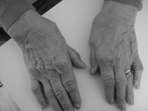
Typically, patients have pain localised to the base of the thumb, often radiating to the thenar eminence and the metacarpophalangeal (MCP) joint.13 The pain in basal thumb arthritis is often activity related, particularly after excessive use involving forceful pinch. Daily activities such as grasping large objects, unscrewing jar tops, turning doorknobs and writing can exacerbate symptoms.14,15 As the arthritis progresses, pain may become constant. In addition to pain, weakness or loss of motion are commonly reported.12,16 These symptoms are a protective mechanism to prevent excessive use of a damaged joint. In the later stages of the disease when instability and subluxation of the joint occurs, patients report an inability to abduct the thumb, weakness of pinch grip and a deterioration of hand function.17,18 As a result, the thumb cannot be brought far enough away from the index finger to grasp large objects. Collapse of the thumb may eventually occur, producing a swan‐neck deformity with progressive adduction of the first metacarpal and compensatory hyperextension of the MCP joint (fig 1).
Figure 1 Clinical deformity of basal thumb arthritis. Patient consent was obtained for publication of figures.
Associated conditions
Other conditions may also be associated with basal thumb arthritis, such as carpal tunnel syndrome, scaphotrapezial arthrosis, trigger digits, MCP hyperextension and tenosynovitis of the wrist.16 Florack et al19 noted that 43% of their patients who underwent surgery for arthritis to the base of the thumb had carpal tunnel syndrome. The prevalence of compression neuropathy was found to be higher in women and in patients with diabetes. The basal joint is in close proximity to the carpal tunnel and hence inflammation in one site can spread to a neighbouring site. In addition, the skeletal changes that occur at the basal joint can affect the dimensions of the carpal tunnel, leading to reduced volume for the median nerve.16 It is therefore important to inquire about these conditions to make a comprehensive diagnosis and tailor the treatment accordingly.
How is this condition diagnosed?
A detailed history and clinical examination is all that is normally needed to clinch the diagnosis. Radiographs are helpful in staging the disease and for patient education, as discussed in the next section. Nerve conduction studies are recommended, if there are relevant sensory problems.20
Inspection of the hand of a patient with advanced disease may show dorsoradial prominence of the thumb metacarpal base. This may be due to a combination of subluxation, joint inflammation and osteophyte formation.21,22
Tenderness is usually well localised over the joint, and this can be reproduced with thumb and finger pressure applied directly over the affected joint. Crepitus evident on examination implies erosion of the articular cartilage.21,22
Provocative manoeuvres may be helpful in localising symptoms to the basal joint with degenerative changes or synovitis. The“grind” test is performed by rotating the thumb metacarpal base while applying axial compression (ie, compression along the plane of the metacarpal bone).12 When the test is positive, the resulting pain is suggestive of degenerative disease. The “distraction” or “torque” test is performed by rotating the thumb metacarpal base while applying gentle axial traction. A positive test is indicative of synovitis associated with milder disease as a result of traction on an inflamed joint capsule.12,21,22 These tests, however, are more commonly used in a specialist hand surgery setting.
Radiological examination
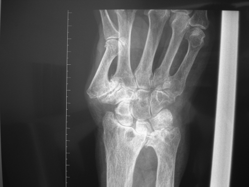
Radiographs of the thumb in three planes are helpful in confirming the diagnosis (fig 2). The standard views will rule out other osseous abnormalities and areas of arthritis.23 Basal joint stress views can also be useful in assessing the laxity of the basal joint.23 This involves a postero‐anterior view of both thumb trapeziometacarpal joints taken simultaneously as the patient pushes the radial aspect of one thumb tip against the other forcefully. This tends to cause the base of the metacarpal to sublux laterally if the joint is lax. There is no indication for magnetic resonance imaging, tomography or ultrasonography in the routine evaluation of basal joint disease.24
Figure 2 Radiographic changes of stage IV basal thumb arthritis.
Eaton and Glickel13 have described a method of staging pathological changes in the basal joint on the basis of the appearance of standard radiographic and stress views. Table 1 shows the radiographic changes which are divided into four stages. This radiographic staging system is helpful in preoperative planning and patient education.
Table 1 Classification system for basal joint arthritis of the thumb.
| Stage | Radiographic changes |
|---|---|
| I | Normal TM joint contours with <1/3 joint subluxation |
| II | TM joint narrowing and osteophytes or loose bodies <2 mm. Instability apparent on stress views. ⩾1/3 joint subluxation |
| III | TM joint narrowing, subchondral sclerosis, osteophytes or loose bodies >2 mm. ⩾1/3 joint subluxation |
| IV | Advanced disease of TM and scaphotrapezial joint |
TM, trapeziometacarpal.
Treatment options
Conservative
The mainstay of initial treatment of basal thumb arthritis of any stage is activity modifications, rest, non‐steroidal anti‐inflammatory drugs, and thenar intrinsic and extrinsic muscle strengthening exercises.25
Examples of activity modification include less forceful pinching, alternate hand use and switching to larger‐diameter writing instruments.
The hand is rested by immobilising the base of the thumb in a splint.26 Patients may already be wearing a splint obtained from a chemist, which is often a standard wrist immobiliser that leaves the thumb free to move, thereby exacerbating the symptoms. The most effective splint for immobilising and resting the basal joint is a well‐fitted custom‐made splint fashioned by a qualified hand therapist. This is usually in the form of a long opponens or thumb spica splint.26 Initially, splinting is usually continuous for 3 or 4 weeks, and in combination with non‐steroidal anti‐inflammatory drugs. If there is improvement after this, the splint can be worn during the day and the patients gradually wean themselves from the splint over the course of 1 month, or make intermittent use thereafter when the thumb is symptomatic or under heavy load. If there is no improvement of symptoms after 2 months, surgical options can be discussed.24,26
Corticosteroid injections of the trapeziometacarpal joint can be useful when conservative measures have not worked.20,22 Pain relief can be effective and last from a few days to several months. Repetitive injections, however, have a diminishing effect and can theoretically weaken the capsular support of the joint and further compromise the articular cartilage.22,27
Box 1 gives a treatment protocol that can be used as a guide in treating patients with basal thumb arthritis. It is important to realise, however, that there is a poor correlation between radiographic severity of degenerative disease and clinical symptomatology.12,13,22 Hence, patients with minimal joint space narrowing may have disabling pain, and conversely asymptomatic patients with stage IV disease are diagnosed incidentally on radiographic studies.
Recently, numerous studies have investigated the potential role of chondroprotective agents in repairing articular cartilage and decelerating the degenerative process. The reports of limited clinical experience with two of these agents, glucosamine and chondroitin sulphate, as well as the accompanying publicity in the popular media, have generated controversy.28 The authors think that if patients do not wish to pursue potential surgical options, and after all the best available medical treatments have been considered, then it is not unreasonable for patients to try other complementary treatments to control their symptoms of pain. In the meantime, patients should be cautioned by their doctors as to the lack of information on these forms of drugs.
Surgical
The indications for surgery in patients with basal joint arthritis are persistent pain and functional disability after failed conservative treatment in the compliant patient.24,25 Staging can then provide a rationale for the selection of reconstructive surgical options.13
Various procedures are available to a hand surgeon. These range from soft tissue procedures in the early stage of disease before joint destruction23 to excision of the trapezium for advanced stages of the disease.29 The choice of procedure is often tailor made to suit the patients' symptoms and expectations.
Conclusion
Basal thumb arthritis is a common condition seen in hand clinics across the UK, and is often associated with other pathological conditions such as carpal tunnel syndrome and scaphotrapezial arthritis. A detailed history and examination is necessary and is normally all that is needed to make the diagnosis. Radiographs are useful for confirming the diagnosis and staging the disease to plan for surgery.
All patients presenting with this condition should have a period of conservative management before pursuing surgical options. A variety of surgical procedures are available to treat the condition when conservative measures have failed, to control symptoms and improve hand function.
Box 1: A treatment protocol used in treatment of basal thumb arthritis
Early disease (stage I) with intermittent symptoms and minimal restriction of activities of daily living and leisure
Activity modification
Splintage
Non‐steroidal anti‐inflammatory drugs (NSAIDs)
Intermediate disease (stage II or III) with restriction of activities of daily living and leisure
Splintage
NSAIDs
Corticosteroid injections
Surgery
Advanced disease (stage IV) with marked restriction of activities of daily living and leisure, continuous use of analgesia and repeated consultations
Trial period of splintage, NSAIDs and steroid injections
Surgery
Key references
Pellegrini VD Jr. The basal articulations of the thumb: pain, instability, and osteoarthritis. In: Peimer CA, ed. Surgery of the hand and upper extremity. New York: McGraw‐Hill, 1996:1019–39.
Burton RI. Basal joint arthrosis of the thumb. Orthop Clin North Am 1973;4:331–48.
Pellegrini VD Jr. Osteoarthritis at the base of the thumb. Orthop Clin North Am 1992;23:83–102.
Glickel SZ. Clinical assessment of the thumb trapeziometacarpal joint. Hand Clin 2001;17:185–95.
Barron OA, Glickel SZ, Eaton RG. Basal joint arthritis of the thumb. J Am Acad Orthop Surg 2000;8:314–23.
Multiple‐choice questions (true (T)/false (F)); answers at the end of references
Arthritis of the carpometacarpal (CMC) joint is a common condition affecting predominantly men aged >50 years.
CMC joint arthritis is often associated with other conditions such as carpal tunnel syndrome.
The range of motion and the compressive forces acting across the CMC joint predispose it to degeneration.
The diagnosis of CMC joint arthritis is made radiologically.
The indication for surgery is a patient with stage IV disease.
Abbreviations
CMC - carpometacarpal
MCP - metacarpophalangeal
Answer
1. F; 2. T; 3. T; 4. F; 5. F.
Footnotes
Competing interests: None.
References
- 1.Armstrong A L, Hunter J B, Davis T R C. The prevalence of degenerative arthritis of the base of the thumb in post‐menopausal women. J Hand Surg [Br] 199419340–341. [DOI] [PubMed] [Google Scholar]
- 2.Xu L, Strauch R J, Atheshian G A.et al Topography of the osteoarthritic thumb carpometacarpal joint and its variations with regard to gender age and osteoarthritic stage. J Hand Surg [Am] 199823454–464. [DOI] [PubMed] [Google Scholar]
- 3.Eaton R G, Lane L B, Littler J W.et al Ligament reconstruction for the painful thumb carpometacarpal joint: a long‐term assessment. J Hand Surg [Am] 19849692–699. [DOI] [PubMed] [Google Scholar]
- 4.Eaton R G, Littler J W. Ligament reconstruction for the painful thumb carpometacarpal joint. J Bone Joint Surg [Am] 1973551755–2666. [PubMed] [Google Scholar]
- 5.Pellegrini V D., Jr The basal articulations of the thumb: pain, instability, and osteoarthritis. In: Peimer CA, ed. Surgery of the hand and upper extremity. New York: McGraw‐Hill, 19961019–1039.
- 6.Kirk J A, Ansell B M, Bywaters E G L. The hypermobility syndrome: musculoskeletal complaints associated with generalized joint hypermobility. Ann Rheum Dis 196726419–425. [DOI] [PMC free article] [PubMed] [Google Scholar]
- 7.Cannon R, Dowd G, William D.et al A long‐term study following Bennett's fracture. J Hand Surg [Br] 198611426–431. [DOI] [PubMed] [Google Scholar]
- 8.Griffiths J. Fractures at the base of the first metacarpal bone. J Bone Joint Surg [Br] 196446712–719. [PubMed] [Google Scholar]
- 9.Papp S R, Athwal G S, Pichora D R. The rheumatoid wrist. J Am Acad Orthop Surg 20061465–77. [DOI] [PubMed] [Google Scholar]
- 10.Taleisnik J.The wrist. New York: Churchill Livingstone, 1985421–436.
- 11.Pellegrini V D., Jr Osteoarthritis of the trapeziometacarpal joint: the pathophysiology of articular cartilage degeneration. I. Anatomy and pathology of the aging joint. J Hand Surg 199116967–974. [DOI] [PubMed] [Google Scholar]
- 12.Burton R I. Basal joint arthrosis of the thumb. Orthop Clin North Am 19734331–348. [PubMed] [Google Scholar]
- 13.Eaton R G, Glickel S Z. Trapeziometacarpal osteoarthritis: staging as a rationale for treatment. Hand Clin 19873455–469. [PubMed] [Google Scholar]
- 14.Lasserre C, Pauzat D, Derennes R. Osteoarthritis of the trapeziometacarpal joint. J Bone Joint Surg [Br] 194931534–536. [PubMed] [Google Scholar]
- 15.Marmor L, Peter J. Osteoarthritis of the carpometacarpal joint of the thumb. Am J Surg 1969117632–636. [DOI] [PubMed] [Google Scholar]
- 16.Melone C P, Beavers B, Isani A. The basal joint pain syndrome. Clin Orthop Relat Res 198722058–67. [PubMed] [Google Scholar]
- 17.Burton R I. Basal joint implant arthroplasty in osteoarthritis. Indications, techniques, pitfalls, and problems. Hand Clin 19873473–487. [PubMed] [Google Scholar]
- 18.Swanson A B. Disabling arthritis at the base of the thumb: treatment by resection of the trapezium and flexible (sillicone) implant arthroplasty. J Bone Joint Surg [Am] 197254456–471. [PubMed] [Google Scholar]
- 19.Florack T M, Miller R J, Pelligrini V D.et al The prevalence of carpal tunnel syndrome in patients with basal joint arthritis of the thumb. J Hand Surg [Am] 199217624–630. [DOI] [PubMed] [Google Scholar]
- 20.Pellegrini V D., Jr Osteoarthritis at the base of the thumb. Orthop Clin North Am 19922383–102. [PubMed] [Google Scholar]
- 21.Pomerance J F. Painful basal joint arthritis of the thumb. Part I: anatomy, pathophysiology, and diagnosis. Am J Orthop 199524401–408. [PubMed] [Google Scholar]
- 22.Glickel S Z. Clinical assessment of the thumb trapeziometacarpal joint. Hand Clin 200117185–195. [PubMed] [Google Scholar]
- 23.Eaton R G, Littler J W. Ligament reconstruction for the painful thumb carpometacarpal joint. J Bone Joint Surg [Am] 1973551655–1666. [PubMed] [Google Scholar]
- 24.Barron O A, Glickel S Z, Eaton R G. Basal joint arthritis of the thumb. J Am Acad Orthop Surg 20008314–323. [DOI] [PubMed] [Google Scholar]
- 25.Pomerance J F. Painful basal joint arthritis of the thumb. Part II: treatment. Am J Orthop 199524466–472. [PubMed] [Google Scholar]
- 26.Swigart C R, Eaton R G, Glickel S Z.et al Splinting in the treatment of arthritis of the first carpometacarpal joint. J Hand Surg 19992486–91. [DOI] [PubMed] [Google Scholar]
- 27.Burton R I, Pelligrini V D. Surgical management of basal joint arthritis of the thumb. Part II: ligament reconstruction with tendon interposition arthroplasty. J Hand Surg [Am] 198611324–332. [DOI] [PubMed] [Google Scholar]
- 28.Towheed T E, Maxwell L, Anastassiades TP et a l. Glucosamine therapy for treating osteoarthritis. Cochrane Database Syst Rev 2005(2)CD002946. [DOI] [PMC free article] [PubMed]
- 29.Gervis W H. Excision of the trapezium for osteoarthritis of the trapeziometacarpal joint. J Bone Joint Surg [Br] 194931537–539. [PubMed] [Google Scholar]


