Abstract
Patients commonly present to their general practitioner with swellings of the hand. These include a variety of diagnoses with certain lesions (for example, ganglion) being more common than others. Some may even be familiar as they are often site specific. For those that are not routinely seen, or for those that look suspicious, referral to a hand surgeon is usually customary. This article aims to provide general practitioners with clear and concise information and terms of reference on the common hand swellings that they may encounter.
Swellings of the hand are commonly encountered in a general practice setting and include a multitude of diagnoses. They may arise from any tissue in the hand including skin, subcutaneous fat, muscle, nerves, vessels, tendon, bone and cartilage. Fortunately, most are benign, asymptomatic and may not require surgical intervention. Ganglions, epidermoid inclusion cysts, giant cell tumours of the tendon sheath, and swellings associated with arthropathy comprise the majority of lesions. These are usually familiar to the general practitioner as they are often site‐specific, making the diagnosis straightforward in most cases. However, a malignant swelling may have a benign appearance and growth behaviour that belies its true aggressiveness, and a missed diagnosis can have a disastrous consequence.
This article presents the characteristics of a variety of hand swellings in terms of presentation, natural history and management. If the diagnosis is in doubt, or malignancy is suspected, early referral is warranted.
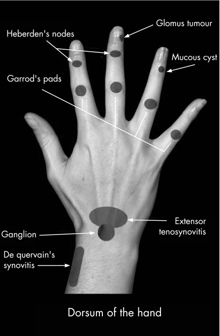
SWELLINGS TO THE DORSAL SURFACE OF THE WRIST AND HAND (FIG 1)
Figure 1 Common swellings found on the dorsum of the hand and wrist.
Ganglion
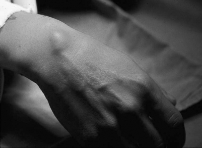
Ganglia are the most common swellings encountered in the hand. They are found in relation to joints and tendons in various parts of the body and approximately two thirds of wrist ganglia lie on the dorsal surface. They usually lie over the scapho‐lunate ligament, just distal to the radius (fig 2). The ganglion is more evident with the wrist in mild palmar flexion. The swelling is not usually lobulated unless the ganglion is large. There may be a history of the swelling varying in size and the lesion will trans‐illuminate if sufficiently close to the surface. Reassurance and, if necessary, aspiration is the treatment of choice. Surgery has a high recurrence rate (one third of cases) and spontaneous resolution is common if conservative treatment is applied.
Figure 2 A ganglion located on the dorsum of the wrist.
Rheumatoid extensor tenosynovitis
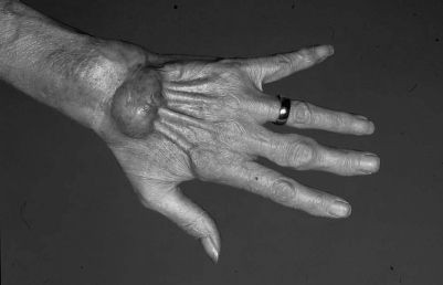
In the established case, rheumatoid tenosynovitis is usually obvious with the presence of other stigmata of the condition. However, synovitis under the extensor retinaculum can easily be mistaken for a dorsal ganglion as the swelling lies in a similar position (though it is commonly broader and flatter). Its relationship to the extensor tendons can often be demonstrated by the “Tuck” sign (fig 3). Maximal digital extension produces an infolding of the swelling at its distal edge, the site where the tenosynovium is attached to the extensor tendons. The opportunity for histological confirmation of an early rheumatoid process should not be missed.
Figure 3 Extensor tenosynovitis of the hand.
Metacarpal bossing
This is a small bony lump (usually at the second or third carpometacarpal joint) that lies somewhat distal and radial to the classic site for a ganglion. This commonly arises following trauma to the hand but may also occur spontaneously. The development of the lesion may be associated with a period of local discomfort and the digital extensor tendons may “snap” over the lump. Reassurance is preferred to surgery unless persistent pain or clicking occurs.
De Quervain's syndrome (stenosing tenovaginitis)
This condition presents with pain and thickening to the first extensor compartment which often produces a fusiform swelling over the radial styloid. Occasionally crepitus may be present on movement of the thumb. Resisted extension to the thumb usually provokes pain and is probably a more reliable provocative test than the use of Finkelstein's manoeuvre. The latter involves flexing the thumb across the palm and deviating the wrist ulnarly. The manoeuvre often causes discomfort to a normal wrist and it is therefore wise to compare the unaffected side. Finklestein's test is not specific for De Quervain's syndrome and it is commonly positive in patients with osteoarthritis at the base of the thumb or patients with non‐union of the scaphoid.
De Quervain's syndrome may result from unaccustomed vigorous activity (in radial or ulnar deviation) and rest in a splint for three weeks usually leads to a resolution of symptoms. A steroid injection is often curative but surgical decompression of the first extensor compartment may be required in resistant cases.
Keratoacanthoma
Patients, usually the elderly, present with a rapidly growing dome‐shaped swelling with a central keratin filled crater. It is often found on sun exposed areas such as the dorsum of the hands. The lesion is thought to arise from hair follicle cells. The natural history is for spontaneous regression (after 3–6 months) and, if left untreated, it leaves a depressed scar. Excision is preferred to confirm the diagnosis and to exclude malignancy.
Squamous cell carcinoma
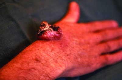
These common lesions occur mainly in older age groups, in areas of sun damaged skin (actinic keratoses) and also in the immunosuppressed. Classically, patients present with an enlarging keratotic nodule with an everted edge, usually on the dorsum of the hand (fig 4). Such lesions may be mistaken for a keratoacanthoma. Ulceration may also be present. They are painless unless invasion of the deeper structures has occurred. Bleeding or a non‐healing wound are often the reason for patients attending the practice surgery. Lymph nodes may be palpable either as a result of secondary spread or because of infection. Excision is the treatment of choice.
Figure 4 Squamous cell carcinoma.
Malignant melanoma
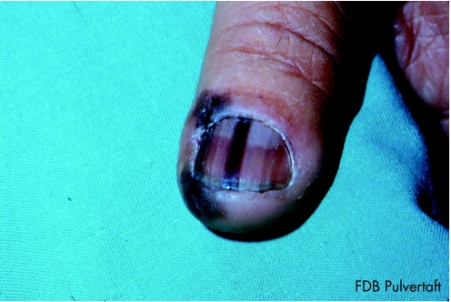
Changes in a pre‐existing pigmented lesion, whether it be a change in colour, size, outline or be associated with local itching, ulceration and bleeding, should arouse suspicion of a malignant melanoma. The tumour is not often seen in the hand but a delay in referral contributes to a poor prognosis. An amelanotic melanoma has no pigmentation and may present as a granulomatous outgrowth, which mimics the appearance of a pyogenic granuloma. Some melanomas arise in the palms and beneath the fingernails—subungual (fig 5). These may be mistaken for a chronic paronychia. Early referral for surgical excision is imperative.
Figure 5 Subungual malignant melanoma of the thumb.
Heberden's nodes
These are nodular deformities at the distal interphalangeal joint of the fingers (usually the middle and index) and are commonly seen in the elderly. The deformity is created by osteophyte formation and synovial thickening at the margin of an osteoarthritic joint. Significant functional problems are rarely encountered though patients may suffer with stiffness and pain. Surgical intervention is seldom indicated unless there is severe pain or instability. In such cases, a joint fusion may be appropriate.
Dupuytren's nodules
These firm and often distinct nodules lie in the distal part of the palm, in the palmar fascia or over the proximal phalanx where they may be adherent to the underlying flexor tendon sheath. They are usually located at the base of the ring finger but can involve any digit. Symptoms are few, but tenderness over the nodules, or discomfort on heavy use, may occur. There may be associated flexion contractures of the metacarpophalangeal or proximal interphalangeal joints. In addition, there may be thickening of the subcutaneous tissues over the dorsal surface of the proximal interphalangeal joints (Garrod's pads). These can be cosmetically unsightly, provoking a need for medical advice. Patients with Garrod's pads have a constitutional tendency to develop Dupuytren's disease and the thickened tissues are consistent with a Dupuytren process. Reassurance is preferred and only a minority require surgery. Patients with contracture of the fingers should be referred for consideration of surgical release if the hand cannot be laid flat on a table.
Glomus tumour
This is a benign vascular tumour and is characterised by a symptom triad of paroxysmal pain, trigger point tenderness and cold intolerance. They are usually solitary and can be found in the nail bed or pulps of the fingers. Examination may or may not reveal a swelling (especially if located beneath the nail) but if present, it has a slight blue discolouration. Ridging of the nail may also be apparent and x rays may demonstrate erosion of the underlying phalanx. Pain can be reproduced by gentle pressure of the affected area. Excision is usually curative.
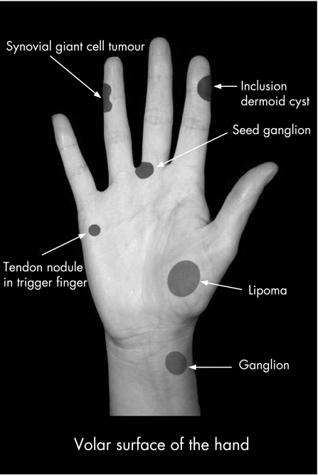
SWELLINGS TO THE VOLAR SURFACE OF THE WRIST AND HAND (FIG 6)
Figure 6 Common swellings involving the volar surface of the hand and wrist.
Ganglion
These swellings are intimately related to the radial artery and usually arise between the radial styloid and the flexor carpi radialis tendon from either the radiocarpal or trapezioscaphoid joints. Management is the same for dorsal ganglia with surgery restricted to large lesions with disabling symptoms. Many ganglia resolve spontaneously over time if left untreated.
Trigger finger
The site of triggering is almost invariably at the level of the metacarpophalangeal joints and is usually associated with nodularity of the tendon rather than stenosis of the flexor tendon sheath. In the acute stage, patients may present with discomfort in the palm during movement of the involved digit. This then progresses to the characteristic history of the finger locking in flexion and having to be passively drawn into extension with a painful “click”. Examination may demonstrate a palpable lump on the flexor tendon as it moves proximodistally in flexion and extension. Seventy per cent resolve with steroid injections though surgical release may be required in recurrent cases.
Lipoma
These swellings may arise at any site in the hand but when they do occur they are found most frequently in the thenar or hypothenar areas. They may be soft or solid in consistency, lobulated and may be attached to deeper structures such as muscle. Diagnosis is commonly made by magnetic resonance imaging. Excision is offered in symptomatic cases.
SWELLINGS OF THE FINGERS
Seed ganglion
These arise from the flexor tendon sheath. They lie either over the proximal phalanx of the fingers or in the distal palm. These swellings are small in size, firm in consistency and do not excurse with the tendon, although there may be an element of mobility in a lateral direction. The swelling can cause discomfort, particularly when gripping firm objects. Excision of the ganglion with a small segment of sheath is curative. Recurrence is rare.
Mucous cysts
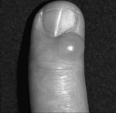
The cyst is a ganglion which usually arises from the distal interphalangeal joint. The lesion is a “blow‐out” of an osteoarthitic joint and is commonly found on the dorsum of the finger, to one side of the midline (fig 7). The index and middle fingers are often affected. The cyst develops in the superficial tissues around the edge of the extensor tendon and the overlying skin is commonly thinned. Occasionally, there may be pressure to the germinal nail matrix producing deformation of the nail. The area is at risk for inadvertent injury and the thin skin may be breached to produce a sinus that discharges intermittently. Excision is usually curative although a minority recur.
Figure 7 Classic presentation of a mucous cyst.
Inclusion epidermoid cyst
Those that are found in the hand are usually the result of trauma. A penetrating injury drives epidermal elements deep into the soft tissues where they continue to grow. This forms a smooth, spherical swelling which is attached to the skin yet mobile over the underlying structures. Most cysts are found on the volar aspect of the hand, around the fingertips, but sometimes develop in surgical scars in the palm. They are usually painless but may attain a size that interferes with function. In such cases surgical excision can be performed, but the entire cyst wall must be excised if recurrence is to be avoided.
Pyogenic granuloma
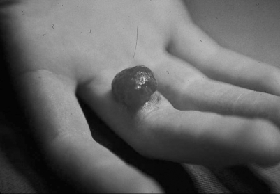
This presents as a florid, friable overgrowth of granulation tissue usually at a site of minor trauma such as scratch or cut (fig 8). They are not painful but patients are often distressed by its rapid growth and bleeding on minimal contact. The diagnosis is often obvious though histological confirmation should be sought in all cases to differentiate between a pyogenic granuloma and the rarer amelanotic melanoma.
Figure 8 Pyogenic granuloma.
Synovial giant cell tumour of the joint or tendon sheath
These swellings originate from the flexor tendon sheath or synovium of the interphalangeal joints. They are usually asymptomatic and thus patients may present late with circumferential lesions and those that involve the entire flexor sheath. They are firm on palpation and have a characteristic lobular appearance. Local recurrence is not unusual after surgical excision.
Enchondroma with pathological fracture
These benign tumours commonly involve the hand skeleton and, in particular, the base of the phalanges. Radiographs demonstrate cortical thinning with slow expansion of bone. Forceful hand activities may abruptly produce pain and swelling, as the weakened bone fractures. Curettage and bone graft is preferred in most cases. However, this may have to be delayed if a fracture has occurred to allow healing of the bone and to prevent instability.
Rheumatoid nodules
Rheumatoid nodules are commonly seen overlying the olecranon at the elbow (fig 9). However, these lesions can also occur at other pressure points in the pulps of the fingers and thumb. The nodules can be excised if they affect hand function, but they often recur.
Figure 9 Rheumatoid nodules—pathognomonic for the disease.
Gout
This condition is characterised by hyperuricaemia and the deposition of monosodium urate crystals in the tissues (tophi). In the acute stage, this provokes an inflammatory reaction that may cause pain and swelling of the finger joints suggestive of an infective arthritis. Large tophi may occur in the pulps of the fingers in chronic cases. Management is usually conservative.
SUMMARY
The above is simply a guide on the common hand swellings and the list is by no means exhaustive. Our aim is to provide general practitioners with clear and concise information and terms of reference. The information needs to be coupled with an adequate history and examination. The history is often pertinent when patients present with a hand swelling. The examination is equally important and will often, in isolation, permit accurate diagnosis. Where the diagnosis remains in doubt, referral to a hand surgeon is necessary.
Footnotes
Funding: None
Competing interests: All authors declare that the answer to the questions on your competing interest form are all No and therefore have nothing to declare
Patient consent for publication of the figures: All images were taken with the patients' verbal consent for use in teaching and publication.


