

Abstract
Bone hydatid disease lacks a typical clinical appearance and image characteristics on x ray or CT are similar to those of tuberculosis, metastases and giant cell tumour or bone cysts. However, MRI does show distinctive diagnostic features of bone hydatid disease, especially in the spine. Until recently, treatment of osseous hydatid disease has been entirely surgical. Effective chemotherapy using benzimidazoles, particularly mebendazole, albendazole and combination treatments, has now been achieved in experimental studies and clinical practice. However, most of these drugs are still in the experimental stage or are in the early stages of clinical use.
Cystic echinococcosis (CE) and alveolar echinococcosis (AE) result from infestation with Echinococcus granulosus (EG) and Echinococcus multilocularis, respectively. Although 12 different species have been identified, only these two are known to affect man, the latter being extremely rare and not affecting bone.1 This article reviews the appearances and characteristics of bone disease due to EG.
EPIDEMIOLOGY
Hydatid disease is prevalent throughout much of the world. CE occurs on every continent except Antarctica and may be transmitted in arctic, temperate and tropical regions. Human infestation with CE is caused by the larval stage of EG and is cosmopolitan in distribution in north and northwestern China, parts of South America, East Africa, Australia, Central Asia, the Mediterranean littoral (including North Africa) and Russia. CE is also endemic in parts of Western Europe and southern USA.2 The geographic distribution of hydatid disease is shown in fig 1.3 By 2002, two countries, Iceland and New Zealand, and one island‐state, Tasmania, had already declared that hydatid disease had been eliminated from their territories. Other hydatid elimination programmes implemented in South America (Argentina, Chile, Uruguay), in Europe (mid‐Wales, Sardinia) and in East Africa (northwest Kenya), showed varying degrees of success, but some were considered as having failed. The reasons for the eventual success of certain hydatid control programmes and the problems encountered in others are analysed and discussed and recommendations for likely optimal approaches considered. The application of new control tools, including use of a hydatid vaccine, is also considered.4

Figure 1 Geographic distribution of hydatid disease. Black stripes indicate areas in which hydatid disease is endemic in North Africa, the Middle and Far East, the United States and Canada.
Hydatid disease may develop in almost any part of the body. The overall incidence of organ infestation is greatest in the liver (50–77%) and lungs (8.5–43%).5 Involvement of the central nervous system occurs in about 3% of all hydatid disease.6,7 The incidence of bone echinococcosis disease is much lower, about 0.5–4% of the total reported cases, and is most commonly seen in the spine (from 50% to >60%), followed by the femur, tibia, humerus, skull, and ribs.8,9,10,11,12
LIFE CYCLE
A parasitic tapeworm, Echinococcus, causes hydatid disease. The complete life cycle was not documented until the 19th century13 and involves two hosts. The definitive host is usually a dog but may be some other carnivore. The adult worm of the parasite lives in the proximal small bowel of the definitive host, attached by hooklets to the mucosa. Eggs are released into the host's intestine and excreted in the faeces.14,15 Sheep are the most common intermediate hosts. They ingest the ovum while grazing on contaminated ground. The ovum loses its protective chitinous layer as it is digested in the duodenum. The released hexacanth embryo, or oncosphere, passes through the intestinal wall into the portal circulation and develops into a cyst within the liver. When the definitive host eats the viscera of the intermediate host, the cycle is completed. Humans may become intermediate hosts through contact with a definitive host (usually a domesticated dog) or ingestion of contaminated water or vegetables.15 One of the main features is that in bone, growth is a very slow process,16 but in the human liver, cysts grow to 1 cm during the first 6 months and 2–3 cm annually thereafter, depending on host tissue resistance.3,15,17
PATHOLOGY
Primary hydatid disease of bone, due to EG, occurs when a blood‐borne scolex settles in bone. In bone involvement, pericyst formation does not occur, thereby allowing aggressive proliferation in an irregular branching fashion along the line of least resistance, especially the bone canals.10,18 The parasite replaces the osseous tissue between trabeculae due to the slow growth of multiple vesicles. With time, the parasite reaches and destroys the cortex, with subsequent spread of the disease to surrounding tissues.10,18,19 Extraosseous cysts may calcify, whereas intraosseous disease rarely demonstrates calcification.18
Because of the features of bone growth just described above, the result is fragmentation and conglomeration of daughter cysts rather than a single one. Slow resorption of the trabeculae, without cortical expansion, occurs as a result of pressure. If the cortex is breeched, then normal uniform expansion occurs in the surrounding soft tissues. Articular cartilage and intervertebral discs offer little resistance to growth.1
HYDATID CYST STRUCTURE
The hydatid cyst has three layers: (1) the outer pericyst, composed of modified host cells that form a dense and fibrous protective zone; (2) the middle laminated membrane, which is acellular and allows the passage of nutrients; and (3) the inner germinal layer, where the scolices (the larval stage of the parasite) and the laminated membrane are produced. The middle laminated membrane and the germinal layers form the true wall of the cyst, usually referred to as the endocyst, although the acellular laminated membrane is occasionally referred to as the ectocyst.14,15 Daughter vesicles (brood capsules) are small spheres that contain the protoscolices and are formed from rests of the germinal layer. Before becoming daughter cysts, these daughter vesicles are attached by a pedicle to the germinal layer of the mother cyst. At gross examination, the vesicles resemble a bunch of grapes. Daughter cysts may grow through the wall of the mother cyst, particularly in bone disease.
Cyst fluid is clear or pale yellow, has a neutral pH, and contains sodium chloride, proteins, glucose, ions, lipids, and polysaccharides. The fluid is antigenic and may also contain scolices and hooklets. When vesicles rupture within the cyst, scolices pass into the cyst fluid and form a white sediment known as hydatid sand.14
CLINICAL APPEARANCES AND CHARACTERISTICS
Chaussier reported the first hydatid disease in spine in 1807, and then the disease was regularly reported. Its clinical characteristics, diagnosis and therapy, however, are still not definitive.20,21,22
Patients usually present because of pain, swelling or pathological fracture. In addition, spinal disease may present with compromised neurology, manifested with severe back pain, weakness and sphincter disturbances. This cestode parasitic infestation, uncommonly involving the nervous system, presents with varied clinical manifestation, at times causing diagnostic dilemmas.23 Because of the lack of a characteristic clinical appearance, hydatid disease is usually difficult to distinguish from tuberculous spondylitis, chronic osteomyelitis, aneurysmal bone cysts, giant‐cell tumours, solitary cysts, neurofibromatosis, fibrocystic disease, chondrosarcoma and tuberculosis.
Serological assay
Serological tests are important in differential diagnosis. The immunoreaction of the human body is related to the hydatid cyst's integrity, growth vigour and location. Immunoreaction is heavier in ruptured hydatid cysts and lower when intact. Rupture can result in severe allergic shock and even death. Serological tests are frequently negative when the hydatid cyst is ageing, calcified or dead. Lightowlers et al24 found immunoreaction was intense in bone hydatid cyst disease and deduced that this was due to a lack of a fibrous capsule and cyst contiguity with human tissue. At present, the serologic examinations used in diagnosing hydatid cyst disease are classified into two categories. One is detection of antigen from the hydatid fluid and protoscoleces, and the main antigenic components are Ag 5 and Ag B25; the other is detection of the antibody in the blood serum of patients. Specific antibody examinations are used for diagnosis including the following: the Casoni test, indirect haemagglutination (IHA), counterimmunoelectrophoresis (CIE), enzyme‐linked immunosorbent assay (ELISA) and gold‐labelled antibody.26,27 The Casoni test, IHA and CIE are called the three items of hydatid cyst examination. They show high false positive results and poor sensitivity because the native antigens are prepared directly from hydatid cyst fluid without purification. ELISA is mainly used as a confirmatory test because of its high specificity and sensitivity.26,27,28 Ramzy et al29 confirm the final diagnosis rate of detecting IgG1 is 97.7% by ELISA. Baveja et al30 report the specificity of IgG, IgM and IgA combination detection in patients' plasma is 98.0%.
The so‐called eight tests of immunodiagnosis include ELISA and the gold‐labelled method, which detect four types of antigen including the following: the antigen of the cyst fluid, the antigen of the cephalomere, the half purified antigen of cyst fluid, and the antigen of alveolar echinococcosis. This eight‐test battery provides the most sensitive and specific serological test, with up to 92.6% sensitivity and 91.9% specificity.31 Detection of the specific IgG subclass antibodies of IgG1 and IgG4 isotypes in human CE and AE is also of potential significance32 and may provide the possibility of detection in early disease as well as in chronic infections. It may also provide a better correlation with hydatid patient prognosis following medical treatment.33
Assessment of post‐treatment disease activity among 28 patients with CE was insensitive using detection of CE‐specific total IgG antibody. At diagnosis, concentrations of CE‐specific total IgG, IgG1 and IgG2 antibodies were significantly elevated compared with IgG3 and IgG4 antibodies by ELISA using crude horse hydatid cyst fluid as antigen. During post‐treatment follow up, the IgG2 antibody response provided the best correlate of disease activity.34
For cerebrospinal hydatid disease, the diagnosis may impose some problems35 by serological assay. Serological misdiagnosis may occur as a result of the blood–brain barrier—about 40 KD molecule mass can pass through it freely, however, the main hydatid antigen is 60 KD.36
The value of imaging in diagnosis
Early diagnosis is uncommon and is primarily based on x ray findings. The x ray appearance is that of cystic or irregular destruction of bone. In a few cases there are arcuate lines of calcification in the “capsule” wall, or there may be multicystic destruction of bone. The above‐mentioned characteristics often cause misdiagnosis such as bone cyst and giant cell tumour of bone. In the spine, the appearance is of an irregular destruction of vertebral body and, sometimes, intervertebral space narrowing. The x ray appearance of spinal echinococcosis is non‐specific—for example, the non‐specific sacral destruction shown in the radiograph in fig 2A. Findings on x ray include monolocular, bilocular, or multilocular cysts. Monolocular cysts, as in this case, are rarely observed and are characterised by oval or polycyclic non‐specific lacunae of variable sizes. Progression of the disease takes place in two forms: formation of diverticuli, and exogenous vesiculation.37,38,39,40 Potential complications include pathological fracture, infection, and fistulisation of the abscess.

Figure 2 (A) Radiograph showing the destructive region in the sacrum. (B) The sacral bone destruction combined with sequestrum on computed tomographic (CT) scan. (C) Sacral bone destruction on three‐dimensional CT scan reconstruction. (D) Magnetic resonance image (MRI) showing the occupying lesion in the spinal canal and in the anterior sacrum.
Pluchino et al report that using plain x ray, osseous changes are found in 27% of cases and “moth eaten areas” with surrounding sclerosis are typical.41 But Liu et al42 report their initial diagnostic results by x ray; 15 cases are diagnosed as tuberculosis, 6 cases as metastases, 2 cases as a chordoblastoma, 1 case as a psoas abscess, 2 cases as bone cysts, 2 cases as bone giant cell tumours, and in the remaining 9 cases no specific diagnosis was conclusively made.
Most computed tomographic (CT) scans are non‐specific for bone hydatid disease—for example, the sacrum is destroyed as shown in fig 2B, and in fig 2C with a three‐dimensional CT scan reconstruction. A typical CT appearance for a CE cyst (fig 3) is of a round or ovoid space‐occupying lesion with a “double layered arcuate calcification”. This appearance can be considered as specific for hydatid cyst caused by CE infestation rather than any other cyst forming disease. Vertebrae and vertebral arches were destructed multicystically and expansively. CT images can also recognise rupture of the endocyst by showing a folded detached endocyst, but the typical films are rarely found. In our 25 cases of spinal hydatid disease, 13 cases were scanned by CT and only in 5 cases (23.1%) were they diagnosed correctly.
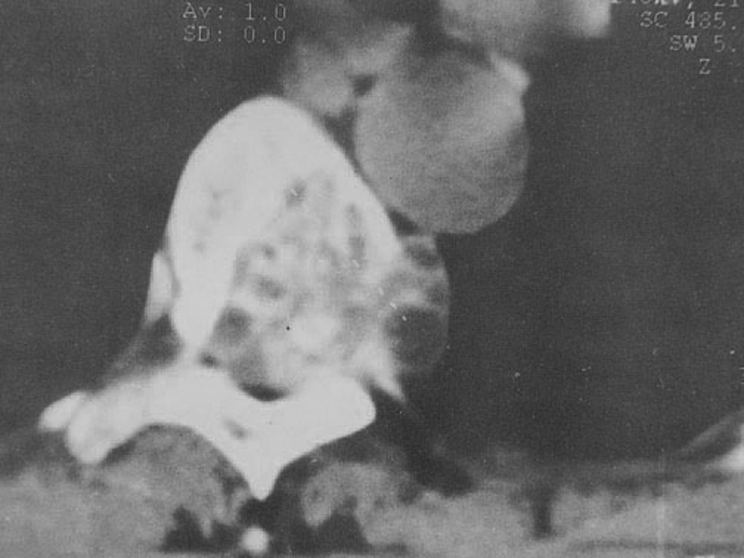
Figure 3 CT appearance of a typical cystic echinococcosis (CE) cyst as a round or ovoid space‐occupying lesion, and double layer arcuate calcification.
Magnetic resonance imaging (MRI) is the most helpful technique for diagnosing hydatid cyst disease.43,44 Typically, the sacrum is destroyed by a cystic, eccentric, expansive, hydatid lesion (fig 2D). One characteristic is that the T1 weighted (W) image signal of the parent cyst is similar to muscle and higher than in the daughter cyst (fig 4). The signal of the secondary cyst is similar to water. Both the parent and the daughter cysts show a high signal in the T2W image sequence and the shape of a rose or a wheel due to spaces or septi of the daughter cyst (fig 5). A typical multilocular MR appearance is seen in figs 6A and 6B. The multilocular appearance is another characteristic of hydatid cyst disease. When the cyst is ruptured or infected, the signal is enhanced because of increased protein in the cyst fluid and there may be an indistinct or vague edge (fig 7). In addition, MRI is more reliable than CT because it shows not only the typical signs of a pericystic wall and its multilocular character, but it also has a special value in showing the relationship of the lesion to the surrounding organs.45

Figure 4 MRI showing that the T1 weighted image signal of the parent cyst is similar to muscle and higher than in the secondary cyst. The signal of the secondary cyst is similar to water, whether located in the parent cyst, overlying it or lying adjacent to it.
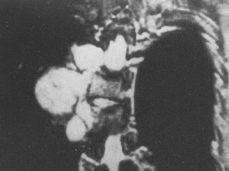
Figure 5 The capsule has a low signal. Vertebral body destruction, with the cysts protruding into the thoracic cavity with both the parent and secondary cysts showing high signals in T2 weighted image and with secondary cyst false intervals giving the appearance or shapes of roses or wheels.

Figure 6 (A) Transverse multilocular MR views of spinal hydatid disease. (B) Sagittal views of a multiple cyst of spinal hydatid disease.
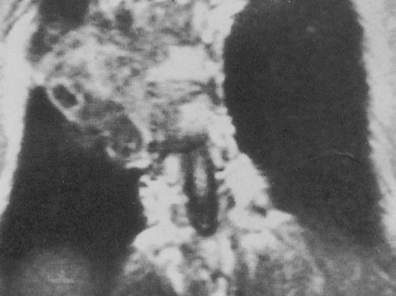
Figure 7 Rupture of a concurrent infected thoracic vertebral hydatid lesion showing a signal with indistinct edge in the T1 weighted image, viewed in the coronal plane.
More recently magnetic resonance hydrography (MRH) has been added to the arsenal of investigations and is particularly helpful in assessing accurately the number of daughter cysts and the extent of the disease. Three cases of spinal hydatid disease were reported in 69 hydatid diseases by the MRH method.46
SURGICAL TREATMENT
Early diagnosis is uncommon and is primarily based on x ray findings. Because patients usually present at an advanced stage of the disease, treatment is therefore difficult and recurrence is common.
From 1964, Alldred and Nisbet et al47 advocated wide surgical excision when treating disease in long bones but followed a more conservative approach when treating disease in the axial skeleton. Booz,48 Hooper and McLean49 and Duran et al50 advocated thorough mechanical curettage to remove macroscopic cysts and “chemical” sterilisation of the scolices using formalin, 0.5% silver nitrate or hypertonic saline, any major defect being filled with autogenous bone graft.
In 2001 Yildiz et al12 reported 15 cases of bone hydatidosis treated by curettage, swabbing with povidine iodine and filling the defect with polymethylmethacrylate (PMMA) in 10 patients. Three of these had a recurrence after 5 years, but seven had no signs of relapse during a mean follow‐up of 52 months. The combination of antihelminthic treatment, wide resection and the use of PMMA gave the best outcome in the treatment of bone hydatidosis. For the spinal hydatid disease, the operative procedure of choice is laminectomy. However, decompression with anterior vertebrectomy and fusion also gives good results.20,51,52,53 In the series by Pamir et al,53 neurological improvement was seen in 63% of the cases and recurrence in 18%. Overall, a recurrence rate of 30–40% is described.54 Liu et al42 reported 11 relapses in 37 bone hydatid disease cases, a recurrence rate of 43.83%. There is a correlation between cyst localisation and recurrence.
In a retrospective study in 2005, 12 patients (60%) relapsed out of a total of 20 cases with spinal hydatidosis. Of these, 18 cases were exposed posteriorly. An additional fusion with instrumentation was performed after removing the involved posterior elements in eight patients. An anterior approach was used in two patients: a total corpectomy was performed and a bone graft was added. Two weeks later, a posterior decompression and fixation with instrumentation was done.8
Recently treatment of osseous hydatid disease has been entirely surgical, the aims being removal of the cyst and surrounding bone, replacement of bone defects with bone grafts or a prosthesis, avoidance of secondary infection, and prevention of recurrence. Unfortunately these goals are rarely achieved completely in this relentless disease. Most “scolecidal” agents do not kill all microscopic daughter cysts, and therefore recurrence is likely, and surgery is often only palliative. Although long‐term survival is possible, the disease is not easy to eradicate and may be impossible to cure.39,55,56 Effective chemotherapy would therefore be of great benefit.
CHEMOTHERAPY
The efficacy of chemotherapeutic treatment is related to the drug absorption, metabolism and bioavailability in the blood and cysts; therefore the review of chemotherapy includes both bone echinococcosis and other hydatid diseases, so we cannot discuss them separately. The treatment experience of other hydatid diseases might be presumed to be suitable for bone hydatid disease.
Benzimidazoles
Chemotherapy with benzimidazoles, particularly mebendazole and albendazole, has been used for the treatment of human echinococcosis since 1974, when Heath and Chevis57 first reported that the benzimidazole derivative mebendazole could kill the germinal membrane of echinococcus in mice by limiting glucose uptake. The first report, in 1977, of the use of high‐dose mebendazole by Bekhti et al58 was encouraging. The variable success is thought to be due to the insolubility of mebendazole with its consequent poor absorption leading to low concentrations in serum and cysts.59
Albendazole is a relatively new compound offering the distinct advantages of better absorption and higher levels of the active metabolite albendazole sulfoxide (AlbSO) in the cysts and in blood.60 More recently, albendazole, because of its higher level in blood plasma (250 mg/l) than that of mebendazole (70–90 mg/l), is now regarded by the World Health Organization as the agent of first choice to treat hydatid disease.61,62
According to 27 publications, 666 CE patients have been treated by benzimidazoles, indicating that mebendazole and albendazole are not completely effective overall, with a total success rate of approximately 25–30% of cases. In 40–50% of cases there is an improvement and in 25–30% of cases the treatments have been found to be ineffective.63 Benzimidazoles are the most commonly used postoperative adjuvant chemotherapy drug.8
Albendazole liposome
Albendazole liposome (Alb‐L), entrapment of albendazole within lipomes (10 mg/kg daily for 3 months), may alter albendazole metabolism and increase the drug bioavailability. Liu Dapeng et al42 used it postoperatively for 3 months to prevent relapse.
Praziquantel
Li et al64 reported 126 CE and 14 AE patients treated by praziquantel orally at a dosage of 25–50 mg/kg per day for 1–6 months. Approximately 90% of the patients improved or become stable. Urrea‐Paris65 investigated the therapeutic effect of praziquantel against the different developmental stages of the EG metacestode, and demonstrated that the drug is only useful in the early young cyst, but has no effect against the mature cyst.
Combination treatments
Albendazole + praziquantel
In 21 patients with cystic hydatid diseases treated orally with albendazole 10 mg/kg combined with praziquantel 25 mg/kg daily for 1 month before surgery, the AlbSO concentrations in both blood and cyst fluid collected after final medication and during the operation were higher in the combined treatment group than in the group of 26 patients treated with only albendazole 10–20 mg/kg. Furthermore, live protoscoleces in the cyst fluid have been found in two out of 21 and 13 out of 26 cases.66 Yasway67 reported four patients with cystic hydatid disease who had been treated with albendazole 400 mg for 2–5 treatment courses combined with praziquantel 50 mg/kg once daily (albendazole 400 mg daily on monthly courses plus praziquantel 50 mg/kg in different regimens). After 3 months of using the combined treatment, cysts disappeared completely in three patients, while in the fourth patient a reduction in cysts size by more than 75% after 2 months combination treatment was observed.
A preliminary study67 on the treatment of 10 CE patients using a combination of praziquantel (40 mg/kg per day) and albendazole (800 mg/day) has been reported. After 2–3 months the cysts had disappeared or were substantially reduced in size and number. Another report by Urrea68 has shown that the effect of albendazole and praziquantel combination therapy against the protoscoleces is better than that of albendazole or praziquantel treatment alone. However, a case report of a 53‐year‐old man with a severe destructive lesion of the L4 vertebral body caused by an EG hydatid cyst, treated continuously and long‐term with albendazole, followed by albendazole combined with praziquantel after surgery, did not achieve eradication of the disease.69
Albendazole combined with cimetidine
In patients treated with a combination of albendazole and cimetidine, the AlbSO concentrations in cyst fluid and bile were twice that found in CE patients treated with albendazole alone; this suggests that cimetidine might play a role in possibly increasing albendazole's bioavailability and therapeutic effect.32 Luder et al found that cimetidine could alter the metabolism of mebendazole, resulting in an increase of the drug concentration in the blood.70
Other combination treatments
Del Estal et al71 reported that the surfactant sodium taurocholate could enhance the absorption constant of albendazole and result in an increase of its bioavailability. Jung et al reported that dexamethasone could increase the plasma concentration of AlbSO by over 50%.72
Other drugs
Other drugs against hydatid disease are only in the experimental stage. These are: poly L‐lactid albendazole; polyethylene glycol mebendazole; oxfendazole, a benzimidazole derivative; nitazoxanide; isoprinosine; mitomycin C; cyclosporin A (CyA); α‐difluoromethylornithine (DFMO); ivermectin and albendazole combination therapy; amphotericin B; matrine, a quinolizidine alkaloid; albendazole combined with AlbSO; albendazole combined with dipeptide methyl ester; albendazole combined with levamisole.
SUMMARY
Hydatid disease is prevalent throughout much of the world, but the incidence of bone echinococcosis is low. Bone echinococcosis is often misdiagnosed. Radiographic and CT images lack disease‐specific characteristics whereas MRI images offer a greater chance of direct diagnosis. To diagnostic correctly, serological assay methods used for diagnosis not only included the Casoni test, IHA, CIE and CFT, but also included eight tests of immunodiagnosis for a high sensitivity and specificity. The case history is important in the diagnosis, especially if the patient has had contact with dogs or sheep or has come from a livestock farm. Up to now, the treatment of osseous hydatid disease has been entirely surgical, but recurrence is likely. Effective chemotherapy would therefore be of great benefit using benzimidazoles,, particularly mebendazole, albendazole and combination treatments. Great achievement has been attained in both experimental studies and clinical practice. However, most drugs or these combined treatments have been used just before surgery, and there are no data dealing with the long‐term follow up results. The antihydatid effect of albendazole combined with other drugs is worth further study.
ACKNOWLEDGEMENTS
We are grateful to Dr Michael L Harding for revising the manuscript and all the work he has done with his wife Penny in China from 2005 to 2007.
Abbreviations
Alb‐L - albendazole liposome
AlbSO - albendazole sulfoxide
AR - alveolar echinococcosis
CE - cystic echinococcosis
CIE - counterimmunoelectrophoresis
CT - computed tomography
EG - Echinococcus granulosus
ELISA - enzyme‐linked immunosorbent assay
IHA - indirect haemagglutination
MRH - magnetic resonance hydrography
MRI - magnetic resonance imaging
PMMA - polymethylmethacrylate
Footnotes
Conflict of interest: none stated
Patient consent to publish figures 2–7 was received.
References
- 1.Szypryt E P, Morris D L, Mulholland R C. Combined chemotherapy and surgery for hydatid bone disease. J Bone Joint Surg Br 198769141–144. [DOI] [PubMed] [Google Scholar]
- 2.Gemmell M A, Lawson J R, Roberts M G. Towards global control of cystic and alveolar hydatid diseases. Parasitol Today 19873144–151. [DOI] [PubMed] [Google Scholar]
- 3.Pedrosa I, Saiz A, Arrazola J.et al Hydatid disease: radiologic and pathologic features and complications. Radiographics 200020795–817. [DOI] [PubMed] [Google Scholar]
- 4.Craig P S, Larrieu E. Control of cystic echinococcosis/hydatidosis: 1863–2002. Adv Parasitol 200661443–508. [DOI] [PubMed] [Google Scholar]
- 5.Kammerer W S. Echinococcosis affecting the central nervous system. Semin Neurol 199313144–147. [DOI] [PubMed] [Google Scholar]
- 6.Charles R W, Govender S, Naidoo K S. Echinococcal infection of the spine with neural involvement. Spine 19881347–49. [DOI] [PubMed] [Google Scholar]
- 7.Iplikcioglu A C, Kokes F, Bayar A.et al Spinal invasion of pulmonary hydatidosis: computed tomographic demonstration. Neurosurgery 199129467–468. [PubMed] [Google Scholar]
- 8.Herrera A, Martinez A A, Rodriguez J. Spinal hydatidostal. Spine 2005302439–2444. [DOI] [PubMed] [Google Scholar]
- 9.Sapkas G S, Stathakopoulos D P, Babis G C.et al Hydatid disease of bones and joints. 8 cases followed for 4–16 years. Acta Orthop Scand 19986989–94. [DOI] [PubMed] [Google Scholar]
- 10.Torricelli P, Martinelli C, Biagini R.et al Radiographic and computed tomographic findings in hydatid disease of bone. Skeletal Radiol 199019435–439. [DOI] [PubMed] [Google Scholar]
- 11.Beggs I. The radiology of hydatid disease. AJR Am J Roentgenol 1985145639–648. [DOI] [PubMed] [Google Scholar]
- 12.Yildiz Y, Bayrakci K, Altay M.et al The use of polymethylmethacrylate in the management of hydatid disease of bone. J Bone Joint Surg Br 2001831005–1008. [DOI] [PubMed] [Google Scholar]
- 13.Lewis J W, Jr, Koss N, Kerstein M D. A review of echinococcal disease. Ann Surg 1975181390–396. [DOI] [PMC free article] [PubMed] [Google Scholar]
- 14.King C H. Cestodes (tapeworms). In: Mandell GL, Bennett JE, Dolin R. Principles and practice of infectious diseases. 4th ed. New York: Churchill Livingstone, 19952544–2553.
- 15.Lewall D B. Hydatid disease: biology, pathology, imaging and classification. Clin Radiol 199853863–874. [DOI] [PubMed] [Google Scholar]
- 16.Kumar R, Cornah M S, Morris D L. Hydatid cyst—a rare cause of pathological fracture: a case report. Injury 198415284–285. [DOI] [PubMed] [Google Scholar]
- 17.von Sinner W N. New diagnostic signs in hydatid disease; radiography, ultrasound, CT and MRI correlated to pathology. Eur J Radiol 199112150–159. [DOI] [PubMed] [Google Scholar]
- 18.Ogut A G, Kanberoglu K, Altug A.et al CT and MRI in hydatid disease of cervical vertebrae. Neuroradiology 199234430–432. [DOI] [PubMed] [Google Scholar]
- 19.Braithwaite P A, Lees R F. Vertebral hydatid disease: radiological assessment. Radiology 1981140763–766. [DOI] [PubMed] [Google Scholar]
- 20.Pamir M N, Ozduman K, Elmaci I. Spinal hydatid disease. Spinal Cord 200240153–160. [DOI] [PubMed] [Google Scholar]
- 21.Lam K S, Faraj A, Mulholland R C.et al Medical decompression of vertebral hydatidosis. Spine 1997222050–2055. [DOI] [PubMed] [Google Scholar]
- 22.Tekkok I H, Benli K. Primary spinal extradural hydatid disease: report of a case with magnetic resonance characteristics and pathological correlation. Neurosurgery 199333320–323. [PubMed] [Google Scholar]
- 23.Rumana M, Mahadevan A, Nayil Khurshid M.et al Cestode parasitic infestation: intracranial and spinal hydatid disease—a clinicopathological study of 29 cases from South India. Clin Neuropathol 20062598–104. [PubMed] [Google Scholar]
- 24.Lightowlers M W, Gottstein B. Echinococcosis/hydatidosis; antigens, immunological and molecular diagnosis. In: Thompson RCA, Lymbery AJ, eds. Echinococcus and hydatid disease. Wallingford: CAB International, 1995355–410.
- 25.Barbieri M, Fernandez V, Gonzalez G.et al Diagnostic evaluation of a synthetic peptide derived from a novel antigen B subunit as related to other available peptides and native antigens used for serology of cystic hydatidosis. Parasite Immunol 19982051–61. [DOI] [PubMed] [Google Scholar]
- 26.Zhong X G, Huang Y G, Tang Z R.et al Immunological diagnosis of echinococcus disease [in Chinese]. Acta Academic Medicine Shantou 200114212–214. [Google Scholar]
- 27.Han X M. Development of immunological methods in diagnosis echinococcus disease [in Chinese]. Chinese Reports of Endemic Diseases 19971287–90. [Google Scholar]
- 28.Yao F R, Qiao J Y, Yang J H.et al Comparative study of primarily and postoperation relapse of echinococcus disease by four types of immune examinations [in Chinese]. Chin J Parasit Dis Con 2002153–4. [Google Scholar]
- 29.Ramzy R M, Helmy H, El Zayyat E A.et al An enzyme‐linked immunosorbent assay for detection of IgG1 antibodies specific to human cystic echinococcosis in Egypt. Trop Med Int Health 19994616–620. [DOI] [PubMed] [Google Scholar]
- 30.Baveja U K, Basak S, Thusoo T K. Immunodiagnosis of human hydatid disease. J Commun Dis 199729313–319. [PubMed] [Google Scholar]
- 31.Fu Y, Feng X H, Wen H. Observation in clinical application of the 8‐ tests immunodiagnos for human hadatidosis [in Chinese]. Acta Academic Medicine Xinjiang 200023242–243. [Google Scholar]
- 32.Wen H, Craig P S. Immunoglobulin G subclass responses in human cystic and alveolar echinococcosis. Am J Trop Med Hyg 199451741–748. [DOI] [PubMed] [Google Scholar]
- 33.Huang B C, Jia F J, Fu T X. Determination of serum specific IgG4 for diagnosing hydatidosis [in Chinese]. Chin J Parasit Dis Con 200114283–284. [Google Scholar]
- 34.Lawn S D, Bligh J, Craig P S.et al Human cystic echinococcosis: evaluation of post‐treatment serologic follow‐up by IgG subclass antibody detection. Am J Trop Med Hyg 200470329–335. [PubMed] [Google Scholar]
- 35.El‐Arousy M H, Ismail S A. Cerebrospinal echinococcosis: serodiagnosis using different hydatid cyst fluid antigens. J Egypt Soc Parasitol 200535193–204. [PubMed] [Google Scholar]
- 36.Chen X H, Wen H, Zang Z X. An analysis for immuno‐misdiagnosis of human hydatid disease [in Chinese]. 200220121–122. [PubMed] [Google Scholar]
- 37.Kalinova K, Proichev V, Stefanova P.et al Hydatid bone disease: a case report and review of the literature. J Orthop Surg (Hong Kong) 200513323–325. [DOI] [PubMed] [Google Scholar]
- 38.Zlitni M, Ezzaouia K, Lebib H.et al Hydatid cyst of bone: diagnosis and treatment. World J Surg 20012575–82. [DOI] [PubMed] [Google Scholar]
- 39.Markakis P, Markaki S, Prevedorou D.et al Echinococcosis of bone: clinico‐laboratory findings and differential diagnostic problems. Arch Anat Cytol Pathol 19903892–94. [PubMed] [Google Scholar]
- 40.Saidi F. Hydatid cysts of bone. In: Saidi F, ed. Surgery of hydatid disease, 1st ed. London: WB Saunders, 1976135–138.
- 41.Pluchino F, Lodrini S. Multiple primitive epidural spinal hydatid cysts: case report. Acta Neurochir (Wien) 198159(3–4)257–262. [DOI] [PubMed] [Google Scholar]
- 42.Liu D, Xie Z R, Zhang R.et al Treatment and diagnosis of bone hydatid disease [in Chinese]. Chin J Orthop 200424403–406. [Google Scholar]
- 43.Islekel S, Ersahin Y, Zileli M.et al Spinal hydatid disease. Spinal Cord 199836166–170. [DOI] [PubMed] [Google Scholar]
- 44.Singh S, Korah I P, Gibikote S V.et al Sacral hydatidosis: value of MRI in the diagnosis. Skeletal Radiol 199827518–521. [DOI] [PubMed] [Google Scholar]
- 45.Wang J, Chen H. Vertebral hydatid disease: MRI diagnosis [in Chinese]. Chinese J Med Imaging 200210101–102. [Google Scholar]
- 46.Wang J, Jia W X, Li C W.et al Clinical application of magnetic resonance hydrography on hydatid disease diagnosis. Acta Academic Medicine Xinjiang 2005281079–1082. [Google Scholar]
- 47.Alldred A J, Nisbet N W. The management of hydatid disease of bone and joint. J Bone Joint Surg Br 196446260–267. [PubMed] [Google Scholar]
- 48.Booz M K. The management of hydatid disease of bone and joint. J Bone Joint Surg Br 197254698–709. [PubMed] [Google Scholar]
- 49.Hooper J, McLean I. Hydatid disease of the femur. Report of a case. J Bone Joint Surg Am 197759(7)974–976. [PubMed] [Google Scholar]
- 50.Ferrandez H D, Gomez‐Castresana F, Lopez‐Duran L.et al Osseous hydatidosis. J Bone Joint Surg Am 197860685–690. [PubMed] [Google Scholar]
- 51.Sharma N K, Chitkara N, Bakshi N.et al Primary spinal extradural hydatid cyst. Neurol India 20035189–90. [PubMed] [Google Scholar]
- 52.Awasthy N, Chand K. Primary hydatid disease of the spine: an unusual case. Br J Neurosurg 200519425–427. [DOI] [PubMed] [Google Scholar]
- 53.Pamir M N, Akalan N, Ozgen T.et al Spinal hydatid cysts. Surg Neurol 19842153–57. [PubMed] [Google Scholar]
- 54.Turtas S, Viale E S, Pau A. Long‐term results of surgery for hydatid disease of the spine. Surg Neurol 198013468–470. [PubMed] [Google Scholar]
- 55.Natarajan M V, Kumar A K, Sivaseelam A.et al Using a custom mega prosthesis to treat hydatidosis of bone: a report of 3 cases. J Orthop Surg (Hong Kong) 200210203–205. [DOI] [PubMed] [Google Scholar]
- 56.Mnaymneh W, Yacoubian V, Bikhazi K. Hydatidosis of the pelvic girdle—treatment by partial pelvectomy. A case report. J Bone Joint Surg Am 197759538–540. [PubMed] [Google Scholar]
- 57.Heath D D, Chevis R A F. Mebendazole and hydatid cysts. Lancet 1974ii218–219. [DOI] [PubMed] [Google Scholar]
- 58.Bekhti A, Schaaps J P, Capron M.et al Treatment of hepatic hydatid disease with mebedazole: preliminary results in four cases. BMJ 1977ii1047–1051. [DOI] [PMC free article] [PubMed] [Google Scholar]
- 59.Morris D L, Gould S E. Serum and cyst concentrations of mebendazole and flubendazole in hydatid disease. BMJ (Clin Res Ed) 1982285175. [DOI] [PMC free article] [PubMed] [Google Scholar]
- 60.Morris D L, Dykes P W, Dickson B.et al Albendazole in hydatid disease. BMJ (Clin Res Ed) 1983286103–104. [DOI] [PMC free article] [PubMed] [Google Scholar]
- 61.Chen Geng, Shi Dazhong The treatment status for hydatid disease. Endemic disease. Bulletin 2001
- 62.Xin Ying, Zhang Rui The development of drug treatment for hydatid disease [in Chinese]. Health Profession Education 200518(23)127–130. [Google Scholar]
- 63.Wen H, Yang W G, Vuiton D.et al Status and development of diagnosis and treatment for human echinococcosus [in Chinese]. Acta Academic Medicine Xinjiang 199619183–184. [Google Scholar]
- 64.Li J, Zhou P F, Yang W G. Investigation of the treatment for liver alveolar hydatidosis [in Chinese]. Acta Academice Medicine Xinjiang 199316238 [Google Scholar]
- 65.Urrea‐Paris M A, Moreno M J, Casado N.et al Relationship between the efficacy of praziquantel treatment and the cystic differentiation in vivo of Echinococcus granulosus metacestode. Parasitol Res 20028826–31. [DOI] [PubMed] [Google Scholar]
- 66.Cobo F, Yarnoz C, Sesma B.et al Albendazole plus praziquantel versus albendazole alone as a pre‐operative treatment in intra‐abdominal hydatisosis caused by Echinococcus granulosus. Trop Med Int Health 19983462–466. [DOI] [PubMed] [Google Scholar]
- 67.Yasawy M I, al Karawi M A, Mohamed A R. Combination of praziquantel and albendazole in the treatment of hydatid disease. Trop Med Parasitol 199344192–194. [PubMed] [Google Scholar]
- 68.Urrea‐Paris M A, Moreno M J, Casado N.et al In vitro effect of praziquantel and albendazole combination therapy on the larval stage of Echinococcus granulosus. Parasitol Res 200086957–964. [DOI] [PubMed] [Google Scholar]
- 69.El‐On J, Ben‐Noun L, Galitza Z.et al Case report: clinical and serological evaluation of echinococcosis of the spine. Trans R Soc Trop Med Hyg 200397567–569. [DOI] [PubMed] [Google Scholar]
- 70.Luder P J, Siffert B, Witassek F.et al Treatment of hydatid disease with high oral doses of mebendazole. Long‐term follow‐up of plasma mebendazole levels and drug interactions. Eur J Clin Pharmacol 198631443–448. [DOI] [PubMed] [Google Scholar]
- 71.del Estal J L, Alvarez A I, Villaverde C.et al Effect of surfactants on albendazole absorption. J Pharm Biomed Ana 199119(10–12)1161–1164. [DOI] [PubMed] [Google Scholar]
- 72.Jung H, Hurtado M, Medina M T.et al Dexamethasone increases plasma levels of albendazole. J Neurol 1990237279–280. [DOI] [PubMed] [Google Scholar]