

Abstract
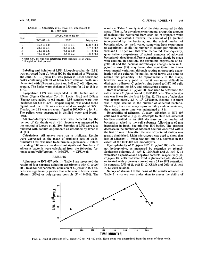
Campylobacter jejuni is able to colonize the human intestinal mucosa and cause disease. For this reason, it was important to investigate mechanisms by which C. jejuni adheres to epithelial cells and intestinal mucus gel. All strains of C. jejuni used were able to adhere to INT 407 epithelial cells and mucus, but high adherence to one substrate did not necessarily indicate comparable adherence to the other. The adherence of C. jejuni to cells was inhibited partially by treating the bacterial cells with proteases or glutaraldehyde or by adding a certain carbohydrate (fucose or mannose) to the medium. The flagellum of C. jejuni was identified as a potential adhesin by comparing adherence of flagellated and aflagellated variants. Shearing of the bacterial cells to remove the flagella reduced bacterial adhesion, whereas immobilization of the flagellum with KCN increased adhesion. Purified flagella showed specific, fucose-resistant binding to epithelial cells but not to intestinal mucus. The presence of a second, nonproteinaceous adhesin was suggested because no single treatment of the bacteria completely inhibited adhesion. Lipopolysaccharide (LPS) was identified as another C. jejuni adhesin. [3H]LPS specifically bound to epithelial cells, and this phenomenon was inhibited by periodate oxidation of the LPS or glutaraldehyde fixation of the epithelial cells. LPS, unlike flagella, was fucose sensitive and inhibited binding of whole bacterial cells to INT 407 cells. LPS was also able to bind to intestinal mucus gel. These data indicate that both flagella and LPS are important in adhesion to the mucosal surface.
Full text
PDF


Selected References
These references are in PubMed. This may not be the complete list of references from this article.
- Attridge S. R., Rowley D. The role of the flagellum in the adherence of Vibrio cholerae. J Infect Dis. 1983 May;147(5):864–872. doi: 10.1093/infdis/147.5.864. [DOI] [PubMed] [Google Scholar]
- Beachey E. H. Bacterial adherence: adhesin-receptor interactions mediating the attachment of bacteria to mucosal surface. J Infect Dis. 1981 Mar;143(3):325–345. doi: 10.1093/infdis/143.3.325. [DOI] [PubMed] [Google Scholar]
- Caldwell M. B., Guerry P., Lee E. C., Burans J. P., Walker R. I. Reversible expression of flagella in Campylobacter jejuni. Infect Immun. 1985 Dec;50(3):941–943. doi: 10.1128/iai.50.3.941-943.1985. [DOI] [PMC free article] [PubMed] [Google Scholar]
- Caldwell M. B., Walker R. I., Stewart S. D., Rogers J. E. Simple adult rabbit model for Campylobacter jejuni enteritis. Infect Immun. 1983 Dec;42(3):1176–1182. doi: 10.1128/iai.42.3.1176-1182.1983. [DOI] [PMC free article] [PubMed] [Google Scholar]
- Cohen P. S., Arruda J. C., Williams T. J., Laux D. C. Adhesion of a human fecal Escherichia coli strain to mouse colonic mucus. Infect Immun. 1985 Apr;48(1):139–145. doi: 10.1128/iai.48.1.139-145.1985. [DOI] [PMC free article] [PubMed] [Google Scholar]
- Freter R., O'Brien P. C. Role of chemotaxis in the association of motile bacteria with intestinal mucosa: fitness and virulence of nonchemotactic Vibrio cholerae mutants in infant mice. Infect Immun. 1981 Oct;34(1):222–233. doi: 10.1128/iai.34.1.222-233.1981. [DOI] [PMC free article] [PubMed] [Google Scholar]
- Izhar M., Nuchamowitz Y., Mirelman D. Adherence of Shigella flexneri to guinea pig intestinal cells is mediated by a mucosal adhesion. Infect Immun. 1982 Mar;35(3):1110–1118. doi: 10.1128/iai.35.3.1110-1118.1982. [DOI] [PMC free article] [PubMed] [Google Scholar]
- Jann K., Schmidt G., Blumenstock E., Vosbeck K. Escherichia coli adhesion to Saccharomyces cerevisiae and mammalian cells: role of piliation and surface hydrophobicity. Infect Immun. 1981 May;32(2):484–489. doi: 10.1128/iai.32.2.484-489.1981. [DOI] [PMC free article] [PubMed] [Google Scholar]
- Jentoft N., Dearborn D. G. Labeling of proteins by reductive methylation using sodium cyanoborohydride. J Biol Chem. 1979 Jun 10;254(11):4359–4365. [PubMed] [Google Scholar]
- Karkhanis Y. D., Zeltner J. Y., Jackson J. J., Carlo D. J. A new and improved microassay to determine 2-keto-3-deoxyoctonate in lipopolysaccharide of Gram-negative bacteria. Anal Biochem. 1978 Apr;85(2):595–601. doi: 10.1016/0003-2697(78)90260-9. [DOI] [PubMed] [Google Scholar]
- LOWRY O. H., ROSEBROUGH N. J., FARR A. L., RANDALL R. J. Protein measurement with the Folin phenol reagent. J Biol Chem. 1951 Nov;193(1):265–275. [PubMed] [Google Scholar]
- Logan S. M., Trust T. J. Molecular identification of surface protein antigens of Campylobacter jejuni. Infect Immun. 1983 Nov;42(2):675–682. doi: 10.1128/iai.42.2.675-682.1983. [DOI] [PMC free article] [PubMed] [Google Scholar]
- Logan S. M., Trust T. J. Structural and antigenic heterogeneity of lipopolysaccharides of Campylobacter jejuni and Campylobacter coli. Infect Immun. 1984 Jul;45(1):210–216. doi: 10.1128/iai.45.1.210-216.1984. [DOI] [PMC free article] [PubMed] [Google Scholar]
- Myhal M. L., Cohen P. S., Laux D. C. Altered colonizing ability for mouse large intestine of a surface mutant of a human faecal isolate of Escherichia coli. J Gen Microbiol. 1983 May;129(5):1549–1558. doi: 10.1099/00221287-129-5-1549. [DOI] [PubMed] [Google Scholar]
- Rollins D. M., Coolbaugh J. C., Walker R. I., Weiss E. Biphasic culture system for rapid Campylobacter cultivation. Appl Environ Microbiol. 1983 Jan;45(1):284–289. doi: 10.1128/aem.45.1.284-289.1983. [DOI] [PMC free article] [PubMed] [Google Scholar]
- Rozee K. R., Cooper D., Lam K., Costerton J. W. Microbial flora of the mouse ileum mucous layer and epithelial surface. Appl Environ Microbiol. 1982 Jun;43(6):1451–1463. doi: 10.1128/aem.43.6.1451-1463.1982. [DOI] [PMC free article] [PubMed] [Google Scholar]