Abstract
Most computer navigation systems used in total hip arthroplasty integrate preoperative pelvic tilt to calculate the anterior pelvic plane assuming tilt is constant; however, the consistency of pelvic tilt after THA has never been proven. Therefore, using a modern comprehensive gait analysis before and after arthroplasty we sought to compare (1) dynamic pelvic tilt changes and (2) pelvic flexion/extension range-of-motion changes. Twenty-one patients who underwent unilateral THA were prospectively studied. Quantitative pelvic tilt changes (in the sagittal plane) and pelvic range of flexion/extension motion relative to a laboratory coordinate system were compared using a computerized video motion system. Mean gait pelvic tilt was 13.9º ± 4.8º (range, 1.73º–23.1º) preoperatively, 12.5º ± 4.5º (range, 1.4º–18.7º) 2 months postoperatively, and 10.5° ± 5.5º (range, –2.36º–19.2º) 12 months postoperatively. A significant proportion (31%) of patients had more than a 5° difference between preoperative and 12-month postoperative measurements and the variability was spread over 20°. Significant dynamic changes in pelvic tilt occurred after THA. While navigation clearly improves the anatomical position of the component during THA, the functional position of the component will not always be improved because of the significant change between preoperative and postoperative pelvic tilt.
Introduction
Computer-assisted navigation systems in total hip arthroplasty have been developed to improve acetabular cup positioning because mechanical acetabular guides for intraoperative alignment are often insufficient to achieve the desired implant orientation [4].
Considering a cup positioning target of 45° ± 10° of cup abduction and 15° ± 10° of anteversion, the result of a previous study reported unacceptable acetabular alignment in 78% of hips when using the mechanical guide, with a significant variation in cup alignment from the desired goal [5]. In prospective randomized studies, computer-assisted systems allowed surgeons to reliably achieve the previously defined cup positioning targets [1, 14]. In fact, in these studies, percentage of proper cup alignment was significantly improved from 44% to 80% of the cases [11, 20].
When using computer-assisted systems, angles are typically measured perioperatively or postoperatively relative to the anterior pelvic plane [11, 13, 20]. The anterior pelvic plane is defined by the two anterior iliac spines and pubic tubercles [16]. It has been presumed the anterior pelvic plane and the vertical plane are superimposable [14], however important intersubject variations of these planes exist in a standing position [2, 3, 7, 8, 11, 13, 14, 20]. The angle between the anterior pelvic plane and the vertical is defined as the pelvic tilt [2, 3, 7, 8, 11, 13, 14, 20]. Previous studies have reported important interindividual variations of the pelvic tilt: from −18º to 3º in 20 patients in one study [8], from −22º to 27º in 84 patients in another [5], and −23º to 14º in 60 patients in another [20]. It has been clearly demonstrated pelvic tilt substantially affects acetabular cup orientation, particularly when using a computer-assisted system that relies on the anterior pelvic plane [3, 13, 15, 20, 25]. While the intrinsic accuracy of computer-assisted systems for cup positioning is close to 1°, an anterior pelvic tilt of 5° may lead to an error of 5° in the final cup anteversion [25, 26].
To improve the accuracy of cup positioning, the integration of the preoperative pelvic tilt into the navigation program has been proposed [3, 20]. A standard preoperative assessment of the pelvic tilt is made on a mediolateral pelvic radiograph in the standing position [2, 3, 15, 20]. Then the pelvic tilt angle is integrated in the software and acetabular alignment is defined as a function of the implant alignment in the pelvis and oriented to the vertical [3, 20]. While this integration should theoretically improve the functional alignment of the cup related to pelvic tilt when using computer-assisted systems, the assumption that individual pelvic tilt is the same before and after THA has not been verified. Under static conditions pelvic tilt before and after THA seems consistent [3, 18]. However, whether the consistency applies to dynamic tilt during activities of daily living has not been confirmed. We hypothesized that dynamic pelvic tilt and range of motion would be consistent before and after THA.
Therefore, using a modern comprehensive gait analysis we sought to compare: (1) dynamic pelvic tilt changes before and after total hip arthroplasty; and (2) pelvis flexion/extension range-of-motion changes before and after arthroplasty.
Materials and Methods
We prospectively followed 21 patients who underwent unilateral THA between September 2005 and January 2006. Due to the absence of previous studies in the literature, we were unable to estimate the expected difference and therefore unable to calculate the number of subjects to include using a formal power analysis. Patients between the ages of 40 and 85 years old undergoing unilateral primary hip surgery for degenerative joint disease were enrolled. We excluded patients with severe deformity such as developmental dysplasia of the hip (Crowe types III or IV), osteomyelitis, septicemia, hip joint infection or other active infection, neurological or musculoskeletal disorders, or disease that might adversely affect normal gait or weight bearing in either lower extremity. Thirteen men and eight women comprising 13 right hips and eight left hips were enrolled. Patients were a mean 63 ± 13 years old (range, 40–85 years) with a mean body mass index (BMI) of 30 ± 6 kg/m2 (range, 21–47 kg/m2). The study protocol was approved by the local institutional review board and informed consent was obtained from all patients.
All patients were operated under general anesthesia by the senior author (MWP) through a single-incision posterior approach for 12 cases and through a two-incision minimally invasive surgical approach with fluoroscopy assistance for the other cases. The same advanced anesthetic and perioperative pain management protocol was used for all patients. The two-incision THAs were performed with intraoperative fluoroscopy using a previously described technique [19]. The THAs were performed through a 6- to 9-cm mini-posterior approach, splitting the gluteus maximus fascia [19]. An uncemented hemispherical acetabular component and an uncemented femoral stem were used for all patients. No drains were used in the wounds. Thromboembolic prophylaxis was performed with foot pumps, compression stockings, early mobilization, and aspirin 325 mg by mouth twice daily for 6 weeks. The preoperative teaching and postoperative rapid rehabilitation program was identical for all patients.
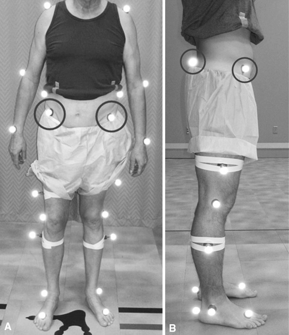
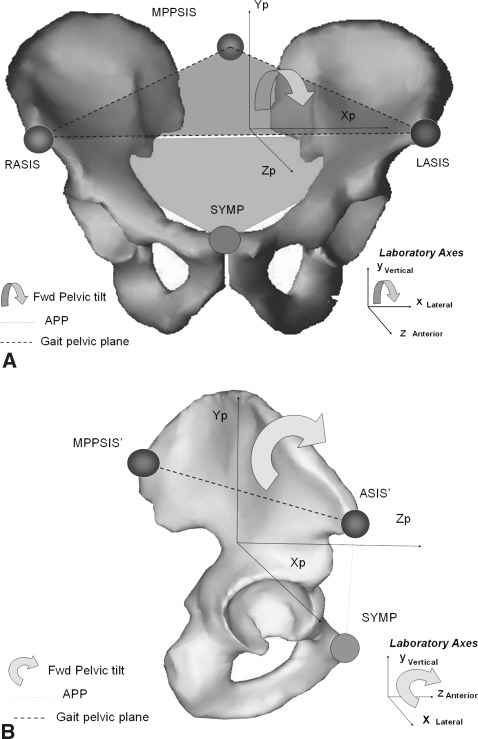
Gait analyses were performed preoperatively and postoperatively at 2 months and 12 months for all patients. Gait measurements were acquired with a computerized video motion analysis system utilizing 10 infrared cameras (EvaRT 4.0; Motion Analysis Corporation, Santa Rosa, CA). Retroreflective markers were placed at bony prominences for establishing anatomic coordinate systems for the pelvis, thigh, shank, and foot by the same physiotherapist for all trials and all patients. For the pelvis, the markers set included markers on the right and left anterior superior iliac spines (ASIS) and the midpoint between the right and left posterior superior iliac spines (PSIS) (Fig. 1A–B). The position of the pelvis was calculated relative to the laboratory coordinate system. Pelvic flexion/extension was defined as the motion of the mediolateral axis of the pelvis (Fig. 2A–B). An additional set of data corresponding to the standing position (static position) was recorded to calibrate the software. After a brief orientation session, the subject was instructed to walk at a self-selected pace on the laboratory walkway. Testing was conducted in a permanent motion analysis laboratory environment with a level vinyl-tiled floor. The 3-D marker coordinates were input to a commercial software program (OrthoTrak 5.0; Motion Analysis Corp.) to calculate the pelvic flexion/extension range of motion.
Fig. 1A–B.
The (A) lateral and (B) frontal views of the patient with the markers set are shown. The pelvis markers set included markers on the right and left anterior superior iliac spines (ASIS) and the midpoint between the right and left posterior superior iliac spines (PSIS). The position of the pelvis was defined by a marker set relative to a laboratory coordinate system.
Fig. 2A–B.
The (A) anteroposterior representation of the gait pelvic plane and the anterior pelvic plane (APP) is shown. The anterior pelvic plane is defined by the three points: left anterosuperior iliac spine (LASIS), the right anterosuperior iliac spine (RASIS), and the symphysis (SYMP). The gait pelvic plane is defined by the three points: LASIS, RASIS, and the midpoint between the right and left posterior superior iliac spines (MPPSIS). The forward pelvic tilt was defined as rotation around the axis defined by the LASIS and the RASIS seen by an observer positioned along a medial-lateral axis of the pelvis. (B) Mediolateral representation of the gait pelvic plane and the anterior pelvic plane (APP). The anterior pelvic plane is defined by the three points: LASIS, RASIS, and SYMP. The gait pelvic plane is defined by the three points: LASIS, RASIS, and the MPPSIS. The forward pelvic tilt was defined as rotation around the axis defined by the LASIS and the RASIS seen by an observer positioned along a mediolateral axis of the pelvis.
Gait cycle periods were selected by heel-strike to heel-strike events from three consecutive trials. All gait events were expressed as a percentage of the gait cycle, independent of the actual time for a stride, to produce a normalized gait cycle.
Pelvic kinematics were obtained for all patients at the three different evaluation times and average gait pelvic tilt was extracted by the same independent observer based on the Fournier analysis. Free-speed walking on a level surface is approximately periodic and, according to Sutherland et al. [22], angular analysis can be performed based on the assumption that angular rotations are periodic waveforms. Thus any periodic waveform can be constructed by superimposing a combination of waveforms that have the proper amplitudes, phases, and harmonics, and the data can be subjected to Fourier analysis, which mathematically resolves the data into these component waveforms [22]. Gait pelvic tilt (GPt) changes were obtained for each patient by comparison of the average (Av) value of the pelvic tilt at each time point (GPt change 1 = Av Pt 2 months − Av PT preoperative/GPt change 2 = Av PT 12 months − Av Pt 2 months/Pt change 3 = Av PT 12 months − Av preoperative).
Pelvic range-of-motion (ROM) values were obtained for all patients at the three different evaluation times from the pelvic kinematics based on the Fourier analysis. According to this model, pelvic ROM can be described as maximum amplitude of the waveform component, ie, the difference between the two extreme values of the pelvic tilt angles during the normalized gait cycle (pelvic ROM = maximum GPt − minimum GPt). All values of gait pelvic tilt and pelvic ROM were expressed in degrees.
To confirm the reliability of the gait pelvic tilt measurement, gait pelvic tilt values of 20 healthy subjects studied at two different time points were compared using a Bland and Altman method. For these 20 subjects, mean gait pelvic tilt change between the two exams was −0.22º ± 1.8º and all the gait pelvic tilt changes were less than 3º. Based on these results, changes greater than 3º were defined as clinical changes and not related to the intrinsic error of the method of evaluation.
Full sets of values from 21 patients were available preoperatively and at 2 months postoperatively and from 19 patients for the final evaluation at 12 months. One patient developed a Guillain-Barré syndrome between the 2-month and 12-month postoperative visit and another patient refused to perform the final evaluation.
Pelvic tilt values, gait pelvic tilt changes, and pelvic ROM at the three different evaluation times were described using means and standard deviations for the entire series. Then individual gait pelvic tilt changes were categorized as the following: less than 5º, between 5° and 10º, and more than 10º. Finally gait pelvic tilt values and pelvic ROM at the different time points were analyzed according to post-hoc comparisons for repeated measurements using a Student-Neuman-Keuls test [21]. Concerning the gait pelvic tilt evaluation, the null hypothesis was defined as no change of the gait pelvic tilt value over the three evaluation time. A p value of 0.05 was considered significant with a 95% confidence interval. We performed all analyses using SPSS software (version 12; SPSS, Inc., Chicago, IL). All calculations assumed two-tailed tests and a significance level of α = 0.05.
Results
The preoperative and 12-month mean gait pelvic tilt differed (p = 0.02), but the tilt did not differ between the preoperative and the 2-month postoperative evaluation (p = 0.06) or between the 2- and 12-month postoperative evaluation (p = 0.22) (Fig. 3). Global mean gait pelvic tilt changes were: −1.5º ± 3.3º (range, −0.6º to −7º) between the preoperative evaluation and the 2-month postoperative evaluation, −1.3º ± 4.8º (range, 0.1º to −12.3º) between the 2-month and the 12-month postoperative evaluation, and −3.01º ± 5.3º (range, 0.4º to −12.8º) between the preoperative and the 12-month evaluation. We observed no differences of the gait pelvic tilt changes among the three evaluation times. Individually between the preoperative and the 2-month evaluation, changes between 5º and 10º were observed for five patients (24%) (Fig. 4). Between the 2-month and 12-month postoperative evaluation, changes between 5º and 10º were observed for four patients (21%) and changes greater than 10º for two patients (11%) (Fig. 4). Between the preoperative and the 12-month evaluation, changes between 5º and 10º were observed for seven patients (37%) and changes greater than 10º for two patients (11%) (Fig. 4). Some changes were only observed between the preoperative and the 2-month postoperative analyses, while some changes were observed between the 2- and 12-month postoperative evaluations.
Fig. 3.
Gait pelvic tilt values for the preoperative evaluation (preoperative), the 2-month evaluation (2 month) and the 12-month evaluation (12 month) are shown. The boundaries of the boxes indicate the 25th and 75th percentiles, and the black lines within the boxes mark the mean values. The whiskers above and below the boxes indicate the ninth and 10th percentiles and the isolated spots represent the outliers.
Fig. 4.
The intraindividual gait pelvic tilt changes are expressed in degrees and categorized as less than 5°, between 5° and 10°, and greater than 10°; between the 2-month and preoperative evaluation (Time 2–Time 1), between the 12-month and 2-month postoperative evaluation and between the 12-month and preoperative evaluation (Time 3–Time 1).
Pelvic ROM decreased from preoperatively to 2 months (p = 0.003) and 12 months (p = 0.0026) and from 2 months to 12 months (p = 0.0025) (Fig. 5). Mean preoperative pelvic ROM was 6.6º ± 3º (range, 2.7º–13.6º). Mean postoperative pelvic ROM was 5.5º ± 2.7º (range, 2.5º–11.8º) for the 2-month evaluation and 4.2º ± 1.8º (range, 1.8º–9.1º) for the 12-month evaluation.
Fig. 5.
Pelvis range of motion for the preoperative evaluation (preoperative), the 2-month evaluation (2 month) and the 12-month evaluation (12 month). The boundaries of the boxes indicate the 25th and 75th percentiles, and the black lines within the boxes mark the mean values. The whiskers above and below the boxes indicate the ninth and 10th percentiles and the isolated dots represent the outliers.
Discussion
Computer-assisted navigation systems in total hip arthroplasty have been developed to improve acetabular cup positioning. These systems are accurate in hitting a fixed target, when considering the pelvis as a fixed bone unit [11, 20]. This implies that the pelvic position is the same before and after THA, but this has never been verified under dynamic conditions [3, 18]. We hypothesized that dynamic pelvic tilt and range of motion would be consistent before and after THA. Therefore, using a modern comprehensive gait analysis we sought to compare (1) dynamic pelvic tilt changes before and after THA and (2) pelvis flexion/extension range-of-motion changes before and after arthroplasty. Our data suggest individual changes greater than 5º for 24% of the patients between the preoperative evaluation and the 2-month evaluation, for 31% of the patients between the 2-month and 12-month evaluation, and for 49% of the patients between the preoperative and the 12-month evaluation. Furthermore, a notable decrease of the pelvic range of motion was observed between the preoperative evaluation and the 12-month postoperative evaluation. According to these results we were unable to verify our hypothesis, defined as no change of the gait pelvic tilt value over the three evaluation times.
One limitation of our study was the absence of a combined static/dynamic evaluation. We did not perform any mediolateral radiographs of the pelvis to calculate the pelvic tilt value and its correlation with the gait pelvic tilt. Thus direct comparisons of the absolute values of the gait pelvic tilt with the previously published pelvic tilt values were not possible. However, as the posterior and anterior part of the pelvis are part of one motion unit, the relatives changes of the gait pelvic tilt can be compared the changes of the pelvic tilt in those previous static studies [24]. Another limitation of our study was the lack of combinative evaluation of the pelvic obliquity, rotation, and flexion/extension motion. Pelvic range of motion is a complex phenomenon but as the sagittal pelvic position is the only variable concerning the pelvis fed into the computer-assisted system and influencing the final cup position we choose to focus on the sagittal range of motion of the pelvis. Furthermore due to the small number of patients, we were unable to evaluate the correlation between the dorsolumbar spine condition, hip range of motion, and the pelvis range of motion. Despite these limitations, this is to our knowledge the first study evaluating dynamic pelvic tilt changes before and after arthroplasty.
Two studies reported static changes of pelvic tilt after THA [3, 18]. The range of changes observed in our study were comparable to the two previous studies evaluating pelvic tilt before and after THA using static methods such as lateral radiograph of the pelvis or CT scan [3, 18]. Nishihara et al. [18] compared the changes in pelvic flexion angles in the same posture (supine, sitting, and standing positions) before and 1 year after THA in 74 patients with a static method of evaluation (combining a 3-D CT scan reconstruction and a standard AP radiograph of the pelvis). The position of the anterior pelvic plane relative to the vertical plane was calculated and defined as the pelvic flexion [18]. The mean ± SD changes were −2º ± 7.5° (range, −26º to −15º) in standing position, −3º ± 5° (range, −14º to −8º) in supine position, and 1º ± 8.7° (range, −25º to −24º) in sitting position [18]. The pelvic flexion changes were lower than 10° for 87% of the patients [18]. The ranges of observed changes in that static study were comparable to our dynamic data. DiGioia et al. [3] reported the results of a study comparing the sagittal pelvic orientation in different positions (standing and sitting) before and after THA in 84 patients. Lateral radiographs of the pelvis in standing and sitting position were performed and the pelvic tilt was calculated [3, 7]. The mean pelvic tilt was 1.2º ± 7.9° (range, −22.5º to 27º) preoperatively and 1.1º ± 8.2° (range, −12.5º to 20º) postoperatively in standing position [3]. That static study reported no differences between preoperative and postoperative pelvic tilt for the entire group [3]. Individual changes were not directly reported but changes in the extreme values suggested individual changes greater than 10º [3]. In these two previous static studies, as in our study, clinically important changes in the pelvic sagittal position were observed in a subset of the patients [3, 18].
Our data demonstrate a decrease in the pelvic range of motion after THA. These changes can be considered a return to a more physiologic gait pattern and have previously been observed [17]. Higher preoperative range of motion of the pelvis can be induced by pain and stiffness in the hip joint before surgery [9]. This alteration in the pattern of motion was previously interpreted as a mechanism to increase effective extension of the hip during stance through increased anterior pelvic tilt and lumbar lordosis [9]. Observations on the frontal trunk and pelvic range of motion before and after arthroplasty have been reported on a group of 12 patients, but nothing concerning the sagittal pelvic range of motion [23]. Therefore we were unable to compare our results with results of previous studies of the literature.
Changes in the pelvic position and pelvic motion were observed for a substantial subset of the studied patients. Complementary comprehensive studies on a larger group of patients are now mandatory to improve the understanding of the pelvic motion after THA. Specifically, future studies should assess the global 3-D aspects of pelvic motion during gait before and after THA in order to clearly estimate the consequences of the pelvic motion for cup positioning. While substantial efforts have been devoted to improve acetabular cup positioning to reduce dislocation, to improve hip range of motion, and to reduce wear after THA, the ideal target for cup position in individual patients remains unclear [1, 6, 10, 12, 14]. Most previous studies of ideal cup position have been performed without considering interindividual variation of the pelvic tilt [1, 6, 10, 12, 14]. The integration of pelvic position into preoperative planning may support the concept of functional acetabular alignment defined by DiGioia et al. [3] as the combination of the implant alignment in bone and pelvic orientation relative to the vertical. This preoperative analysis supports individual anteversion target value planning rather than a 15° ± 5º universal target as recommended by Lewinnek et al. [14]. Our study demonstrated pelvic tilt changes greater than 5° for a substantial subset of patients. Integrating preoperative pelvic tilt in the surgical planning may improve cup positioning, but substantial variations after THA may limit these benefits. While the clinical consequences of these changes on hip stability and implant wear are not known, surgeons should be aware that substantial dynamic pelvic tilt changes after THA do occur, and that the pelvis is not a fixed static bone unit when considering cup positioning. Thus using a computer-assisted system may help to obtain a precise anatomic alignment of the cup but this may result in a maladapted functional alignment of the cup. Complementary studies are now mandatory to find preoperative predictive factors to identify which patient may present a substantial dynamic change and the direction of the change postoperatively. Then, surgeons will be able to define the individual ideal functional position of the cup.
Acknowledgments
We thank Emily Berg for her help in the study coordination, and Kathie Bernhardt and Diana Hansen for their help during the data collection.
Footnotes
Each author certifies that he or she has no commercial associations (eg, consultancies, stock ownership, equity interest, patent/licensing arrangements, etc) that might pose a conflict of interest in connection with the submitted article.
Each author certifies that his or her institution has approved the human protocol for this investigation and that all investigations were conducted in conformity with ethical principles of research, and that informed consent for participation in the study was obtained.
References
- 1.Biedermann R, Tonin A, Krismer M, Rachbauer F, Eibl G, Stockl B. Reducing the risk of dislocation after total hip arthroplasty: the effect of orientation of the acetabular component. J Bone Joint Surg Br. 2005;87:762–769. [DOI] [PubMed]
- 2.Blendea S, Eckman K, Jaramaz B, Levison TJ, Digioia AM 3rd. Measurements of acetabular cup position and pelvic spatial orientation after total hip arthroplasty using computed tomography/radiography matching. Comput Aided Surg. 2005;10:37–43. [DOI] [PubMed]
- 3.DiGioia AM, Hafez MA, Jaramaz B, Levison TJ, Moody JE. Functional pelvic orientation measured from lateral standing and sitting radiographs. Clin Orthop Relat Res. 2006;453:272–276. [DOI] [PubMed]
- 4.DiGioia AM, Jaramaz B, Blackwell M, Simon DA, Morgan F, Moody JE, Nikou C, Colgan BD, Aston CA, Labarca RS, Kischell E, Kanade T. The Otto Aufranc Award. Image guided navigation system to measure intraoperatively acetabular implant alignment. Clin Orthop Relat Res. 1998;355:8–22. [DOI] [PubMed]
- 5.Digioia AM 3rd, Jaramaz B, Plakseychuk AY, Moody JE Jr, Nikou C, Labarca RS, Levison TJ, Picard F. Comparison of a mechanical acetabular alignment guide with computer placement of the socket. J Arthroplasty. 2002;17:359–364. [DOI] [PubMed]
- 6.D’Lima DD, Urquhart AG, Buehler KO, Walker RH, Colwell CW, Jr. The effect of the orientation of the acetabular and femoral components on the range of motion of the hip at different head-neck ratios. J Bone Joint Surg Am. 2000;82:315–321. [DOI] [PubMed]
- 7.Eckman K, Hafez MA, Ed F, Jaramaz B, Levison TJ, Digioia AM, 3rd. Accuracy of pelvic flexion measurements from lateral radiographs. Clin Orthop Relat Res. 2006;451:154–160. [DOI] [PubMed]
- 8.Eddine TA, Migaud H, Chantelot C, Cotten A, Fontaine C, Duquennoy A. Variations of pelvic anteversion in the lying and standing positions: analysis of 24 control subjects and implications for CT measurement of position of a prosthetic cup. Surg Radiol Anat. 2001;23:105–110. [DOI] [PubMed]
- 9.Hurwitz DE, Hulet CH, Andriacchi TP, Rosenberg AG, Galante JO. Gait compensations in patients with osteoarthritis of the hip and their relationship to pain and passive hip motion. J Orthop Res. 1997;15:629–635. [DOI] [PubMed]
- 10.Jolles BM, Zangger P, Leyvraz PF. Factors predisposing to dislocation after primary total hip arthroplasty: a multivariate analysis. J Arthroplasty. 2002;17:282–288. [DOI] [PubMed]
- 11.Kalteis T, Handel M, Bathis H, Perlick L, Tingart M, Grifka J. Imageless navigation for insertion of the acetabular component in total hip arthroplasty: is it as accurate as CT-based navigation? J Bone Joint Surg Br. 2006;88:163–167. [DOI] [PubMed]
- 12.Kennedy JG, Rogers WB, Soffe KE, Sullivan RJ, Griffen DG, Sheehan LJ. Effect of acetabular component orientation on recurrent dislocation, pelvic osteolysis, polyethylene wear, and component migration. J Arthroplasty. 1998;13:530–534. [DOI] [PubMed]
- 13.Lembeck B, Mueller O, Reize P, Wuelker N. Pelvic tilt makes acetabular cup navigation inaccurate. Acta Orthop. 2005;76:517–523. [DOI] [PubMed]
- 14.Lewinnek GE, Lewis JL, Tarr R, Compere CL, Zimmerman JR. Dislocations after total hip-replacement arthroplasties. J Bone Joint Surg Am. 1978;60:217–20. [PubMed]
- 15.McCollum DE, Gray WJ. Dislocation after total hip arthroplasty. Causes and prevention. Clin Orthop Relat Res. 1990;261:159–170. [PubMed]
- 16.McKibbin B. Anatomical factors in the stability of the hip joint in the newborn. J Bone Joint Surg Br. 1970;52:148–159. [PubMed]
- 17.Miki H, Sugano N, Hagio K, Nishii T, Kawakami H, Kakimoto A, Nakamura N, Yoshikawa H. Recovery of walking speed and symmetrical movement of the pelvis and lower extremity joints after unilateral THA. J Biomech. 2004;37:443–455. [DOI] [PubMed]
- 18.Nishihara S, Sugano N, Nishii T, Ohzono K, Yoshikawa H. Measurements of pelvic flexion angle using three-dimensional computed tomography. Clin Orthop Relat Res. 2003;411:140–151. [DOI] [PubMed]
- 19.Pagnano MW, Trousdale RT, Meneghini RM, Hanssen AD. Patients preferred a mini-posterior THA to a contralateral two-incision THA. Clin Orthop Relat Res. 2006;453:156–159. [DOI] [PubMed]
- 20.Parratte S, Argenson JN. Validation and usefulness of a computer-assisted cup-positioning system in total hip arthroplasty. A prospective, randomized, controlled study. J Bone Joint Surg Am. 2007;89:494–499. [DOI] [PubMed]
- 21.Sloan DA, Donnelly MB, Schwartz RW, Strodel WE. The Objective Structured Clinical Examination. The new gold standard for evaluating postgraduate clinical performance. Ann Surg. 1995;222:735–742. [DOI] [PMC free article] [PubMed]
- 22.Sutherland DH, Olshen R, Biden EN, Wyatt MP. Modeling and prediction regions for motion data. In: The Development of Mature Walking. Oxford, UK/Philadelphia, PA: Blackwell/Lippincott; 1988:24–32.
- 23.Vogt L, Brettmann K, Pfeifer K, Banzer W. Walking patterns of hip arthroplasty patients: some observations on the medio-lateral excursions of the trunk. Disabil Rehabil. 2003;25:309–317. [DOI] [PubMed]
- 24.Vogt L, Portscher M, Brettmann K, Pfeifer K, Banzer W. Cross-validation of marker configurations to measure pelvic kinematics in gait. Gait Posture. 2003;18:178–184. [DOI] [PubMed]
- 25.Wolf A, Digioia AM 3rd, Mor AB, Jaramaz B. Cup alignment error model for total hip arthroplasty. Clin Orthop Relat Res. 2005;437:132–137. [DOI] [PubMed]
- 26.Wolf A, DiGioia AM, 3rd, Mor AB, Jaramaz B. A kinematic model for calculating cup alignment error during total hip arthroplasty. J Biomech. 2005;38:2257–2265. [DOI] [PubMed]


