Abstract
The objective of this study was to determine if inflammatory tolerance and enhancement of innate immune function could be induced by the gram-positive cell wall component peptidoglycan (PGN). Male mice (C57BL6/J or C3H/HeJ, 8-12 weeks of age) were given intraperitoneal injections of 1mg PGN on 2 consecutive days. The mice were then challenged with lipopolysaccharide (LPS) or live Pseudomonas aeruginosa (1 × 10⁸ colony-forming units) 2 days after the second pretreatment. Mice pretreated with PGN had diminished plasma concentrations of TNFα and IFNγ and elevated concentrations of IL-10 in response to a subsequent LPS or Pseudomonas challenge when compared to untreated controls. Bacterial clearance was improved in mice pretreated with PGN, and mortality in response to a subsequent Pseudomonas challenge was significantly attenuated. PGN pretreatment of LPS-unresponsive mice (C3H/HeJ) verified that the effect of PGN pretreatment was not due to any LPS contamination. We have previously demonstrated that PGN pretreatment induced resistance to a Gram-positive bacterial challenge. The present study extends those results by showing that exposure to the Gram-positive bacterial cell wall component peptidoglycan also induces cross-tolerance to LPS and non-specifically enhances innate immune function in that PGN-pretreated mice had increased resistance to Gram-negative bacterial challenge.
Keywords: mice, peptidoglycan, tolerance, bacterial infection, TLR4
1. Introduction
Injection of the Gram-negative bacterial cell wall molecule lipopolysaccharide (LPS) can initiate a dramatic proinflammatory response that results in profound hemodynamic derangements and may even progress to shock and death. However, animals that survive a sublethal exposure to LPS are resistant to subsequent challenges with normally lethal doses of LPS during the first few days following the initial exposure [1]. Although the cytokine profile in LPS tolerant animals is similar to that seen in septic patients, innate immune function in the LPS tolerant state actually seems improved in that bacterial clearance is enhanced and mortality is suppressed after a Gram-negative bacterial challenge [2]. We have previously shown that induction of LPS tolerance not only diminishes the inflammatory response to subsequent immune challenges but also appears to non-specifically enhance the effectiveness of the innate immune system to clear both Gram-negative and Gram-positive bacterial challenges [3-5]. Further, exposure to LPS confers resistance to inflammation and injury induced by other challenges including myocardial infarction [6], neural ischemia [7], and gastric insults [8].
Over the last decade, significant advances have been made in our understanding of the process employed by innate immune cells to detect, and react to, a myriad of microbial organisms. It is now known that innate detection of a wide spectrum of microbes relies upon recognition of a relatively small number of molecules that are common to broad classifications of microorganisms. These common, conserved pathogen-associated molecular patterns (PAMPs) act as ligands for toll-like receptors and other pattern-recognition receptors (PRRs) of immune cells, and this interaction initiates both the inflammatory response and the innate immune response to the invading microorganisms. For example, an innate immune response against some bacterial infections is initiated in part by LPS, a TLR4 ligand in Gram-negative bacterial cell walls [9]. Recent findings demonstrate that different PRR's are activated by gram-positive bacterial molecules when compared to activation by gram-negative bacterial molecules and it is likely that, during an infection, multiple PRRs are activated by different bacterial molecules. How the host responds to different PRR ligands and how PRR activation may influence responses to infection are basic questions that may ultimately provide insight into the pathogenesis of sepsis. We have previously demonstrated that pretreatment with peptidoglycan, derived from the cell wall of Gram-positive bacteria, resulted in enhanced resistance to challenge with the Gram-positive bacteria Staphylococcus aureus (Murphey, In Press, Crit Care Med). In the present study, we were interested in determining if tolerance induced by peptidoglycan was specific for S. aureus or Gram positive bacteria, or if PGN-induced tolerance was associated with a nonspecific enhancement of innate immune function in response to a live bacterial challenge similar to that observed after LPS pretreatment. The aim of this study was to determine if peptidoglycan (PGN) could induce tolerance to a subsequent Gram-negative bacterial challenge. We pretreated mice with PGN and studied the response of the mice to a subsequent challenge with live Pseudomonas aeruginosa, a bacterium frequently associated with nosocomial infections and a major pathogen in critically ill patients.
2. Materials and Methods
2.1 Tolerance Model
Male, 6-8 week old wild type C57BL/6J, C3H/HeJ (TLR4 mutant), and C3H/HeOuJ (TLR4 intact) mice were purchased from the Jackson Laboratory (Bar Harbor, ME). Mice were housed in a monitored, light-dark cycled environment and provided with standard lab chow and water ad libitum. Peptidoglycan (PGN), derived from Staphylococccus aureus, was purchased from Sigma Chemical (St. Louis, MO). Mice were injected with PGN (1mg in 0.1ml of saline i.p.) once daily for 2 consecutive days. Control mice received normal saline (0.1 ml) in the same regimen. All mice were injected at the same time each morning. All studies were approved by the Institutional Animal Care and Use Committee (IACUC) at the University of Texas Medical Branch and met National Institutes of Health guidelines for the use of experimental animals in research.
2.2 Gram-negative bacterial challenge
In the initial group of mice, LPS (E. coli-derived 0111.B4, Sigma Chemical, St. Louis, MO) was injected (100 μg i.v.) 48 hrs after the PGN pretreatment. For subsequent groups, live Pseudomonas aeruginosa (strain 19660, American Type Culture Collection, Rockville, MD) was inoculated into tryptic soy broth and allowed to replicate overnight in a shaking incubator at 37°C. The resulting bacterial culture was washed with 10ml of sterile 0.9% saline. Viable numbers of colony-forming units (cfu) were determined by plating serial dilutions overnight on tryptic soy agar. Bacteria were suspended in sterile 0.9% saline at a final concentration of 1 × 10⁹ cfu/ml. Mice were challenged with 0.1 ml of this suspension (1 × 10⁸ cfu; i.v.) 2 days after the second dose of PGN.
2.3 Bacterial Clearance
The mice were sacrificed under isoflurane 6 hours after intravenous injection of Pseudomonas aeruginosa. Lungs, spleens, and liver tissue samples were aseptically excised, weighed and homogenized in sterile saline using sterile tissue grinders. Serial dilutions of tissue homogenates were plated on tryptic soy agar and incubated overnight at 37°C. Bacterial colony-forming units were counted to assess bacterial burden.
2.4 Enzyme-linked Immunosorbent Assay (ELISA)
Blood samples were collected 6 hours after the Pseudomonas challenge and plasma cytokine concentrations were measured by ELISA. IFN-γ, IL-10, and TNFα ELISA kits (eBiosciences, San Diego, CA) were used to measure cytokine concentrations in plasma according to the manufacturer's instructions. Briefly, standards or experimental samples were added to 96-well plates coated with monoclonal antibody against the cytokine of interest and incubated for 2 hours. After washing, horseradish peroxidase-conjugated, cytokine-specific antibody was added to each well, incubated for 2 hours, and washed. Substrate solution (TMB, Sigma Chemical, St. Louis, MO) was added and incubated for 30 minutes, and the reaction was terminated by the addition of stop solution (2N H2SO4). Cytokine concentrations were determined by measuring optical density at 450 nM using a microtiter plate reader (Dynatech Laboratories, Chantilly, VA).
2.5 Analysis of splenocyte phenotype
48 hours after the second PGN injection, spleens were aseptically excised and transferred to 6-well culture plates containing RPMI 1640. The spleens were minced and passed through sterile mesh. Erythrocytes were lysed (erythrocyte lysis kit, R&D Systems). The remaining splenocytes were resuspended in PBS and incubated for 30 minutes at 4°C with fluorescence-labeled antibodies including anti-F4/80, anti-mouse neutrophil (clone7/4), and anti-ly6-C/G (Caltag Laboratories); FcγIII/IIR (BD Pharmingen); and anti-TLR4 (Imgenex). Isotype-matched antibodies were used as controls. Cells were washed with PBS and resuspended in 1% paraformaldehyde before analysis by a FACScan flow cytometer (BD Biosciences). Data were analyzed and graphed by WinMidi software.
2.6 Data Analysis
Results are presented as mean ± SEM. Multiple group data were analyzed by ANOVA and post-hoc Tukeys test while comparisons between 2 groups were performed by unpaired t test. Survival curves were analyzed by log-rank test. A value of p<0.05 was considered statistically significant.
3. Results
3.1 Pretreatment with peptidoglycan was associated with a decreased plasma IFNγ and TNFα and an increased plasma IL-10 response to subsequent challenge with LPS or Pseudomonas aeruginosa
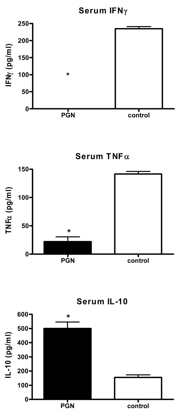
Administration of PGN alone resulted in elevated plasma concentrations of TNFα and IL-10, but not IFNγ, at 6 hours after injection (Table 1). In the absence of further challenge, those cytokines were not detectable 48 hours later. However, mice challenged with LPS 48 hours after pretreatment with PGN had attenuated induction of plasma IFNγ and TNFα when compared to saline pretreated control mice (Table 1). Plasma IL-10 concentrations were significantly higher in PGN-pretreated animals than in control animals after challenge with LPS (Table 1). Similarly, mice pretreated with PGN prior to a live bacterial challenge with Ps. aeruginosa had attenuated induction of plasma IFNγ and TNFα when compared to saline treated mice (Fig. 1). Plasma IL-10 concentrations were significantly higher in pretreated animals than in control animals after challenge with Pseudomonas. (Fig. 1).
Table 1.
Plasma cytokine concentration after i.p. injection of PGN alone, or after PGN followed by LPS challenge.
| Pretreatment | Challenge | IFNγ
(pg/ml) |
IL-10
(pg/ml) |
TNFα
(pg/ml) |
|---|---|---|---|---|
| PGN – 6hrs1 | None | ND | 418 ± 135 * | 123 ± 44 * |
| Saline – 6hrs | None | ND | ND | ND |
| PGN – 48hrs2 | None | ND | ND | ND |
| Saline – 48hrs | None | ND | ND | ND |
| PGN – 48hrs3 | LPS | 22 ± 17 * | 1850 ± 473 * | 103 ± 36 |
| Saline – 48hrs | LPS | 140 ± 76 | 594 ± 154 | 148 ± 49 |
PGN (or saline) was injected and blood samples collected either 6 or 48 hours later without further challenge.
PGN (or saline) was injected and blood samples collected either 6 or 48 hours later without further challenge.
In the third group, LPS was injected 48 hours after PGN pretreatment and blood collected 6 hours after the LPS challenge. Data shown are Mean ± SEM. ND = Not detectable. Lower limit of detection for IFNγ assay was 8 pg/ml; 30pg/ml for the IL-10 assay; and 8 pg/ml for the TNFα assay.
= p<0.05 vs. respective saline control, N = 5 per group.
Fig. 1.
Mice were pretreated with peptidoglycan (PGN) for 2 consecutive days and subsequently challenged with live Pseudomonas aeruginosa 2 days after the second PGN dose. Blood samples were collected 6 hours after the Pseudomonas challenge, and plasma concentrations of IFNγ, TNFα, and IL-10 were determined by ELISA. (* indicates p<0.05 vs control, N= 5 per group).
3.2 Pretreatment with peptidoglycan was associated with enhanced clearance of a Pseudomonas aeruginosa challenge
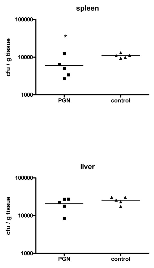
Mice were injected with PGN on 2 consecutive days. 48 hr after the second pretreatment, the mice were challenged with an i.v. injection of 1 × 10⁸ cfu Pseudomonas bacteria and sacrificed 6 hr later for sample collection. Mice pretreated with PGN had approximately 2-fold lower concentrations of bacterial colony-forming units in spleen tissue when compared to control animals (Fig. 2). Bacterial growth in liver tissue did not differ between the pretreated and control animals.
Fig. 2.
Mice were pretreated with peptidoglycan (PGN) prior to challenge with live Pseudomonas aeruginosa. The mice were sacrificed 6 hours later. The number of bacteria cultured from the spleens of mice was significantly lower in the PGN-pretreated group than in saline-pretreated controls (*indicates p<0.05 vs. control, N = 5 per group).
3.3 Pretreatment with peptidoglycan was associated with an increase in number of macrophages and an increase in TLR4 cell surface expression
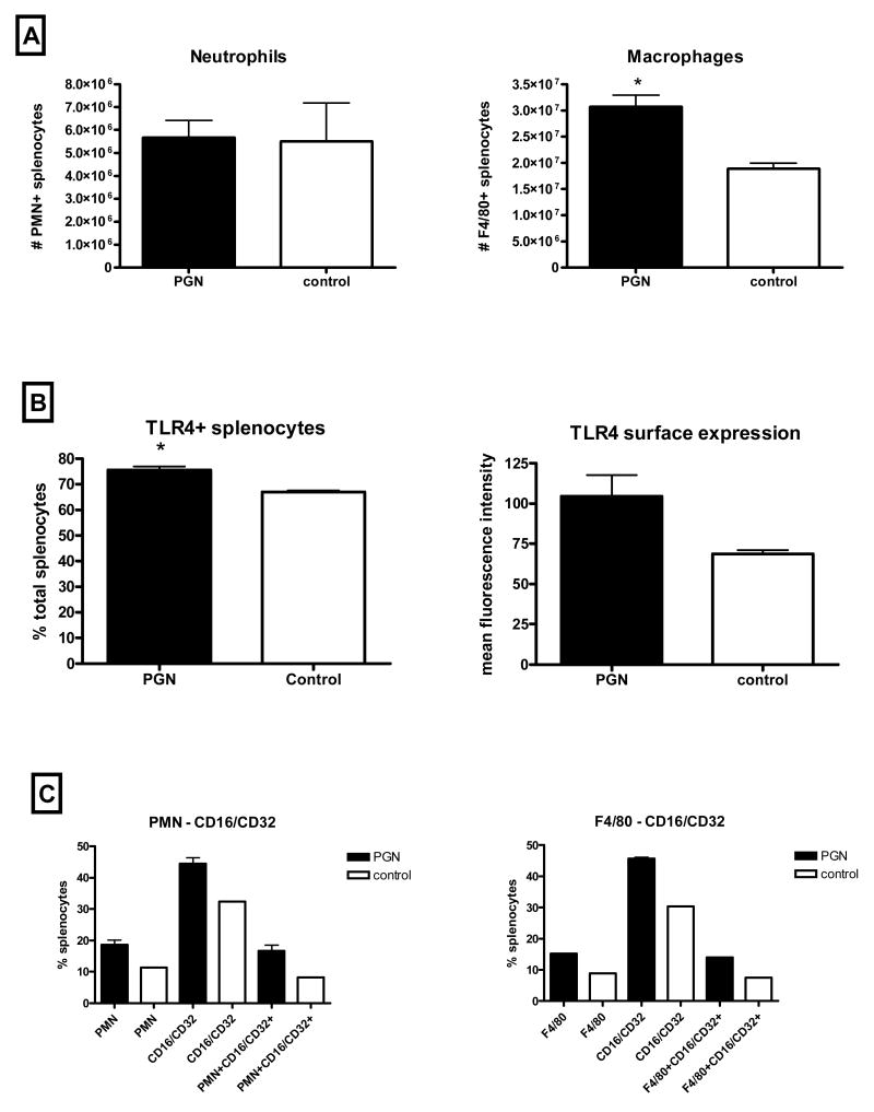
Mice were injected with PGN on 2 consecutive days. 48 hr after the second pretreatment, the mice were sacrificed and spleen samples were homogenized for flow cytometric analysis. Pretreatment with PGN was associated with significantly elevated numbers of macrophages, but not neutrophils, when compared to control mice (Fig. 3A). The percent of total splenocytes with cell surface expression of TLR4 was increased in pretreated mice when compared to control mice. Additionally, surface expression of TLR4 appeared to be increased on a per cell basis based upon higher mean fluorescence of individual cells (Fig. 3B). Both macrophages and neutrophils tended to have higher cell surface expression of FcγIII/IIR and TLR4 in mice that had been pretreated compared to control mice (Fig. 3C).
Fig. 3.
Mice were pretreated with peptidoglycan (PGN). The mice were sacrificed 48 hours later. Spleen homogenates from pretreated mice had more macrophages, and higher cell surface expression of TLR4 and FcγIII/IIR than those from control mice (* indicates p<0.05, N = 3 per group, experiment repeated with similar results).
3.4 Pretreatment with peptidoglycan was associated with decreased mortality after a Pseudomonas aeruginosa challenge
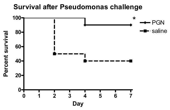
Mice were injected with PGN on 2 consecutive days. 48 hr after the pretreatment, the mice were challenged with an i.v. injection of 1 × 10⁸ cfu of Pseudomonas bacteria and observed for mortality over the next 7 days. One of the 10 mice that had been injected with PGN prior to the Pseudomonas challenge died during the observation period, compared to 60% mortality in control mice by day 4 (Figure 4).
Fig. 4.
Mice were pretreated with peptidoglycan (PGN) prior to challenge with live Pseudomonas aeruginosa. The mice were observed for mortality for 7 days after the challenge. Mice that were pretreated had significantly better survival after the Pseudomonas challenge than control mice (p<0.05, N = 10 per group).
3.5 Enhancement of bacterial clearance was independent of signaling through the TLR4 receptor
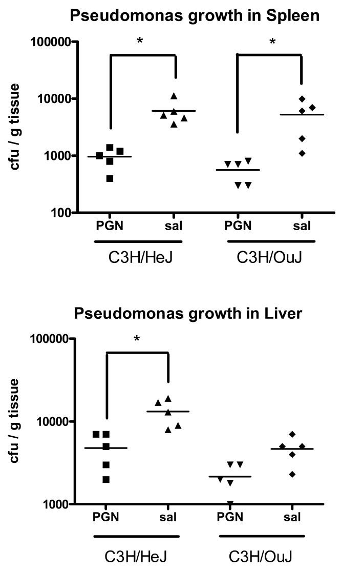
LPS-unresponsive, TLR4 mutant C3H/HeJ mice, or TLR4 intact C3H/HeOuJ mice were injected with PGN for 2 consecutive days. The mice were challenged with an i.v. injection of 1 × 10⁸ cfu of Pseudomonas aeruginosa bacteria 48 hr after the second PGN injection and killed 6 hr later for sample collection. As was demonstrated in C57BL/6 mice, bacterial counts in spleen tissues were lower in C3H/HeJ mice pretreated with PGN when compared to saline-pretreated controls. Liver bacterial counts were also significantly lower in PGN-pretreated C3H/HeJ mice compared to saline-pretreated controls (Figure 5).
Fig. 5.
TLR4 mutant C3H/HeJ mice, or TLR4 intact C3H/HeOuJ mice, were pretreated with peptidoglycan (PGN) prior to challenge with live Pseudomonas aeruginosa. The mice were sacrificed 6 hours later. Mice that were pretreated with PGN had significantly decreased numbers of bacterial-forming colonies in spleen and liver tissue homogenates when compared to control mice (* indicates p<0.05, N = 5 per group).
4. Discussion
These studies demonstrate that, similar to LPS from Gram-negative bacteria, a component of the cell wall of Gram-positive bacteria can reduce inflammation and induce enhancement of the innate immune response to a subsequent bacterial challenge. We have previously shown that pretreatment with peptidoglycan (PGN) derived from Staphylococcus aureus improved the resistance of mice to a challenge with live S. aureus (In Press, Crit Care Med). The present study extends those findings by demonstrating that the enhancement of innate immune function was not specific to that bacterium or to Gram positive bacteria, and suggests that the mechanism does not involve targeting of PGN specifically. In this study, S. aureus-derived PGN induced tolerance to a subsequent Pseudomonas aeruginosa challenge that was demonstrated by a diminished proinflammatory cytokine (TNFα) response; diminished Th1 cytokine response (IFNγ); and an increased Th2 cytokine response (IL-10). Despite the predominant Th2 response, PGN-pretreated animals demonstrated enhanced bacterial clearance after the Pseudomonas challenge. These results were duplicated in LPS-resistant C3H/HeJ mice, suggesting both that TLR4 signaling was not necessary for those effects and that tolerance induction was not a result of LPS contamination of the peptidoglycan (PGN).
Inducible inflammatory tolerance was first described in 1947 by Beeson who noted that consecutive administrations of fractionated bacterial suspensions to rabbits were associated with progressively diminished physiologic responses [10]. Since those observations, purified LPS has been reported to induce tolerance not only to subsequent injections of LPS but also to a variety of other stimuli and injuries, including tolerance to CpG DNA [11], TNFα and IL-1β [12], ethanol intoxication [13] and an attenuation of inflammation and injury after a variety of ischemic insults [6-8]. Similarly, tolerance to LPS can be induced by a variety of other pretreatments, including cytokines [12,14], heat shock proteins [15], and other bacterial peptides including lipoproteins and lipoteichoic acid [16-18].
Tolerance induced by components of gram-positive bacterial cell walls has not been studied as extensively as tolerance induced by LPS. Other components of gram-positive bacteria include lipoteichoic acid (LTA), which is a polymer anchored in the outer cytoplasmic membrane by a lipid moiety and, as such, is the Gram-positive bacterial equivalent to LPS from Gram-negative bacteria. LPS-binding protein and CD14 facilitate recognition of LTA, although cell stimulation occurs through TLR2 rather than TLR4 [19]. In cell culture, exposure of macrophages to either LTA or PGN results in suppression of cytokine release after restimulation with the same molecule [18,20]. In the case of LTA, tolerance induction was dependent upon TLR2 [18]. We have tested pretreatment with lipoteichoic acid followed by challenge with Staphylococcus aureus and found a similar tolerance induction, albeit not as strong as that induced by PGN (unpublished observations). In other animal models, pretreatment with LTA reduced cardiac injury induced by ischemia/reperfusion similar to the protection provided by pretreatment with LPS [21]. There are few studies of gram-positive tolerance induction and subsequent effects on innate immune function. Ausobsky et al tested muramyl dipeptide, a component of peptidoglycan, in an in vivo model and reported an attenuation of mortality after challenge with Klebsiella [22]. O'Brien et al demonstrated tolerance and enhanced clearance of a Gram-negative bacterial challenge in mice pretreated with bacterial lipoprotein, a molecule derived from both Gram-positive and Gram-negative bacteria [23].
The first step in host defense is recognition of microbial organisms. The innate immune system must recognize a vast number of different pathogens and distinguish them from self. In the absence of ability to specifically recognize pathogens, the innate immune system relies upon recognition of LPS, lipoteichoic acid, peptidoglycan, and other bacterial molecules such as flagellin or unique nucleic acids which are highly conserved across broad classes of bacteria and, as such, provide the innate immune system with an effective method to detect a diverse multitude of microbial organisms. A consequence of the interaction of these pathogen-associated ligands with Toll-like and other innate receptors on macrophages, monocytes, and neutrophils is up-regulation of inflammatory gene expression and subsequent release of inflammatory cytokines and chemokines. Peptidoglycan, consisting of glycan strands cross-linked by peptides, is the major component of the cell wall of gram-positive bacteria. PGN administration effectively enhanced bacterial clearance not only in C57BL6/J mice but also in TLR4 mutant C3H/HeJ mice when compared to their respective controls, suggesting that the enhancement of immune function occurred through mechanisms other than TLR4 signaling. Detection and activation of immune cells in response to PGN primarily occurs through alternate pattern recognition receptors including NOD1 and NOD2 [24,25], CD14, and a family of peptidoglycan recognition proteins (PGRP's) [26] and it remains possible that the mechanism of PGN-induced tolerance worked through these receptor signaling pathways.
How tolerance induction exerts a positive effect on bacterial clearance is not entirely clear. The number of phagocytic cells was increased after tolerance induction, and surface expression of complement receptor 3 and FcγIII/IIR have been shown to be increased on cells from tolerant animals [17]. Both of those molecules have roles in phagocytosis, so tolerance enhancement of bacterial clearance may be due to increased numbers and activity of phagocytic cells. Recent work confirmed that phagocytosis is significantly improved after induction of tolerance by LPS [27]. Further, surface expression of TLR4 was increased in animals pretreated with PGN in this study. Interestingly, TLR4 was not increased in mice after LPS pretreatment in one of our previous studies, suggesting that this finding was associated, but not causally related to enhanced bacterial clearance.
The clinical relevance of tolerance induction is unclear. Trauma patients may have had an initial proinflammatory response associated with their primary injury. Often, the onset of sepsis in these patients is associated with anti-inflammatory cytokine profiles, and leukocytes isolated from septic patients have a suppressed response to inflammatory stimuli. Sepsis due to Gram-positive infections has been associated with a lesser degree of macrophage hyporesponsiveness to inflammatory stimuli than that seen in patients with Gram-negative sepsis [28]. From a clinical perspective the phenomenon of inducible inflammatory tolerance is of obvious interest because attenuation of the proinflammatory response may have important benefits, especially if it can be achieved while maintaining or even enhancing the innate immune clearance of pathogenic organisms. We observed that pretreatment of mice with either Gram-negative (LPS) or Gram-positive molecules (PGN) resulted in a suppressed proinflammatory response to a subsequent bacterial challenge that was reminiscent of the non-responsiveness of immune cells from septic patients. At the same time, however, this was associated with enhanced innate immune function as manifested by accelerated bacterial clearance and decreased mortality. Therefore, tolerance induced by LPS or PGN differs significantly from clinical cases of sepsis. It remains to be seen whether exposure to other bacterial PAMP's or endogenous ligands has any effect, beneficial or otherwise, on subsequent innate immune function.
Acknowledgments
The authors would like to acknowledge the technical expertise of Kimberly Palkowetz who performed the flow cytometry analyses. This work was supported by grants from the NIH-NIGMS and Shriners of North America
Footnotes
Publisher's Disclaimer: This is a PDF file of an unedited manuscript that has been accepted for publication. As a service to our customers we are providing this early version of the manuscript. The manuscript will undergo copyediting, typesetting, and review of the resulting proof before it is published in its final citable form. Please note that during the production process errors may be discovered which could affect the content, and all legal disclaimers that apply to the journal pertain.
Reference List
- 1.Greisman SE, Young EJ, Carozza FA., Jr Mechanisms of endotoxin tolerance. V. Specificity of the early and late phases of pyrogenic tolerance. J Immunol. 1969;103:1223–1236. [PubMed] [Google Scholar]
- 2.Lehner MD, Ittner J, Bundschuh DS, van R N, Wendel A, Hartung T. Improved innate immunity of endotoxin-tolerant mice increases resistance to Salmonella enterica serovar typhimurium infection despite attenuated cytokine response. Infect Immun. 2001;69:463–471. doi: 10.1128/IAI.69.1.463-471.2001. [DOI] [PMC free article] [PubMed] [Google Scholar]
- 3.Murphey ED, Fang G, Varma TK, Sherwood ER. Improved bacterial clearance and decreased mortality can be induced by LPS tolerance and is not dependent upon IFN-gamma. Shock. 2007;27:289–295. doi: 10.1097/01.shk.0000245024.93740.28. [DOI] [PubMed] [Google Scholar]
- 4.Varma TK, Durham M, Murphey ED, Cui W, Huang Z, Lin CY, Toliver-Kinsky T, Sherwood ER. Endotoxin priming improves clearance of Pseudomonas aeruginosa in wild-type and interleukin-10 knockout mice. Infect Immun. 2005;73:7340–7347. doi: 10.1128/IAI.73.11.7340-7347.2005. [DOI] [PMC free article] [PubMed] [Google Scholar]
- 5.Murphey ED, Fang G, Sherwood ER. Endotoxin pretreatment improves bacterial clearance and decreases mortality in mice challenged with Staphylococcus aureus. Shock. 2008;29:512–518. doi: 10.1097/shk.0b013e318150776f. [DOI] [PubMed] [Google Scholar]
- 6.Meng X, Brown JM, Ao L, Shames BD, Banerjee A, Harken AH. Reduction of infarct size in the rat heart by LPS preconditioning is associated with expression of angiogenic growth factors and increased capillary density. Shock. 1999;12:25–31. doi: 10.1097/00024382-199907000-00004. [DOI] [PubMed] [Google Scholar]
- 7.Lastres-Becker I, Cartmell T, Molina-Holgado F. Endotoxin preconditioning protects neurones from in vitro ischemia: role of endogenous IL-1beta and TNF-alpha. J Neuroimmunol. 2006;173:108–116. doi: 10.1016/j.jneuroim.2005.12.006. [DOI] [PubMed] [Google Scholar]
- 8.Konturek PC, Brzozowski T, Konturek SJ, Taut A, Kwiecien S, Pajdo R, Sliwowski Z, Hahn EG. Bacterial lipopolysaccharide protects gastric mucosa against acute injury in rats by activation of genes for cyclooxygenases and endogenous prostaglandins. Digestion. 1998;59:284–297. doi: 10.1159/000007505. [DOI] [PubMed] [Google Scholar]
- 9.Poltorak A, He XL, Smirnova I, Liu MY, Van Huffel C, Du X, Birdwell D, Alejos E, Silva M, Galanos C, Freudenberg M, Ricciardi-Castagnoli P, Layton B, Beutler B. Defective LPS signaling in C3H/HeJ and C57BL/10ScCr mice: Mutations in Tlr4 gene. Science. 1998;282:2085–2088. doi: 10.1126/science.282.5396.2085. [DOI] [PubMed] [Google Scholar]
- 10.Beeson PB. Tolerance to bacterial pyrogens: I. Factors influencing its development. J Exp Med. 1947;86:29–38. doi: 10.1084/jem.86.1.29. [DOI] [PMC free article] [PubMed] [Google Scholar]
- 11.Abe M, Tokita D, Raimondi G, Thomson AW. Endotoxin modulates the capacity of CpG-activated liver myeloid DC to direct Th1-type responses. Eur J Immunol. 2006;36:2483–2493. doi: 10.1002/eji.200535767. [DOI] [PubMed] [Google Scholar]
- 12.Ferlito M, Romanenko OG, Ashton S, Squadrito F, Halushka PV, Cook JA. Effect of cross-tolerance between endotoxin and TNF-alpha or IL-1beta on cellular signaling and mediator production. J Leukoc Biol. 2001;70:821–829. [PubMed] [Google Scholar]
- 13.Bautista AP, Spitzer JJ. Cross-tolerance between acute alcohol intoxication and endotoxemia. Alcohol Clin Exp Res. 1996;20:1395–1400. doi: 10.1111/j.1530-0277.1996.tb01139.x. [DOI] [PubMed] [Google Scholar]
- 14.Murphey ED, Traber DL. Protective effect of tumor necrosis factor-alpha against subsequent endotoxemia in mice is mediated, in part, by interleukin-10. Crit Care Med. 2001;29:1761–1766. doi: 10.1097/00003246-200109000-00018. [DOI] [PubMed] [Google Scholar]
- 15.Aneja R, Odoms K, Dunsmore K, Shanley TP, Wong HR. Extracellular heat shock protein-70 induces endotoxin tolerance in THP-1 cells. J Immunol. 2006;177:7184–7192. doi: 10.4049/jimmunol.177.10.7184. [DOI] [PubMed] [Google Scholar]
- 16.Sato S, Nomura F, Kawai T, Takeuchi O, Muhlradt PF, Takeda K, Akira S. Synergy and cross-tolerance between toll-like receptor (TLR) 2- and TLR4-mediated signaling pathways. J Immunol. 2000;165:7096–7101. doi: 10.4049/jimmunol.165.12.7096. [DOI] [PubMed] [Google Scholar]
- 17.Wang JH, Doyle M, Manning BJ, Blankson S, Wu QD, Power C, Cahill R, Redmond HP. Cutting edge: bacterial lipoprotein induces endotoxin-independent tolerance to septic shock. J Immunol. 2003;170:14–18. doi: 10.4049/jimmunol.170.1.14. [DOI] [PubMed] [Google Scholar]
- 18.Lehner MD, Morath S, Michelsen KS, Schumann RR, Hartung T. Induction of cross-tolerance by lipopolysaccharide and highly purified lipoteichoic acid via different Toll-like receptors independent of paracrine mediators. J Immunol. 2001;166:5161–5167. doi: 10.4049/jimmunol.166.8.5161. [DOI] [PubMed] [Google Scholar]
- 19.Schroder NW, Morath S, Alexander C, Hamann L, Hartung T, Zahringer U, Gobel UB, Weber JR, Schumann RR. Lipoteichoic acid (LTA) of Streptococcus pneumoniae and Staphylococcus aureus activates immune cells via Toll-like receptor (TLR)-2, lipopolysaccharide-binding protein (LBP), and CD14, whereas TLR-4 and MD-2 are not involved. J Biol Chem. 2003;278:15587–15594. doi: 10.1074/jbc.M212829200. [DOI] [PubMed] [Google Scholar]
- 20.Nakayama K, Okugawa S, Yanagimoto S, Kitazawa T, Tsukada K, Kawada M, Kimura S, Hirai K, Takagaki Y, Ota Y. Involvement of IRAK-M in peptidoglycan-induced tolerance in macrophages. J Biol Chem. 2004;279:6629–6634. doi: 10.1074/jbc.M308620200. [DOI] [PubMed] [Google Scholar]
- 21.Zacharowski K, Frank S, Otto M, Chatterjee PK, Cuzzoorea S, Hafner C, Pfeilschifter J, Thiemermann C. Lipoteichoic acid induces delayed protection in the rat heart - A comparison with endotoxin. Arterioscler Thromb Vasc Biol. 2000;20:1521–1528. doi: 10.1161/01.atv.20.6.1521. [DOI] [PubMed] [Google Scholar]
- 22.Ausobsky JR, Cheadle WG, Brosky BG, Polk HC., Jr Muramyl dipeptide increases tolerance to shock and bacterial challenge in mice. Br J Surg. 1984;71:151–153. doi: 10.1002/bjs.1800710225. [DOI] [PubMed] [Google Scholar]
- 23.O'Brien GC, Wang JH, Redmond HP. Bacterial lipoprotein induces resistance to Gram-negative sepsis in TLR4-deficient mice via enhanced bacterial clearance. J Immunol. 2005;174:1020–1026. doi: 10.4049/jimmunol.174.2.1020. [DOI] [PubMed] [Google Scholar]
- 24.Dziarski R. Peptidoglycan recognition proteins (PGRPs) Mol Immunol. 2004;40:877–886. doi: 10.1016/j.molimm.2003.10.011. [DOI] [PubMed] [Google Scholar]
- 25.Girardin SE, Boneca IG, Carneiro LA, Antignac A, Jehanno M, Viala J, Tedin K, Taha MK, Labigne A, Zahringer U, Coyle AJ, DiStefano PS, Bertin J, Sansonetti PJ, Philpott DJ. Nod1 detects a unique muropeptide from gram-negative bacterial peptidoglycan. Science. 2003;300:1584–1587. doi: 10.1126/science.1084677. [DOI] [PubMed] [Google Scholar]
- 26.Mullaly SC, Kubes P. The role of TLR2 in vivo following challenge with Staphylococcus aureus and prototypic ligands. J Immunol. 2006;177:8154–8163. doi: 10.4049/jimmunol.177.11.8154. [DOI] [PubMed] [Google Scholar]
- 27.Wheeler DS, Lahni PM, Denenberg AG, Poynter SE, Wong HR, Cook JA, Zingarelli B. Induction of endotoxin tolerance enhances bacterial clearance and survival in murine polymicrobial sepsis. Shock ePub. 2008 doi: 10.1097/shk.0b013e318162c190. PMID: 18431464. [DOI] [PMC free article] [PubMed] [Google Scholar]
- 28.Munoz C, Carlet J, Fitting C, Misset B, Bleriot JP, Cavaillon JM. Dysregulation of in vitro cytokine production by monocytes during sepsis. J Clin Invest. 1991;88:1747–1754. doi: 10.1172/JCI115493. [DOI] [PMC free article] [PubMed] [Google Scholar]


