Abstract
Background
Type III growth disturbance (T3GD) following reduction for developmental dysplasia of the hip (DDH) is a relatively rare, but potentially devastating complication. This study evaluated the long-term outcomes of patients treated for DDH who developed a T3GD hip compared to those who didn't, with an emphasis on possible risk factors.
Methods
A case-control design was used. All radiographs of a consecutive set of patients with DDH were evaluated. Twenty-two patients (29 hips) developed T3GD. The control group consisted of 57 patients (72 hips) without any sign of growth disturbance. Variables examined included age at reduction, type of reduction and serial radiographic parameters reflecting pre- and post-reduction status. Average age at final follow up was 26 years in the T3GD group and 34 years in the control group.
Results
Evidence of T3GD was first noticed radiographically at 11 months after reduction and healing of the epiphysis occurred an average of 8.5 months later. Univariate analysis demonstrated no increased risk of T3GD related to age at presentation, presence or absence of the ossific nucleus, type of reduction, initial acetabular index or Smith’s centering ratios. However, the Tönnis grade was significantly associated with an increased risk of T3GD. Tönnis grade 4 hips (high-degree dislocations) had 3.43 times greater risk of developing T3GD compared to those with lower dislocations. At maturity, 90% of the T3GD hips were classified as Severin III/IV, compared to 35% of the controls. At last follow-up, 7 of the 29 T3GD hips (32%) had undergone total hip replacement at an average age of 39 years (range 19 to 57 years).
Conclusions
T3GD remains the most severe and devastating complication after treatment of DDH in children. In most cases, poor acetabular development and flattening of the femoral head lead to early degenerative changes in the hip joint. The risk increases in high-degree dislocations, independent of the treatment performed.
INTRODUCTION
Growth disturbance of the capital femoral epiphysis, commonly referred to as avascular necrosis, is one of the major complications in the treatment of development dislocation the hip.1–10 The reported prevalence varies from 0 to 73 percent and some investigators believe the difference among series depends on the rigor of the diagnosis and the variability in the length of follow-up.1,5,9,11–19 The cause remains unknown, but is thought to be due to vascular or mechanical damage.3,6,7,9,20–22 Interestingly, several cases of growth disturbance have been described in the non- dislocated hip of treated children,3,7,9,12,21,23–25 but growth disturbance does not occur in children with untreated dislocations.26
Factors associated with all types of growth disturbance of the proximal femur include: Age at reduction,2,3,6,7,9,20–22,27,28 absence of the ossified nucleus,21,22,29,30 height of the dislocation,31 an inverted labrum32 or soft-tissue interposition,32,33 the use of preoperative traction,1,2,5,7–9,21,24 the type of reduction,1,6,9,20,22,26,28,34 forceful reduction,5,19 not achieving a concentric reduction,35,36 casting in abduction more than 60 degrees or in full internal rotation, 2,3,6,7,9,21,27 and adductor tenotomy.27 Most likely, growth disturbances are the result of multiple, simultaneous factors.6
According to the systems of Bucholz and Ogden3 and Kalamchi and MacEwen,8 type III growth disturbance (T3GD) is characterized by severe damage to the femoral head and the central part of the physis. This damage is characterized by symmetrical growth retardation of the femoral neck, relative overgrowth of the greater trochanter, and abnormal growth of the whole epiphysis. Identical findings were classified more recently by Kruczynski as type V.12 Prevalence of T3GD has been estimated to range from 14 to 30 percent.9,37 Reported sequelae of T3GD include joint incongruence, femoral head eccentricity, marked coxa vara, acetabular dysplasia, osteoarthritis of the hip joint and limb-length inequality.12 However, we are not aware of any studies providing quantitative data and long-term follow-up to support the relationship between T3GD and these outcomes. The purpose of this study is to describe the outcomes of T3GD, focusing on acetabular development and the prevalence of osteoarthritis, relative to similarly treated hips that did not develop a growth disturbance.
MATERIAL AND METHODS
Patient Selection
We reviewed the charts of 229 children with DDH treated by either closed or open reduction at the University of Iowa Hospitals and Clinics from 1938 to 1990. Of these, 29 hips in 22 patients had a T3GD: Six patients had bilateral disease with bilateral T3GD, one had bilateral disease but unilateral T3GD, and sixteen had unilateral disease and unilateral T3GD. These hips comprise the case group. The control group was composed of patients gathered previously for a study of acetabular development after initial reduction for DDH.31 This group (selected from the same patient population as the cases) included patients who had no further treatment prior to skeletal maturity and no growth disturbances. Seventy-two hips in 57 patients were used as controls. The remaining patients in the initial cohort did not have follow-up past maturity.
Treatment
Closed reduction was achieved through gentle manipulation under general anesthesia, applying traction with the hip and knee flexed while the greater trochanter was pushed anteriorly. The children then wore a hip-spica plaster cast for three months. Concentric reduction of the femoral head was maintained after plaster cast removal with an abduction brace at night and napping for several months.9 Open reduction was indicated when a congruent, stable, closed reduction under anesthesia and arthrographic control was not possible, or when extreme abduction was necessary to maintain reduction. Open reduction was performed through an anteromedial approach38 followed by a plaster cast in the human position for three months. After the cast was removed, patients wore an abduction brace full time for two months, and then at night and during napping hours for a period of one or two years.
Radiographic Evaluation
The diagnosis of T3GD was determined using the classification system of Bucholtz-Ogden.3,8 All cases were confirmed by two authors who were not the treating physicians (C.A.F. and J.A.M.). The time to first appearance of T3GD was recorded in months after reduction. The following measurements and classifications were recorded for both the cases and controls: Age at reduction, type of reduction, pre-reduction Tönnis grade of dislocation,29 the presence and characteristics of the ossific nucleus, serial measures of the Smith centering ratios39 and the acetabular index40 from reduction until skeletal maturity. Additionally, the skeletal maturity film was evaluated for the following: Acetabular index, acetabular width and depth,29 femoral head sphericity index,41,42 Severin22 and Stulberg33 classifications, acetabular roof orientation42 (upsloping, horizontal or downsloping), presence of coxa magna, and the presence of degenerative joint disease using the Boyer classification.43
Radiographs were taken at different times post-reduction; therefore, not all patients had radiographs at all follow-up periods of interest in this study. This is common in any study of clinical practice. Since missing data can lead to biased estimates of risk (due to the use of incomplete data or the exclusion of cases with incomplete data), values for missing data are commonly imputed.44 To partly correct for this source of bias, all analyses were performed on a dataset that included both observed and imputed values for the acetabular index, acetabular floor thickness, and lateral and superior centering ratios. Using a linear spline function, we estimated missing values of given predictors based on the magnitude and pattern of each individual patient’s existing measurements.45 Simultaneously, the function adjusted these values to estimate what would have been observed if all radiographs had been taken at the same time relative to the reduction, namely, six, twelve and eighteen months, and two through seven years post-reduction. This function creates a complete series of data for each patient, with measurements corresponding to a standardized follow-up protocol. This procedure was used in a previous paper.31
Statistical Analyses
Cases and controls were compared using a combination of parametric and non-parametric tests as appropriate, including Fisher’s exact test, odds ratios, and Student’s t-tests. Alpha was set at 0.05.
RESULTS
Cases with Type III Growth Disturbance
Of the 22 patients in the case group, 20 were female (87%), 7 had bilateral DDH (33%), and the left hip was affected in 7 of the 14 patients with unilateral disease. The initial treatment was open reduction in 12 hips (41%) and the remainder were treated by closed reduction. The average age at reduction was 21 months: 7 patients were treated at less than twelve months of age (32%), 9 between 12 and 24 months (41%), and 6 after 25 months of age (28%). The growth disturbance was first noted, on average, at 11 months post-reduction (range 5-19 months). Only one of the patients with bilateral DDH did not develop bilateral growth disturbance. The average time until healing was 8.5 months after the appearance of the growth disturbance (range: 3-84 months). One patient underwent an acetabular shelf and various derotation osteotomy procedures at age ten years for acetabular dysplasia, and a second patient underwent a trochanteric arrest at age seven for femoral dysplasia. Only data prior to these procedures was used for this evaluation, resulting in an average age at final follow-up of 26 years (range: 19-57 years).
Controls
Of the 57 control cases (71 hips), 47 were female (82%) and 43 had a unilateral dislocation (75%). The left hip was involved in 28 of the unilateral cases (65%). Initial treatment consisted of open reduction in 24 (33%) and closed reduction in the remainder. Average patient age at reduction was 16 months: 21 patients (37%) were treated before the age of 12 months, 26 between 13 and 24 months (46%), and 10 after the age of 24 months (18%). The average age at last follow-up was 34 years (range: 18-60 years).
CASE/CONTROL COMPARISONS
Pre-Reduction to Skeletal Maturity
Table 1 summarizes the case and control-group comparisons. There was no difference in the prevalence of T3GD dependent on the age at reduction (p=0.09) or type of reduction (p<0.49). However, the odds of developing T3GD were 3.36 (p=0.008, 95% CI 1.36 to 8.30) in Tönnis grade 4 hips compared to grades 2 and 3. Of the 37 hips without an ossific nucleus at reduction, 9 (24%) developed T3GD, compared to 19 of 63 hips (30%) without a nucleus (p<0.65). There was no difference in the acetabular index (AI) between the two groups until 5 years after reduction, when the improvement slowed for the T3GD hips relative to that in the control hips. At that time, the average AI was 25.69 degrees in the T3GD cases compared to 22.74 degrees in the controls (p=0.03). By 7 years after reduction, the AI inT3GD hips averaged 26.59 degrees compared to 20.73 degrees in the control hips (p=0.001).
TABLE 1.
Characteristics of Cases and Controls
| Type of Reduction | Type III Cases | Controls |
|---|---|---|
| Open | 12 (41%) | 25 (34%) |
| Closed | 17 (59%) | 48 (66%) |
| Ossific Nucleus prior to Reduction | ||
| Present | 20 (68%) | 44 (60%) |
| Not Present | 9 (32%) | 29 (40%) |
| Age at reduction | ||
| <12mo | 9 (31%) | 27 (37%) |
| 12-24 | 12 (41%) | 32 (44%) |
| 25+ | 8 (28%) | 14 (19%) |
| Tönnis grade at reduction | ||
| Grade 2 or 3 | 10 (34%) | 47 (64%) |
| 4 | 19 (66%) | 26 (36%) |
In looking at the relationship between the femoral head and the acetabulum over time, there was no difference in the lateral Smith centering ratio until 3 years after reduction; from that point on, the T3GD hips consistently had a significantly larger ratio than did the control hips (all p-values <0.05). Likewise, the superior ratio in the cases was significantly smaller than that in the controls beginning at three years after reduction (all p-values <0.05). In these hips, a smaller ratio reflects the collapse of the femoral head (bringing the metaphysis closer to Hilgeinreiner’s line) and not a greater degree of superior subluxation.
Skeletal Maturity
At skeletal maturity, 3 (10%) of the T3GD hips were Severin I or II, compared to 47 (65%) of the controls; 20 (69%) were Severin III compared to 16 (21%); and 6 (21%) were Severin IV compared to 10 (14%) of the controls (p=0.0001). The acetabular sourcil was downsloping in only 3 (10%) of the T3GD cases compared to 39 (53%) of the controls (p=0.0002). Coxa magna was present in 15 (52%) of the T3GD cases and 19 (26%) of the controls (p=0.02). On average, the acetabulum was shallower in the T3GD cases (14.66 versus 17.44, p<0.008)) and the femoral head spherical index was also smaller (22.48 versus 26.69, p<0.003). Signs of degenerative joint disease (grades 1-2) were already present in 7 (24%) of the T3GD cases but in none of the controls (p<0.001).
Final Follow-Up
At final follow-up, the average age of the T3GD group was 26 years (range: 19-57 years), and the control group averaged 34 years (range: 18-73 years). Of the T3GD hips, 7 (24%) have undergone total hip replacement compared to 7 (10%) of the control group (p<0.02). Of the T3GD Severin II hips, none have yet undergone total hip replacement, compared to 1 (15%) of the Severin III hips, and 6 (43%) of the Severin IV hips. In terms of the Stulberg classification, none of the Stulberg II hips have undergone total hip replacement, compared to 4 (19%) of the Stulberg III and 3 (75%) or the Stulberg IV hips.
DISCUSSION
Growth of the proximal end of the femur is a complex process likely determined by genetic factors, pressures across the hip joint, joint nutrition and vascular circulation.18,21,46,47 Disturbance of one or several of these processes during treatment of DDH could lead to a growth abnormality of the proximal femur.
Several treatment factors have been hypothesized to cause a growth disturbance including age at reduction,2,6,7,9,48–50 absence of the ossified nucleus,27,48,49,51–53 height of the dislocation,38 an inverted labrum36 or soft-tissue interposition,35,36 the use of preoperative traction,1,2,5,7–9,21,24,26 type of reduction,1,6,20,22,26,28,34 forceful reduction,5,19 not achieving a concentric reduction,35,36 casting in abduction more than 60 degrees or in full internal rotation,2,3,6,7,9,21,27 and adductor tenotomy.27
Some controversy exists about the age of the child at the time of treatment and the development of a growth disturbance. Most authors agree that the younger the patient, the worse the prognosis, due to the fragility of the femoral epiphysis (composed mostly of cartilage and end-arteriolar network without anastomotic circulation).7,8,27,29,30,54 In addition, Segal et al.,52 Tönnis 55 and Clarke et al.51 proposed that treatment should be delayed until the appearance of the ossified nucleus and an effective collateral circulation is established. However, this concept has recently been challenged.49 With the data available from the current study, we did not find a significant difference in the risk of T3GD due to the age at reduction or the presence or absence of the femoral ossific nucleus.
The prevalence of T3GD is difficult to estimate, and likely varies due to the diversity of treatments used to treat DDH. Soft-tissue interposition after closed reduction in DDH has been related to an increased risk of growth disturbances.35,36 Increased pressure in the hip joint is thought to occlude cartilaginous canals or venous outflow, leading to growth disturbances of the femoral epiphysis.27,51 Damage to the central part of the physis could lead to a definitive growth arrest or impair the development of the whole femoral head. Some investigators report an increase risk of growth disturbances after closed reduction,7,9,34 and others after surgical treatment, either by anteromedial20,26 or anterolateral approaches.56–59 Conversely, closed and open reduction have been associated with the same rates of growth disturbance in other reports.36,38 In this series, we were unable to find any significant difference in the prevalence of T3GD between closed or open reduction by the anteromedial approach.
Several early radiographic signs are thought to predict later appearance of T3GD. We found the ossified epiphysis always showed an anarchical and complete fragmentation with a subsequent broadening and collapsing pattern. When the epiphysis was absent, we saw a delay of ossification and subsequently, the same pattern described previously. But the most precocious and consistent findings were seen in the physis and metaphysis, such as metaphyseal irregularity, osteolysis or cyst formation and medial beaking of the femoral neck. We could not detect bridging bone across the physis. Phy-seal growth arrest was followed by appositional periostic bone around the neck of the femur and normal growth of the greater trochanter, leading to a short, broad femoral neck and a reversal of the epiphyseal-trochanter relationship. These changes were observed within the first year after treatment, and healing time was also quickly established. However, long-term and careful follow-up is necessary because there may be pattern changes over time, as previously described.38,50
Another controversial point is the height of the dislocation at the time of reduction. Cooperman et al.34 in a 30-year follow-up, and Gibson et al.57 pointed out that the high degree of initial displacement was not a factor in subsequent growth disturbance. But in this study, the severity of anatomical dislocation was a strong predictor of T3GD. Patients with Tönnis grade IV dislocation were at a three-times greater risk of developing this complication. Because of this, we suggest that other alternative treatments, combining an open reduction by antero-lateral approach and a femoral shortening osteotomy, should be considered in the case of a high dislocation. Combined procedures are usually indicated in older children,13,18,58,60–62 but in special circumstances might be applied in those younger than two years.64
Acetabular remodeling, measured by the acetabular index, proceeded normally in this series until approximately five years after reduction, when development slowed in the T3GD hips. The sourcil was upsloping or horizontal in 90% of the T3GD hips. On the other hand, the superior Smith ratio was not different between T3GD hips and the controls within the first three years post-reduction, but from that point it became smaller, quantifying the collapse and hypertrophy of the femoral head and assuming a false subluxation pattern. The latter is represented by the development of true coxa magna, a smaller femoral head index and the relative shallowness of the acetabulum. Because the majority of acetabular development occurs by eight years of age,32,63,64 the deformed femoral epiphysis alters its normal growth and tends to deteriorate over time.
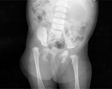
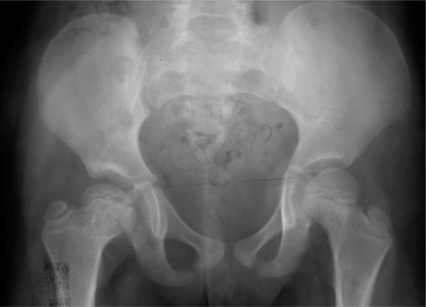
The results of this study demonstrate that T3GD leads to acetabular dysplasia even if a concentric reduction was obtained at the time of reduction. At skeletal maturity, 90% of the T3GD hips were classified as Severin III or IV. There is also strong evidence that residual acetabular dysplasia leads to early degenerative joint disease.4,6,9,25,31–34 Signs of osteoarthritis were already present in 24 % of the T3GD cases at the time of skeletal maturity. In addition, the Severin classification was a strong predictor of total hip replacement: None of the T3GD-Severin II hips underwent total hip replacement compared to 15 percent and 43 percent of the T3GD-Severin III and IV hips, respectively. The Stulberg classification was also predictive of degenerative joint disease and subsequent total hip replacement: None of the Stulberg II hips developed osteoarthritis, compared to 19 percent of the Stulberg III and 75 percent of the Stulberg IV hips. Therefore, residual acetabular dysplasia, as assessed by the Severin and Stulberg classifications, predicts total hip replacement. Figures 1 through 5 summarize the treatment and outcomes of a patient who underwent closed reduction at six months of age for a Tönnis grade 3 dislocation.
Figure 1.
Pre-reduction radiograph of a 6-month-old female with a Tonnis grade 3 dislocation of the right hip.
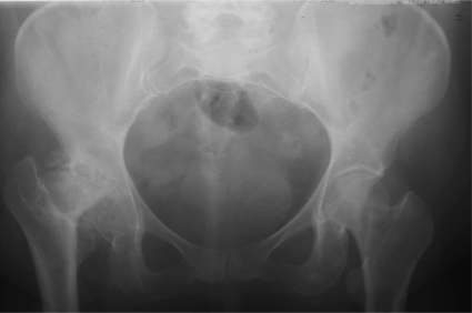
Figure 5.
Radiograph of the patient at age 45, immediately prior to a left total hip replacement. Note extensive degenerative joint disease.
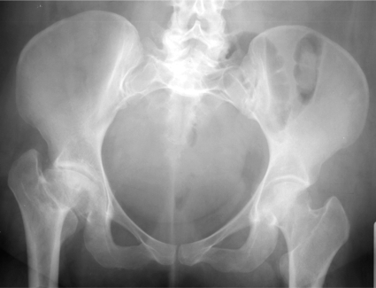
At skeletal maturity her hip was classified as Severin III and by adulthood (Figure 4), it was classified as Severin IV and Stulberg IV. Figure 5 shows the hip immediately prior to total hip arthroplasty, with severe osteoarthritis causing pain and functional deficits.
Figure 4.
Patient at 35 years of age. There is a significant deformity of the femoral head and neck, with a decreased articular-trochanteric distance. Signs of early degenerative joint disease include decreased joint space, sclerosis and cyst formation.
Figure 2.
Patient at 10 months post reduction. There is evidence of delay of the ossific nucleus on the right side. Note a small defect in the ossific nucleus of the left, non-affected hip.
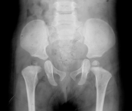
Figure 3.
Patient at 5 years post reduction. Note the femoral head deformity, horizontal epiphyseal line, and the abnormal metaphysis on the right side. The acetabulum is developing normally. Note horizontilization of the left femoral epiphysis indicating a mild case of type II growth disturbance in the passively treated non-affected hip.
Relative narrowness and insufficiency of the acetabulum plus early cessation of hip remodeling and coxa magna observed in the T3GD cases may have indicated the need for pelvic osteotomy.6,65 Some investigators suggest these procedures should be performed at an early age, while the femoral head has its highest remodeling potential.1,27,66 Depending on joint congruence or incongruence, the age of the patient, and the need to increase the acetabular volume, it seems logical to perform a redirectional osteotomy, or the more commonly performed shelf procedure, or Chiari osteotomy.56,66 Prospective long-term studies are required to validate these indications.
In conclusion, T3GD remains the most severe and devastating complication after treatment of developmental dysplasia of the hip in children. Acetabular dysplasia developed in 90% of the hips in our series. In most of the cases, poor acetabular development and flattening of the femoral head led to early degenerative changes in the hip joint. The risk increases in high-degree dislocations, independent of the treatment performed.
Footnotes
Investigation performed at the Department of Orthopaedics and Rehabilitation, University of Iowa Hospitals and Clinics, Iowa City, Iowa
REFERENCES
- 1.Brougham D. I., Broughton N. S., Cole W. G., Menelaus M. B. Avascular necrosis following closed reduction of congenital dislocation of the hip. Review of influencing factors and long-term follow-up. Journal of Bone & Joint Surgery - British Volume. 1990;72(4):557–62. doi: 10.1302/0301-620X.72B4.2380203. [DOI] [PubMed] [Google Scholar]
- 2.Buchanan J. R., Greer R. B., III Prevention of avascular necrosis during treatment of congenital dislocation of the hip. Surgical Forum. 1978;29:546–9. [PubMed] [Google Scholar]
- 3.Bucholz R. W., Ogden J. A. Sixth Open Scientific Meeting of the Hip Society. St. Louis: C.V. Mosby; 1978. Patterns of ischemic necrosis of the proximal femur in nonoperatively treated congenital hip disease; pp. 43–64. [Google Scholar]
- 4.Cooperman D. R., Wallensten R, Stulberg S. D. Acetabular dysplasia in the adult. Clinical Orthopaedics & Related Research. 1983;(175):79–85. [PubMed] [Google Scholar]
- 5.Crego C, Schwartzmann J. Follow-up study of the early treatment of congenital dislocation of the hip. J Bone and Joint Surg. 1948;30:428–442. [PubMed] [Google Scholar]
- 6.Fisher Rl, Cary J. M. Avascular necrosis complicating congenital dislocation of the hip. Course, prognosis and orthopaedic management. International Orthopaedics. 1978;2:229–240. [Google Scholar]
- 7.Gage J. R., Winter R. B. Avascular necrosis of the capital femoral epiphysis as a complication of closed reduction of congenital dislocation of the hip. A critical review of twenty years’ experience at Gillette Children’s Hospital. Journal of Bone & Joint Surgery - American Volume. 1972;54(2):373–88. [PubMed] [Google Scholar]
- 8.Kalamchi A, MacEwen G. D. Avascular necrosis following treatment of congenital dislocation of the hip. Journal of Bone & Joint Surgery - American Volume. 1980;62(6):876–88. [PubMed] [Google Scholar]
- 9.Malvitz T. A., Weinstein S. L. Closed reduction for congenital dysplasia of the hip. Functional and radiographic results after an average of thirty years. Journal of Bone & Joint Surgery - American Volume. 1994;76(12):1777–92. doi: 10.2106/00004623-199412000-00004. [DOI] [PubMed] [Google Scholar]
- 10.Westin G. W., Ilfeld F. W., Provost J. Total avascular necrosis of the capital femoral epiphysis in congenital dislocated hips. Clinical Orthopaedics & Related Research. 1976;(119):93–8. [PubMed] [Google Scholar]
- 11.Brougham D. I., Broughton N. S., Cole W. G., Menelaus M. B. The predictability of acetabular development after closed reduction for congenital dislocation of the hip. Journal of Bone & Joint Surgery - British Volume. 1988;70(5):733–6. doi: 10.1302/0301-620X.70B5.3192570. [DOI] [PubMed] [Google Scholar]
- 12.Kruczynski J. Avascular necrosis after nonoperative treatment of developmental hip dislocation. Prognosis in 36 patients followed 17-26 years. Acta Orthopaedica Scandinavica. 1995;66(3):239–44. doi: 10.3109/17453679508995532. [DOI] [PubMed] [Google Scholar]
- 13.Morel G. The treatment of congenital dislocation and subluxation of the hip in the older child. Acta Orthopaedica Scandinavica. 1975;46(3):364–99. [PubMed] [Google Scholar]
- 14.Pous J. G., Camous J. Y., el Blidi S. Cause and prevention of osteochondritis in congenital dislocation of the hip. Clinical Orthopaedics & Related Research. 1992;(281):56–62. [PubMed] [Google Scholar]
- 15.Sosna A, Rejholec M. Ludloff’s open reduction of the hip: long-term results. Journal of Pediatric Orthopedics. 1992;12(5):603–6. [PubMed] [Google Scholar]
- 16.Thomas I. H., Dunin A. J., Cole W. G., Menelaus M. B. Avascular necrosis after open reduction for congenital dislocation of the hip: analysis of causative factors and natural history. Journal of Pediatric Orthopedics. 1989;9(5):525–31. doi: 10.1097/01241398-198909010-00005. [DOI] [PubMed] [Google Scholar]
- 17.Weinstein S. L. The medial approach in congenital dislocation of the hip. Israel Journal of Medical Sciences. 1980;16(4):272–5. [PubMed] [Google Scholar]
- 18.Weinstein S. L., Mubarak S. J., Wenger D. R. Developmental hip dysplasia and dislocation: Part I. Instructional Course Lectures. 2004;53:523–30. [PubMed] [Google Scholar]
- 19.Esteve R. Congenital dislocation of the hip. A review and assessment of results of treatment with special reference to frame reduction as compared with manipulative reduction. Journal of Bone & Joint Surgery - British Volume. 1960;42-B:253–63. doi: 10.1302/0301-620X.42B2.253. [DOI] [PubMed] [Google Scholar]
- 20.Mau H, Dorr W. M., Henkel L, Lutsche J. Open reduction of congenital dislocation of the hip by Ludloff’s method. Journal of Bone & Joint Surgery - American Volume. 1971;53(7):1281–8. [PubMed] [Google Scholar]
- 21.Schoenecker P. L., Bitz M, Whiteside L. The acute effect of position of immobilization on capital femoral epiphyseal blood flow. J Bone and Joint Surg. 1978;60:899–903. [PubMed] [Google Scholar]
- 22.Severin E. Contribution to the knowledge of congenital dislocation of the hip joint. Late results of closed reduction and arthrographic studies of recent cases. Acta Chirurgiae Orthopaedicae et Traumatologiae Scandinavica. 1941;84(Supplementum 63) [Google Scholar]
- 23.Cooper W. F. The techniques of selective placement of factory employees with major physical disabilities. Australian Family Physician. 1980;9(2):109–14. [PubMed] [Google Scholar]
- 24.Weiner D. S., Hoyt W. A., O’Dell H W. Congenital dislocation of the hip. The relationship of premanipulation traction and age to avascular necrosis of the femoral head. Journal of Bone & Joint Surgery - American Volume. 1977;59(3):306–11. [PubMed] [Google Scholar]
- 25.Weinstein S. L., Mubarak S. J., Wenger D. R. Developmental hip dysplasia and dislocation: Part II. Instructional Course Lectures. 2004;53:531–42. [PubMed] [Google Scholar]
- 26.Ferguson A. B. Primary open reduction of congenital dislocation of the hip using a median adductor approach. Journal of Bone & Joint Surgery - American Volume. 1973;55(4):671–89. [PubMed] [Google Scholar]
- 27.Salter R. B., Kostuik J, Dallas S. Avascular necrosis of the femoral head as a complication of treatment for congenital dislocation of the hip in young children: a clinical and experimental investigation. Canadian Journal of Surgery. 1969;12(1):44–61. [PubMed] [Google Scholar]
- 28.Weinstein S. L., Ponseti I. V. Congenital dislocation of the hip. Journal of Bone & Joint Surgery - American Volume. 1979;61(1):119–24. [PubMed] [Google Scholar]
- 29.Tonnis D. General radiography of the hip joint. In: Tonnis D, editor. Congenital Dysplasia and Dislocation of the Hip in Children and Adults. New York: Springer; 1987. pp. 100–142.pp. 100–142. [Google Scholar]
- 30.Weinstein S. L. Developmental hip dysplasia and dislocation. In: Weinstein S. L., editor. Pediatric Orthopaedics. Philadelphia: Lippincott Williams & Wilkens; 2001. pp. 905–956.pp. 905–956. [Google Scholar]
- 31.Albinana J, Dolan L. A., Spratt Kf, Morcuende J, Meyer M. D., Weinstein S. L. Acetabular dysplasia after treatment for developmental dysplasia of the hip. Implications for secondary procedures.[see comment] Journal of Bone & Joint Surgery - British Volume. 2004;86(6):876–86. doi: 10.1302/0301-620x.86b6.14441. [DOI] [PubMed] [Google Scholar]
- 32.Lindstrom J. R., Ponseti I. V., Wenger D. R. Acetabular development after reduction in congenital dislocation of the hip. Journal of Bone & Joint Surgery - American Volume. 1979;61(1):112–8. [PubMed] [Google Scholar]
- 33.Stulberg S. D., Cooperman D. R., Wallensten R. The natural history of Legg-Calve-Perthes disease. Journal of Bone & Joint Surgery - American Volume. 1981;63(7):1095–108. [PubMed] [Google Scholar]
- 34.Cooperman D. R., Wallensten R, Stulberg S. D. Post-reduction avascular necrosis in congenital dislocation of the hip. Journal of Bone & Joint Surgery - American Volume. 1980;62(2):247–58. [PubMed] [Google Scholar]
- 35.Hattori T, Ono Y, Kitakoji T, Takashi S, Iwata H. Soft-tissue interposition after closed reduction in developmental dysplasia of the hip. The long-term effect on acetabular development and avascular necrosis. Journal of Bone & Joint Surgery -British Volume. 1999;81(3):385–91. doi: 10.1302/0301-620x.81b3.9021. [DOI] [PubMed] [Google Scholar]
- 36.Weinstein S. L., Ponseti I. V. Congenital dislocation of the hip. Open reduction through a medial approach. J Bone and Joint Surg. 1979;61:119–124. [PubMed] [Google Scholar]
- 37.Morcuende J. A., Meyer M. D., Dolan L. A., Weinstein S. L. Long-term outcome after open reduction through an anteromedial approach for congenital dislocation of the hip.[see comment] Journal of Bone & Joint Surgery - American Volume. 1997;79(6):810–7. doi: 10.2106/00004623-199706000-00002. [DOI] [PubMed] [Google Scholar]
- 38.Morcuende J. A., Meyer M. D., Dolan L. A., Weinstein S. L. Long-term outcome after open reduction through an anteromedial approach for congenital dislocation of the hip. Journal of Bone & Joint Surgery - American Volume. 1997;79(6):810–7. doi: 10.2106/00004623-199706000-00002. [DOI] [PubMed] [Google Scholar]
- 39.Smith W. S., Badgley C. E., Orwig J. B., Harper J. M. Correlation of postreduction roentgenograms and thirty-one-year follow-up in congenital dislocation of the hip. Journal of Bone & Joint Surgery - American Volume. 1968;50(6):1081–98. [PubMed] [Google Scholar]
- 40.Hilgenreiner H. Zur Fruhdiagnose und Fruhbehndlung der angeborenen Huftgelenksverrenkung. Med Klin. 1925;21:1385–1388. 1425–1429. [Google Scholar]
- 41.Fredensborg N. The results of early treatment of typical congenital dislocation of the hip in Malmo. Journal of Bone & Joint Surgery - British Volume. 1976;58(3):272–8. doi: 10.1302/0301-620X.58B3.956242. [DOI] [PubMed] [Google Scholar]
- 42.Stulberg S. D., Harris W. H. Second Open Scientific Meeting of the Hip Society. C.V. Mosby; 1974. Acetabular dysplasia and the development of osteoarthritis of the hip; pp. 89–93.pp. 89–93. [Google Scholar]
- 43.Boyer D. W., Mickelson M. R., Ponseti I. V. Slipped capital femoral epiphysis. J Bone and Joint Surg. 1981;63:85–95. [PubMed] [Google Scholar]
- 44.Harrell F. E., Jr., Lee Kl, Mark D. B. Multivariable prognostic models: issues in developing models, evaluating assumptions and adequacy, and measuring and reducing errors. Stat Med. 1996;15:361–387. doi: 10.1002/(SICI)1097-0258(19960229)15:4<361::AID-SIM168>3.0.CO;2-4. [DOI] [PubMed] [Google Scholar]
- 45.SAS Institute. SAS/ETS User’s Guide. Version 6. North Carolina: Cary; 1993. The Expand procedure. [Google Scholar]
- 46.Chung M. K. The arterial supply of the developing proximal end of the human femur. J Bone and Joint Surg. 1976;58:961–970. [PubMed] [Google Scholar]
- 47.Siffert R. S. Patterns of deformity of the developing hip. Clin Orthop. 1981;160:14–29. [PubMed] [Google Scholar]
- 48.Kim H. T., Kim J. I., Yoo C. I. Acetabular development after closed reduction of developmental dislocation of the hip. Journal of Pediatric Orthopedics. 2000;20(6):701–8. doi: 10.1097/00004694-200011000-00002. [DOI] [PubMed] [Google Scholar]
- 49.Luhmann S. J., Schoenecker P. L., Anderson A. M., Bassett G. S. The prognostic importance of the ossific nucleus in the treatment of congenital dysplasia of the hip. Journal of Bone & Joint Surgery - American Volume. 1998;80(12):1719–27. doi: 10.2106/00004623-199812000-00001. [DOI] [PubMed] [Google Scholar]
- 50.Kim H. W., Morcuende J. A., Dolan L. A., Weinstein S. L. Acetabular development in developmental dysplasia of the hip complicated by lateral growth disturbance of the capital femoral epiphysis. Journal of Bone & Joint Surgery - American Volume. 2000;82-A(12):1692–700. doi: 10.2106/00004623-200012000-00002. [DOI] [PubMed] [Google Scholar]
- 51.Clarke N. M., Jowett A. J., Parker L. The surgical treatment of established congenital dislocation of the hip: results of surgery after planned delayed intervention following the appearance of the capital femoral ossific nucleus. Journal of Pediatric Orthopedics. 2005;25(4):434–9. doi: 10.1097/01.bpo.0000158003.68918.28. [DOI] [PubMed] [Google Scholar]
- 52.Segal L. S., Boal D. K., Borthwick L, Clark M. W., Localio A. R., Schwentker E. P. Avascular necrosis after treatment of DDH: the protective influence of the ossific nucleus.[see comment] Journal of Pediatric Orthopedics. 1999;19(2):177–84. doi: 10.1097/00004694-199903000-00008. [DOI] [PubMed] [Google Scholar]
- 53.Segal L. S., Schneider D. J., Berlin J. M., Bruno A, Davis B. R., Jacobs C. R. The contribution of the ossific nucleus to the structural stiffness of the capital femoral epiphysis: a porcine model for DDH. Journal of Pediatric Orthopedics. 1999;19(4):433–7. doi: 10.1097/00004694-199907000-00003. [DOI] [PubMed] [Google Scholar]
- 54.Keret D, MacEwen G. D. Growth disturbance of the proximal part of the femur after treatment for congenital dislocation of the hip. Journal of Bone & Joint Surgery - American Volume. 1991;73(3):410–23. [PubMed] [Google Scholar]
- 55.Tonnis D. Letter to the Editor. J Pediatr Orthop. 2001;21:135–137. doi: 10.1097/00004694-200101000-00026. [DOI] [PubMed] [Google Scholar]
- 56.Gabuzda G. M., Renshaw T. S. Reduction of congenital dislocation of the hip.[see comment] Journal of Bone & Joint Surgery - American Volume. 1992;74(4):624–31. [PubMed] [Google Scholar]
- 57.Gibson P. H., Benson M. K. Congenital dislocation of the hip. Review at maturity of 147 hips treated by excision of the limbus and derotation osteotomy. Journal of Bone & Joint Surgery - British Volume. 1982;64(2):169–75. doi: 10.1302/0301-620X.64B2.7068735. [DOI] [PubMed] [Google Scholar]
- 58.Klisic P, Jankovic L. Combined procedure of open reduction and shortening of the femur in treatment of congenital dislocation of the hips in older children. Clinical Orthopaedics & Related Research. 1976;(119):60–9. [PubMed] [Google Scholar]
- 59.O’Brien T. Growth-disturbance lines in congenital dislocation of the hip. Journal of Bone & Joint Surgery - American Volume. 1985;67(4):626–32. [PubMed] [Google Scholar]
- 60.Janovec M. [The development of early diagnosis and treatment of congenital hip dislocation in Czechoslovakia] Acta Chirurgiae Orthopaedicae et Traumatologiae Cechoslovaca. 1989;56(4):281–8. [PubMed] [Google Scholar]
- 61.Karakas E. S., Baktir A, Argun M, Turk C. Y. One-stage treatment of congenital dislocation of the hip in older children. Journal of Pediatric Orthopedics. 1995;15(3):330–6. doi: 10.1097/01241398-199505000-00014. [DOI] [PubMed] [Google Scholar]
- 62.Wenger D. R., Lee C. S., Kolman B. Derotational femoral shortening for developmental dislocation of the hip: special indications and results in the child younger than 2 years. Journal of Pediatric Orthopedics. 1995;15(6):768–79. doi: 10.1097/01241398-199511000-00009. [DOI] [PubMed] [Google Scholar]
- 63.Ponseti I. V. Growth and development of the acetabulum in the normal child. J Bone and Joint Surg. 1978;60:575–585. [PubMed] [Google Scholar]
- 64.Ponseti I. V. Morphology of the acetabulum in congenital dislocation of the hip. Gross, histological and roentgenographic studies. Journal of Bone & Joint Surgery - American Volume. 1978;60(5):586–99. [PubMed] [Google Scholar]
- 65.Thomas Cl, Gage J. R., Ogden J. A. Treatment concepts for proximal femoral ischemic necrosis complicating congenital hip disease. Journal of Bone & Joint Surgery - American Volume. 1982;64(6):817–28. [PubMed] [Google Scholar]
- 66.Bar-On E, Huo M. H., Deluca P. A. Early innominate osteotomy as a treatment for avascular necrosis complicating developmental hip dysplasia. Journal of Pediatric Orthopaedics. Part B. 1997;6(2):138–45. doi: 10.1097/01202412-199704000-00010. [DOI] [PubMed] [Google Scholar]


