Abstract
Contemporary locking plates promote biological fixation through indirect reduction techniques and by elevating the plate from the bone. They have improved fixation strength in osteoporotic bone. Periarticular locking plates are rapidly being adopted for bridge plating of periprosthetic femur fractures. When these plates are used for indirect reduction and bridge plating osteosynthesis, fracture union occurs by secondary bone healing with callus formation which is stimulated by interfragmentary motion. In two patients with similar periprosthetic femur fractures treated with periarticular locking plates one fracture healed by ample callus formation while the other resulted in a non-union and had no callus formation six months post-operatively. The case, which progressed to secondar y bone healing with callus formation, exhibited varus migration as a result of loss of fixation. The non-union case retained stable fixation. The difference in outcome may indicate that callus formation was promoted by interfragmentary motion secondary to loss of fixation. Conversely, in absence of fixation failure, callus formation was suppressed by stable fixation with a stiff locking plate construct which reduced interfragmentary motion. These observations suggest that locked plating constructs should be sufficiently flexible when applied for bridge plating of comminuted fractures to promote callus formation.
INTRODUCTION
Periarticular locking plates are increasingly being used for fixation of periprosthetic femur fractures.1–3 They feature fixed-angle screws to improve fixation strength in osteoporotic bone. The improved fixation strength of locking plates has expanded their indication to bridge plating of comminuted fractures.4 In addition to providing sufficiently strong fixation, locking plates have to enable a mechanical environment at the fracture site that facilitates fracture healing. For bridge plating of periprosthetic femur fractures with locking plates, fracture healing occurs by secondary bone healing, whereby callus formation is stimulated by interfragmentary motion.5 In this regard, concerns are emerging that the stiffness of locked plating constructs may have the potential to suppress interfragmentary motion and callus formation.4,6–8
These case reports present two comparable periprosthetic femur fractures treated with periarticular locking plates. One fracture healed by ample callus formation while the other resulted in a non-union after deficient callus formation. Additionally, biomechanical factors that may have contributed to the difference in callus formation are discussed.
CASE 1
Patient one was a 77-year-old female with a past medical history of diabetes mellitus type II, hypertension, hyperlipidemia, osteoarthritis, gastroesophageal reflux disease, and mild mitral valve regurgitation. Her diabetes was adequately controlled with an insulin regimen. She had mild chronic kidney disease but no neuropathy or retinopathy. She was a community ambulator who used a cane for balance. She had no history of tobacco or alcohol use. The patient fell after stepping from a curb and suffered a left periprosthetic supracondylar femur fracture. This was a Rorabeck type II (displaced and prosthesis intact) closed injury and the limb was neurovascularly intact.9
She was taken to the operating room on post injury day two for open reduction internal fixation. A nine hole Synthes AO-LISS plate with eight 5.0 locking screws was utilized. She had no post-operative complications other than requiring a short course of oral antibiotics for a superficial wound infection that cleared. The patient was evaluated in follow-up at 7 weeks and 15 weeks with radiographs. The fracture united with ample callus seen on radiographs. She was weight bearing without pain at the fifteen week follow-up. Close inspection of the radiographs demonstrates increasing varus compared to immediate post operative films suggestive of loss of fixation (Figure 1).
Figure 1.
Post-operative and 6-months follow-up radiographs of case 1. Secondary fracture healing occurred by ample callus formation in the presence of varus migration.
CASE 2
Patient two was a 74-year-old female with a past medical history of diabetes mellitus type II, valvular heart disease, hypertension, osteoarthritis, and obstructive sleep apnea. Her diabetes was adequately controlled with an insulin regimen, with HgA1c ranging from 5.0-6.0. She had no known nephropathy or retinopathy, but did have mild peripheral neuropathy affecting her toes only. She was an independent community ambulator. She had no history of tobacco or alcohol use. The patient fell while walking in her house and suffered a left periprosthetic supracondylar femur fracture. This was a Rorabeck type II closed injury and the limb was neurovascularly intact.9
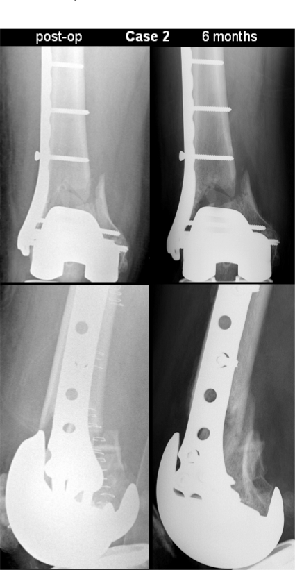
She was taken to the operating room on post injury day one for open reduction internal fixation by the same surgeon as the previous patient. A Smith and Nephew Peri-Loc distal femur plate was used with one 6.5 mm and seven 4.5 mm locking screws. She had no perioperative or wound complications. She was evaluated in follow up at 4, 8, 12 and 16 weeks from injury with radiographs at each of these visits. She had very little callus formation on radiographs. Her alignment was maintained during this follow up period (Figure 2). She had continued pain at the fracture site throughout clinical follow-up and could not tolerate progression of weight bearing past toe touch. She was confirmed to have a non-union by CT scan at 6 months from injury and was indicated for revision fixation with bone grafting. There was no evidence of infection at the time of revision surgery.
Figure 2.
Post-operative and 6-months follow-up radiographs of case 2. The fixation construct provided stable reduction but callus formation at six months remained deficient, requiring revision by bone grafting.
DISCUSSION
Periarticular locking plates have been increasingly used for periprosthetic femoral fractures after TKR, especially in the setting of osteoporosis. These locking plates support biological fixation by plate elevation and yield improved fixation strength in osteoporotic bone.5,10 However, the fixation concept of locked plating is entirely different from conventional plating and requires a revised understanding of plate fixation.4 In the absence of anatomic reduction and interfragmentary compression, locking plate constructs rely on secondary bone healing by callus formation.11
The two cases of periprosthetic fractures described in this report were remarkably similar patients with similar injury mechanisms, fractures and treatments, but very different outcomes. Both patients were female non-smokers of similar age who sustained a Rorabeck type II closed fracture in a fall from standing. The major identifiable difference between the two cases was that case 1 exhibited increasing varus migration due to a loss of metaphyseal fixation which in turn permitted increased interfragmentary motion and medial gap closure. In case 2, alignment was maintained even after 16 weeks, suggesting that implant fixation remained intact. Since the locked plate remained securely fixed to the bone, the construct likely retained its original fixation stiffness throughout the post-operative period limiting interfragmentary motion and continuing to support a small medial gap. This leads to the speculation that increased interfragmentary motion secondary to a loss of fixation promoted callus formation and secondary bone healing in case 1. Conversely, the continuously high construct stiffness in case 2 suppressed callus formation and maintained a medial gap.
This speculation is supported by biomechanical factors known to promote secondary bone healing. Secondary bone healing is induced by interfragmentary motion in the millimeter-range and can be enhanced by passive or active dynamization.12,13 Clinically, secondary bone healing is expected to occur with use of external fixators.5 Monolateral fixators have an axial stiffness in the range of 50 - 400 N/mm.14–16 Ilizarov ring fixators exhibit an initial stiffness of 50 N/mm under small loads that increases to approximately 140N for loads greater than 800 N.14 Their low initial stiffness enables average interfragmentary motion between 1-3 mm in the early post-operative phase under reduced weight bearing conditions to promote callus formation in the acute healing phase.17 The benefit of this interfragmentary motion is well supported by the clinical success of the Ilizarov method18 and by the original work of Goodship and Kenwright.19
While locking plate constructs have been termed internal fixators,20 they can be several-fold stiffer than external fixators. The stiffness of the LISS locking plate tested in an unstable fracture model of the distal femur has been reported to range from 200 N/mm21 to well over 1000 N/mm,22 whereby the large range may be explained by differences in axial loading constraints, bone quality and screw patterns. These reports demonstrate that locked plates can approach the flexibility of an external fixator, but they also can be considerably stiffer, allowing for less than 0.5 mm motion in response to axial one body-weight load bearing.22 The stiffness of locking plate constructs has led to recent speculations that locking plates can potentially act like extremely rigid internal fixators which may run the risk of preventing callus due to their high stiffness and close proximity to the bone.4,6
Clinically, non-unions of periprosthetic femur fractures treated by periarticular locking plates occur at a rate of 0%-13%.1–3 Ricci et al. published a series of 22 patients with periprosthetic supracondylar femur fractures treated with a Locking Condylar Plate.3 Nineteen fractures healed with an average time to union of 12 weeks. Two cases developed infected non-unions and one case had an aseptic nonunion. In a preliminary report, Althausen et al. observed no non-unions in a series of five cases treated with the LISS plate.1 Kregor et al. reported one non-union in 13 periprosthetic femoral fractures treated with the LISS. The mean time to weight bearing was 13 weeks.2
While these reported non-unions can arise from a multitude of factors, the present case report suggests that locked plating constructs may have the potential to suppress interfragmentary motion to a level insufficient for promotion of fracture healing by callus formation. Further research is needed to study callus formation and healing rates of these fractures as a function of the stiffness of the fixation construct.
REFERENCES
- 1.Althausen PL, Lee MA, Finkemeier CG, et al. Operative stabilization of supracondylar femur fractures above total knee arthroplasty: a comparison of four treatment methods. J Arthroplasty. 2003;18:834–9. doi: 10.1016/s0883-5403(03)00339-5. [DOI] [PubMed] [Google Scholar]
- 2.Kregor PJ, Hughes JL, Cole PA. Fixation of distal femoral fractures above total knee arthroplasty utilizing the Less Invasive Stabilization System (L.I.S.S.) Injury. 2001;32(3):SC64–75. doi: 10.1016/s0020-1383(01)00185-1. [DOI] [PubMed] [Google Scholar]
- 3.Ricci WM, Loftus T, Cox C, et al. Locked plates combined with minimally invasive insertion technique for the treatment of periprosthetic supracondylar femur fractures above a total knee arthroplasty. J Orthop Trauma. 2006;20:190–6. doi: 10.1097/00005131-200603000-00005. [DOI] [PubMed] [Google Scholar]
- 4.Egol KA, Kubiak EN, Fulkerson E, et al. Biomechanics of locked plates and screws. J Orthop Trauma. 2004;18:488–493. doi: 10.1097/00005131-200409000-00003. [DOI] [PubMed] [Google Scholar]
- 5.Perren SM. Evolution of the internal fixation of long bone fractures. The scientific basis of biological internal fixation: choosing a new balance between stability and biology. J Bone Joint Surg Br. 2002;84:1093–110. doi: 10.1302/0301-620x.84b8.13752. [DOI] [PubMed] [Google Scholar]
- 6.Kubiak EN, Fulkerson E, Strauss E, et al. The evolution of locked plates. J Bone Joint Surg Am. 2006;88(4):189–200. doi: 10.2106/JBJS.F.00703. [DOI] [PubMed] [Google Scholar]
- 7.Smith WR, Ziran BH, Anglen JO, et al. Locking plates: tips and tricks. J Bone Joint Surg Am. 2007;89:2298–307. doi: 10.2106/00004623-200710000-00028. [DOI] [PubMed] [Google Scholar]
- 8.Uhthoff HK, Poitras P, Backman DS. Internal plate fixation of fractures: short history and recent developments. J Orthop Sci. 2006;11:118–26. doi: 10.1007/s00776-005-0984-7. [DOI] [PMC free article] [PubMed] [Google Scholar]
- 9.Rorabeck CH, Taylor JW. Periprosthetic fractures of the femur complicating total knee arthroplasty. Orthop Clin North Am. 1999;30:265–77. doi: 10.1016/s0030-5898(05)70081-x. [DOI] [PubMed] [Google Scholar]
- 10.Ramotowski W, Granowski R. Zespol. An original method of stable osteosynthesis. Clin Orthop Relat Res. 1991:67–75. [PubMed] [Google Scholar]
- 11.Perren SM. Backgrounds of the technology of internal fixators. Injury. 2003;34:B1–3. doi: 10.1016/j.injury.2003.09.018. [DOI] [PubMed] [Google Scholar]
- 12.Claes LE, Heigele CA, Neidlinger-Wilke C, et al. Effects of mechanical factors on the fracture healing process. Clin Orthop Relat Res. 1998:S132–47. doi: 10.1097/00003086-199810001-00015. [DOI] [PubMed] [Google Scholar]
- 13.Kenwright J, Richardson JB, Goodship AE, et al. Effect of controlled axial micromovement on healing of tibial fractures. Lancet. 1986;2:1185–7. doi: 10.1016/s0140-6736(86)92196-3. [DOI] [PubMed] [Google Scholar]
- 14.Caja V, Kim W, Larsson S, et al. Comparison of the mechanical performance of three types of external fixators: linear, circular and hybrid. Clin Biomech (Bristol, Avon) 1995;10:401–406. doi: 10.1016/0268-0033(95)00014-3. [DOI] [PubMed] [Google Scholar]
- 15.Finlay JB, Moroz TK, Rorabeck CH, et al. Stability of ten configurations of the Hoffmann external-fixation frame. J Bone Joint Surg Am. 1987;69:734–44. [PubMed] [Google Scholar]
- 16.Moroz TK, Finlay JB, Rorabeck CH, et al. External skeletal fixation: choosing a system based on biomechanical stability. J Orthop Trauma. 1988;2:284–96. [PubMed] [Google Scholar]
- 17.Duda GN, Sollmann M, Sporrer S, et al. Inter-fragmentary motion in tibial osteotomies stabilized with ring fixators. Clin Orthop Relat Res. 2002:163–72. doi: 10.1097/00003086-200203000-00025. [DOI] [PubMed] [Google Scholar]
- 18.Ilizarov GA. Clinical application of the tension-stress effect for limb lengthening. Clin Orthop Relat Res. 1990:8–26. [PubMed] [Google Scholar]
- 19.Goodship AE, Watkins PE, Rigby HS, et al. The role of fixator frame stiffness in the control of fracture healing. An experimental study. J Biomech. 1993;26:1027–35. doi: 10.1016/s0021-9290(05)80002-8. [DOI] [PubMed] [Google Scholar]
- 20.Wagner M. General principles for the clinical use of the LCP. Injury. 2003;34:B31–42. doi: 10.1016/j.injury.2003.09.023. [DOI] [PubMed] [Google Scholar]
- 21.Stoffel K, Lorenz KU, Kuster MS. Biomechanical considerations in plate osteosynthesis: the effect of plate-to-bone compression with and without angular screw stability. J Orthop Trauma. 2007;21:362–8. doi: 10.1097/BOT.0b013e31806dd921. [DOI] [PubMed] [Google Scholar]
- 22.Marti A, Fankhauser C, Frenk A, et al. Biomechanical evaluation of the less invasive stabilization system for the internal fixation of distal femur fractures. J Orthop Trauma. 2001;15:482–7. doi: 10.1097/00005131-200109000-00004. [DOI] [PubMed] [Google Scholar]


