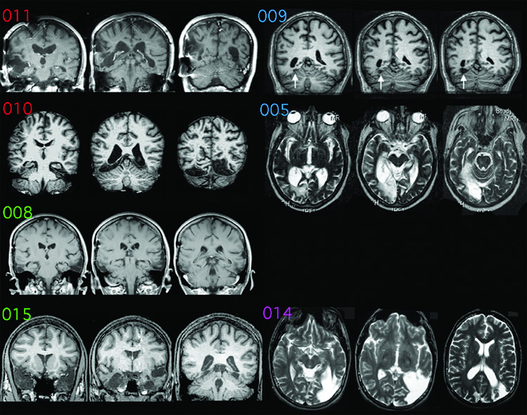
Figure 1.
Magnetic Resonance (MR) images of the prosopagnosic subjects. By convention, right hemispheres are on the left of each image. Scans of subjects 005 and 014 are displayed in axial sections; the others are displayed in coronal sections. The MR images refer to (from top to bottom): left row = bilateral lesions, subjects 011, 010, 008, and 015; right row = unilateral lesions, subjects 009, 005, 014. White arrows for subject 009 show his small infarct. There is no image for subject 001, who had no visible lesion. Letter coloring refers to lesion category: red = bilateral occipitotemporal, green = bilateral anterior temporal, blue = unilateral right occipitotemporal, purple = unilateral left occipitotemporal.

