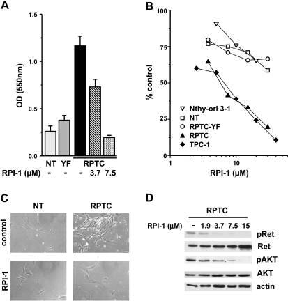
Figure 2.
Effects of RPI-1 on human primary thyrocytes and thyroid cell lines. (A) Abrogation of RET/PTC1 growth-promoting effect. Uninfected thyrocytes (NT) or thyrocytes infected with RET/PTC1 (RPTC) or RET/PTC1-Y451F (YF) were treated with solvent (-) or with the indicated concentrations of RPI-1 for 6 days. Cell density was measured by the SRB colorimetric assay and was expressed as optical density (OD) at 550 nm (mean ± SD). One experiment representative of three is shown. (B) Cell growth inhibition. Primary thyrocytes and thyroid cell lines (follicular epithelial Nthy-ori 3-1 and PTC TPC-1) were exposed to solvent or to different concentrations of RPI-1 for 72 hours. The drug antiproliferative effect was evaluated by the SRB assay. Representative dose-response curves are shown. (C) Reversion of RET/PTC1-transformed morphologic phenotype. Normal human thyrocytes and RPTC thyrocytes were treated with 10 µM RPI-1 for 48 hours and were then photographed under phase-contrast microscope. Original magnification, x100. (D) Inhibition of Ret/ptc1-Y451 and Akt-S473 phosphorylation. RPTC cells were exposed to solvent (-) or to the indicated concentrations of RPI-1 for 24 hours. Western blot analysis of total protein extracts was performed with phospho-specific antibodies recognizing phosphorylated Y451 (pRet) and S473 (pAkt) of Ret/ptc1 and Akt, respectively. The filter was then stripped and reprobed with the respective antiprotein antibodies. Antiactin blot is shown as protein loading control.

