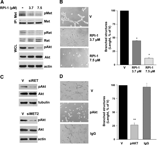
Figure 5.
Effects of concomitant or selective inhibition of Ret/ptc1, Met, and Akt signaling on TPC-1 cell morphogenic pheynotype. (A) Inhibition of Akt activation by the Ret/Met tyrosine kinase inhibitor RPI-1. Cells were exposed to solvent (-) or to the indicated concentrations of RPI-1 for 24 hours. Whole cell lysates (WCL) were analyzed directly or subjected to Met immunoprecipitation (IP) and then analyzed by Western blot analysis. Activation status of the three kinases was detected using antibodies recognizing phosphorylated Ret/ptc1 Y451 (pRet), Met Y1234/Y1235 (pMet), or Akt S473 (pAkt). Blots were then stripped and reprobed with antibodies against the respective proteins. (B) left: Inhibition of cell ability to form branched structures in Matrigel by a 24-hour treatment with RPI-1. Right: Total length quantification of branched structures. (C) left: Inhibition of AKT activation in RET/PTC1- or MET-silenced cells. Western blot analysis was performed on total cell lysates obtained from cells exposed to RET siRNA (siRET) or MET siRNA (siMET2) for 5 or 3 days, respectively. V indicates transfection reagent. (D) left: Inhibition of branching morphogenesis by anti-pS473 Akt antibody (pAkt) intracellular delivery. V indicates protein delivery reagent; IgG, aspecific immunoglobulins. Right: Total length quantification of branched structures. Representative images are shown. Original magnification, x400. *P < .05, **P < .005.

