Abstract
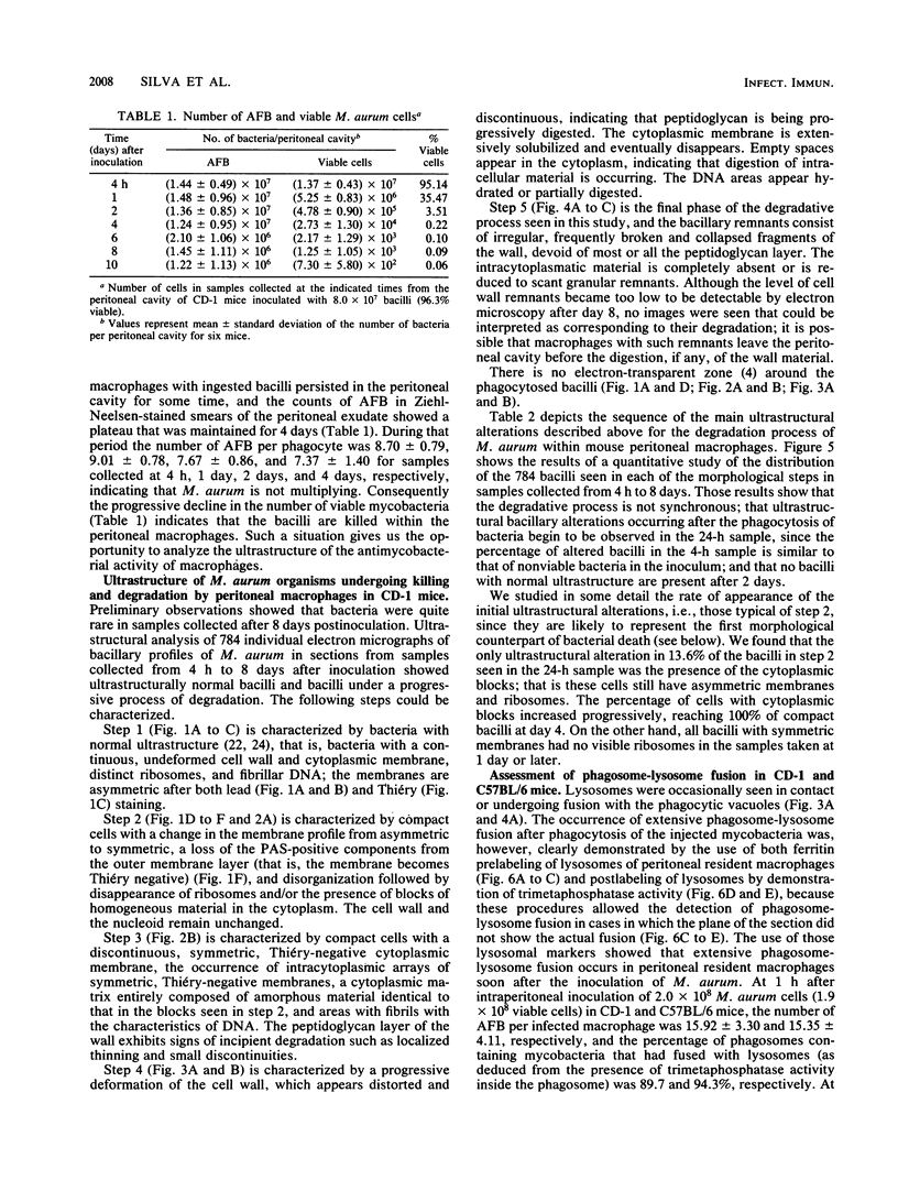
We studied the in vivo killing and degradation of Mycobacterium aurum, a nonpathogenic, acid-fast bacillus, within macrophages after inoculation into the peritoneal cavity of CD-1 mice. The degradative process could be divided in five successive steps that were characterized on ultrastructural and cytochemical grounds and the relative contributions of which were determined by quantitative electron microscopy of samples taken at different times. The main ultrastructural alterations observed during the degradative process were ribosome disaggregation, coagulation of the cytoplasmic matrix, and change in the membrane profile from asymmetric to symmetric, with loss of the polysaccharide components from the outer layer, followed by membrane solubilization and intracellular clearing, followed by digestion of the innermost (peptidoglycan) layer of the cell wall, and at the end of the process, disorganization and collapse of the remaining layers of the cell wall. The correlation between viability and morphology indicated that the first ultrastructural signs of viability loss are cytoplasmic coagulation, change in the membrane geometry, and disappearance of ribosomes. The labeling of lysosomes of peritoneal macrophages with ferritin or by the cytochemical demonstration of inorganic trimetaphosphatase showed that fusion of lysosomes with phagosomes containing mycobacteria occurs in the phagocytes in the mouse peritoneal cavity and is already extensive as soon as 1 h after the inoculation of the bacilli.
Full text
PDF


Images in this article
Selected References
These references are in PubMed. This may not be the complete list of references from this article.
- Babior B. M. Oxygen-dependent microbial killing by phagocytes (first of two parts). N Engl J Med. 1978 Mar 23;298(12):659–668. doi: 10.1056/NEJM197803232981205. [DOI] [PubMed] [Google Scholar]
- Daems W. T., Roos D., van Berkel T. J., van der Rhee H. J. The subcellular distribution and biochemical properties of peroxidase in monocytes and macrophages. Front Biol. 1979;48:463–514. [PubMed] [Google Scholar]
- Draper P., Rees R. J. Electron-transparent zone of mycobacteria may be a defence mechanism. Nature. 1970 Nov 28;228(5274):860–861. doi: 10.1038/228860a0. [DOI] [PubMed] [Google Scholar]
- Elsbach P., Weiss J. Oxygen-dependent and oxygen-independent mechanisms of microbicidal activity of neutrophils. Immunol Lett. 1985;11(3-4):159–163. doi: 10.1016/0165-2478(85)90163-4. [DOI] [PubMed] [Google Scholar]
- Forget A., Skamene E., Gros P., Miailhe A. C., Turcotte R. Differences in response among inbred mouse strains to infection with small doses of Mycobacterium bovis BCG. Infect Immun. 1981 Apr;32(1):42–47. doi: 10.1128/iai.32.1.42-47.1981. [DOI] [PMC free article] [PubMed] [Google Scholar]
- Frehel C., de Chastellier C., Lang T., Rastogi N. Evidence for inhibition of fusion of lysosomal and prelysosomal compartments with phagosomes in macrophages infected with pathogenic Mycobacterium avium. Infect Immun. 1986 Apr;52(1):252–262. doi: 10.1128/iai.52.1.252-262.1986. [DOI] [PMC free article] [PubMed] [Google Scholar]
- Geisow M. J., D'Arcy Hart P., Young M. R. Temporal changes of lysosome and phagosome pH during phagolysosome formation in macrophages: studies by fluorescence spectroscopy. J Cell Biol. 1981 Jun;89(3):645–652. doi: 10.1083/jcb.89.3.645. [DOI] [PMC free article] [PubMed] [Google Scholar]
- Jacques Y. V., Bainton D. F. Changes in pH within the phagocytic vacuoles of human neutrophils and monocytes. Lab Invest. 1978 Sep;39(3):179–185. [PubMed] [Google Scholar]
- Kilburn J. O., Best G. K. Characterization of autolysins from Mycobacterium smegmatis. J Bacteriol. 1977 Feb;129(2):750–755. doi: 10.1128/jb.129.2.750-755.1977. [DOI] [PMC free article] [PubMed] [Google Scholar]
- Klebanoff S. J. Oxygen metabolism and the toxic properties of phagocytes. Ann Intern Med. 1980 Sep;93(3):480–489. doi: 10.7326/0003-4819-93-3-480. [DOI] [PubMed] [Google Scholar]
- Mor N. Intracellular location of Mycobacterium leprae in macrophages of normal and immune-deficient mice and effect of rifampin. Infect Immun. 1983 Nov;42(2):802–811. doi: 10.1128/iai.42.2.802-811.1983. [DOI] [PMC free article] [PubMed] [Google Scholar]
- Petty H. R., Hermann W., McConnell H. M. Cytochemical study of macrophage lysosomal inorganic trimetaphosphatase and acid phosphatase. J Ultrastruct Res. 1985 Jan;90(1):80–88. doi: 10.1016/0889-1605(85)90118-1. [DOI] [PubMed] [Google Scholar]
- Santos Mota J. M., Silva M. T., Carvalho Guerra F. Ultrastructural and chemical alterations induced by dicumarol in Streptococcus faecalis. Biochim Biophys Acta. 1971 Oct 12;249(1):114–121. doi: 10.1016/0005-2736(71)90088-5. [DOI] [PubMed] [Google Scholar]
- Shepard C. C., McRae D. H. A method for counting acid-fast bacteria. Int J Lepr Other Mycobact Dis. 1968 Jan-Mar;36(1):78–82. [PubMed] [Google Scholar]
- Silva M. T. Electron microscopic aspects of membrane alterations during bacterial cell lysis. Exp Cell Res. 1967 May;46(2):245–251. doi: 10.1016/0014-4827(67)90062-6. [DOI] [PubMed] [Google Scholar]
- Silva M. T., Macedo P. M., Costa M. H., Gonçalves H., Torgal J., David H. L. Ultrastructural alterations of mycobacterium leprae in skin biopsies of untreated and treated lepromatous patients. Ann Microbiol (Paris) 1982 Jul-Aug;133(1):75–92. [PubMed] [Google Scholar]
- Silva M. T., Macedo P. M. Electron microscopic study of Mycobacterium leprae membrane. Int J Lepr Other Mycobact Dis. 1983 Jun;51(2):219–224. [PubMed] [Google Scholar]
- Silva M. T., Macedo P. M. The interpretation of the ultrastructure of mycobacterial cells in transmission electron microscopy of ultrathin sections. Int J Lepr Other Mycobact Dis. 1983 Jun;51(2):225–234. [PubMed] [Google Scholar]
- Silva M. T., Macedo P. M. Ultrastructural characterization of normal and damaged membranes of Mycobacterium leprae and of cultivable mycobacteria. J Gen Microbiol. 1984 Feb;130(2):369–380. doi: 10.1099/00221287-130-2-369. [DOI] [PubMed] [Google Scholar]
- Silva M. T., Macedo P. M. Ultrastructure of Mycobacterium leprae and other acid-fast bacteria as influenced by fixation conditions. Ann Microbiol (Paris) 1982 Jul-Aug;133(1):59–73. [PubMed] [Google Scholar]
- Silva M. T., Sousa J. C., Polónia J. J., Macedo P. M. Effects of local anesthetics on bacterial cells. J Bacteriol. 1979 Jan;137(1):461–468. doi: 10.1128/jb.137.1.461-468.1979. [DOI] [PMC free article] [PubMed] [Google Scholar]
- Silva M. T., Sousa J. C. Ultrastructural alterations induced by moist heat in Bacillus cereus. Appl Microbiol. 1972 Sep;24(3):463–476. doi: 10.1128/am.24.3.463-476.1972. [DOI] [PMC free article] [PubMed] [Google Scholar]
- Stolp H., Starr M. P. Bacteriolysis. Annu Rev Microbiol. 1965;19:79–104. doi: 10.1146/annurev.mi.19.100165.000455. [DOI] [PubMed] [Google Scholar]
- VENABLE J. H., COGGESHALL R. A SIMPLIFIED LEAD CITRATE STAIN FOR USE IN ELECTRON MICROSCOPY. J Cell Biol. 1965 May;25:407–408. doi: 10.1083/jcb.25.2.407. [DOI] [PMC free article] [PubMed] [Google Scholar]


