Abstract
We will take a journey from basic pathogenetic mechanisms elicited by viral infections that play a role in the development of type 1 diabetes to clinical interventions, where we will discuss novel combination therapies. The role of viral infections in the development of type 1 diabetes is a rather interesting topic because in experimental models viruses appear capable of both accelerating as well as decelerating the immunological processes leading to type 1 diabetes. Consequently, I will discuss some of the underlying mechanisms for each situation and consider methods to investigate the proposed dichotomy for the involvement of viruses in human type 1 diabetes. Prevention of type 1 diabetes by infection supports the so-called “hygiene hypothesis.” Interestingly, viruses invoke mechanisms that need to be exploited by novel combinatorial immune-based interventions, the first one being the elimination of autoaggressive T-cells attacking the β-cells, ultimately leading to their immediate but temporally limited amelioration. The other is the invigoration of regulatory T-cells (Tregs), which can mediate long-term tolerance to β-cell proteins in the pancreatic islets and draining lymph nodes. In combination, these two immune elements have the potential to permanently stop type 1 diabetes. It is my belief that only combination therapies will enable the permanent prevention and curing of type 1 diabetes.
It is a great honor for me to receive this year's American Diabetes Association Outstanding Scientific Achievement Award, and I would like to express my sincere gratitude to my peers.
What do we know about type 1 diabetes? Well, we can be pretty certain that it is an autoimmune disease. Data from partial pancreas transplants between monozygotic twins showed that the nondiabetic pancreas was rapidly destroyed following transplantation (1) and was accompanied by infiltration of the islets, called insulitis, which is indicative of a strong autoreactive response, in the affected diabetic twin who received the transplant. In addition, autoantibodies to β-cell antigens precede the clinical onset of hyperglycemia and can predict the risk of developing diabetes (2,3). It is, however, still unclear what causes this autoreactivity to begin with. In addition to a strong genetic component, environmental factors, such as viral infections, lifestyle, and nutrition, have been implicated.
One noteworthy and striking observation in human type 1 diabetes is that the degree of islet inflammation is rather mild, that is, only a small percentage of islets are affected, especially in comparison with animal models. Pipeleers and colleagues (4) found that only 3–4% of all islets in pre-diabetic patients are affected by insulitis, a percentage that increased to somewhat higher levels at the time of diabetes diagnosis. Although the pathogenetic implication of this low degree of inflammation is unclear, it might be important in understanding how viral infections, as an additional factor, might contribute to the disease process. Thus, there are many open questions, some of which we will need to answer in order to cure this terrible disease. Usually, and this is also the case for our group, animal models are utilized to better understand these and other immunological processes in type 1 diabetes pathogenesis as well as to define novel interventions. However, translation of at least some of the findings to human type 1 diabetes has been frustrating and ineffective. In this presentation, I will touch on several of the aforementioned issues and delineate present and future strategies that could help improve our mechanistic understanding and translational successes.
Current perspectives in the prevention or cure for type 1 diabetes.
What are our current perspectives of treating or preventing type 1 diabetes and what are the projected timelines? The most basic approach and also ultimate goal is to tackle the disease at its root, to eliminate the cause for type 1 diabetes. This could theoretically occur by genetic modification of genes that predispose an individual to type 1 diabetes, or the products of those genes, as well as by eliminating environmental factors, such as those being studied in The Environmental Determinants of Diabetes in the Young (TEDDY) trial. This has proven to be a very complicated approach, as we have learned over the past two decades that type 1 diabetes is a polygenetic and multifactorial disease (5–9). We now know that many genes, protective as well as enhancing, contribute to the development of type 1 diabetes, which makes it exceedingly difficult to therapeutically modify all of their products in a suitable way. From the studies of George Eisenbarth, it is clear that, in addition to the genetic predisposition, other environmental factors are also implicated in the pathogenesis of type 1 diabetes (2,3). Among them, viral infections are of significant interest, and their potential roles will be discussed later on. It is therefore possible that multiple and unique pathways can lead to type 1 diabetes and that the pathogenesis of the disease is heterogeneous in nature. Thus, eliminating or modifying all of the factors that cause type 1 diabetes will be difficult, unless the field can focus on a few key molecules absolutely essential for type 1 diabetes development.
In our efforts to find a cure, especially for those already afflicted with type 1 diabetes, an unlimited β-cell source is needed, perhaps derived from stem cells, to make islet transplantation more feasible in general (10–21). At present, despite the significant progress in developing functioning β-cells from stem cells, an insufficient mass of functional β-cells can be generated in vitro. However, in the pre-diabetic patient, a number of endogenous β-cells are still functional, thus β-cell augmentation may be one way to achieve success.
Refining current islet transplant protocols is also important for the future success of islet transplantation. At present, islet grafts are lost after 6 months to 4 years due to reemerging autoaggressive immune responses. Recent studies by Monti and Roep (22,23) have demonstrated that indeed the β-cell protein-specific autoimmune response is the main problem in islet graft loss—not the allo-specific immune response—and that the number of preexisting autoreactive T-cells correlates with islet graft loss.
From these investigations, we can conclude that in order to prevent and cure recent-onset type 1 diabetes, there is an absolute need for the permanent suppression of autoaggressive T-cell responses. A possible way in which this might be achieved is through immune-based and/or combinatorial therapies, which are being explored in ongoing clinical trials. The realization from recent studies that autoimmunity in type 1 diabetes can recur despite slowing of disease progression, for example after administration of anti-CD3 to patients with recent-onset type 1 diabetes (24,25) or even after the nonmyeloablative bone marrow transplants that were attempted in Brazil (26), underscores an important dilemma: the necessity for durable and specific immune suppression of islet autoimmunity while simultaneously preserving the patients’ immune function. In my opinion, this will only be possible by strengthening the body's own β-cell–specific, immune regulatory response.
Viral infections as a trigger or enhancer of type 1 diabetes?
Let's venture into an area that has preoccupied us for 10 years now: How do viral infections influence the development of type 1 diabetes? I will first discuss the current evidence in humans and mice that supports the hypothesis that viruses do in fact trigger or enhance the disease process. It is known that viruses, for example Coxsackie B, Rubella, and mumps, can directly infect and lyse β-cells (27–29). This has been demonstrated by Notkins and Yoon (29,30) in 1984, where infection with Coxsackie B4 led to fulminant type 1 diabetes in a child and a mouse model. However, epidemiological evidence indicates that such rapid cases of type 1 diabetes are relatively uncommon. Our own studies published in 2004 (31) have shown that molecular cross-reactivity or “molecular mimicry” between viral and β-cell antigens is unable to precipitate type 1 diabetes in a healthy animal. These data indicate that although viral infections have the potential to lyse β-cells, they do not commonly, in the absence of a vulnerable immune system, mediate onset of type 1 diabetes. If viral infections can be considered unlikely to commonly cause type 1 diabetes in humans by lysing all β-cells rapidly without participation of the immune system, what could their role in the diabetogenic process be?
Let me propose a concept to you that was brought forth by us and which might be called the “fertile-field” hypothesis, a termed coined by my collaborator, Lindsay Whitton (32). It goes like this: We know, as mentioned previously, that genetic predisposition probably accounts for the development of anti-islet autoimmunity, but not type 1 diabetes, in individuals at risk. What if certain viral infections that target the islets or pancreas create a “fertile field” for preexisting islet autoimmunity, resulting in its augmentation, increased β-cell destruction, and ultimately type 1 diabetes? Can we experimentally define the viral factors that can push a pre-diabetic or susceptible animal over the brink and cause type 1 diabetes? I want to share with you now some experimental data that supports this concept.
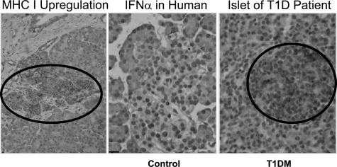
As seen in Fig. 1, which shows data from Francesco Dotta's laboratory (33), major histocompatibility complex (MHC) class I upregulation (left panel) and interferon-α production (right panel) is detected in human islets that otherwise show absolutely no signs of immune infiltration. Although Dotta et al. provide strong evidence for presence of Coxsackie virus proteins in these islets, it is still unclear whether this is always associated with the presence of viral genomes in the islets or β-cells. What is striking about this finding is that, usually, MHC molecules and interferons are only upregulated in the context of significant inflammation or as a direct result of viral infection of a cell. The fact that no insulitis is present in these human islets but elevation of interferon-α and MHC class I expression is observed presents a strong argument for the possibility that a viral infection that has already infected the β-cells and persists. Interferons and MHC class I molecules are usually rapidly downregulated once inflammation ceases or an infection has been cleared, but not when an infection is persistent. Currently, our team and others are attempting to reproduce and extend these findings using freshly frozen human pancreatic specimens from pre-diabetic and diabetic individuals who had accidental deaths (34).
FIG. 1.
Upregulation of MHC class I molecules and interferon-α is found in human islets in the absence of immune infiltration—a viral signature? Histopathology demonstrating MHC class I upregulation (left panel) and interferon-α production (right panel) is detected in human islets that otherwise show absolutely no signs of immune infiltration, similar to healthy islets (control, center panel). IFN, interferon; T1D, type 1 diabetes. (Reprinted and adapted with permission from ref. 33.)
In experimental animal models we can now ask the question, what are the immunological consequences of the increased MHC class I expression on β-cells? Our earlier studies followed the expression level of MHC class I on β-cells in an animal model of virally induced/enhanced diabetes (35). We assessed MHC class I levels in a normal B6 mouse following a viral infection, where no significant numbers of autoreactive CD8+ T-cells were present or activated, and compared them with MHC class I levels in β-cells of a transgenic animal (RIP-GP), which develops type 1 diabetes following a viral infection and in which autoreactive CD8+ T-cells that recognize a protein on β-cells are being generated (36). In the B6 mice, the viral infection lasts for about a week and affects the pancreas, leading to a 10- to 50-fold increase in MHC class I expression on β-cells. Importantly, this increase is transient and recedes to baseline after the virus infection has been cleared. In contrast, in the transgenic RIP mice that develop diabetes, and where the β-cells express a protein that is recognized by autoaggressive CD8+ lymphocytes, MHC class I expression increased more than 100-fold above baseline and remains high. Based on these observations, we asked whether this upregulation of MHC class I molecules plays a role in the recognition and destruction of β-cells by the immune system. This is an important question, since MHC class I restricts CD8+-mediated killing of target cells. Only β-cells that expressed virally induced high levels of MHC class I were killed in vitro by autoreactive CD8+ cytotoxic T-cells. β-Cells with basal levels of MHC class I were invisible to immune recognition. We can conclude from these findings that, under normal physiological circumstances, when there is no inflammation present in islets, β-cells are invisible to CD8+-mediated killing. In contrast, if MHC is being upregulated through inflammation, a viral infection, or both, β-cells become susceptible or “unmasked” to immune-mediated attacks. This is reflected by the rapid development of type 1 diabetes in this virally induced type 1 diabetes animal model.
The upregulation of MHC class I in this model is caused by many inflammatory factors, but the main mediators are interferons. Consequently, the presence of interferons and upregulation of MHC class I is detrimental for the β-cell. Viruses can cause this elevation of interferons and upregulation of MHC class I, and if they are directly present in the pancreas or islets, they can be considered prime suspects in enhancing the diabetogenic process. It is noteworthy that the virus we used, lymphocytic choriomeningitis virus (LCMV), rarely infects β-cells directly, which demonstrates that direct infection of β-cells is not needed. From the findings I have just described, we can make a good case that certain pancreatotropic virus infections do not directly cause, but rather substantially contribute to, the pathogenesis of type 1 diabetes by the inflammatory reaction that they induce by the host.
In summary, we have generated an interesting animal model that emulates what could occur in humans. Viral infections can cause strong inflammation and/or upregulation of MHC class I molecules and interferons. In animal models, for virally induced type 1 diabetes, MHC class I upregulation on β-cells is a prerequisite for their recognition and demise by killer CD8+ T-cells. This is a transient process and recedes to baseline once the virus is cleared. Human islets can also express MHC class I and interferons, and this is seen even in the absence of an inflammatory infiltrate. Thus, infections, although rarely able to directly cause type 1 diabetes, might act as providers of a “fertile field” to precipitate type 1 diabetes in individuals genetically at risk.
Prevention of type 1 diabetes through viral infections.
What about the flipside of this coin, that is, the possibility that viral infections and other types of infectious diseases would actually prevent type 1 diabetes? At first, this seems counterintuitive because of the aforementioned data indicating that viruses can be strong inducers of inflammation. However, emerging experimental evidence from various animal models has taught us otherwise. I will discuss some of the evidence and underlying mechanisms in the following examples.
New unpublished data from us and published work from others (37–39) support the so-called “hygiene hypothesis,” which implies that type 1 diabetes is a disease of the more industrialized countries because lesser numbers of infections occur and the immune system has therefore less opportunity to be properly trained for its main task, which is host defense. For example, in our laboratory, we infected two completely different pre-diabetic type 1 diabetes mouse models, the NOD and the RIP-LCMV (36), with Coxsackie B3. This virus has been shown to systemically infect mice and has been epidemiologically associated with human type 1 diabetes. The Cox B infection usually lasts 10 days and involves the pancreas, but rarely the islets. Interestingly, acceleration of diabetes is only seen in some instances and depends on the timing of infection (40–43), for example, when Coxsackie B3 is given to 10-week-old NOD mice or RIP mice 7 days post triggering of the diabetogenic response. Interestingly, it is important to note that an equally common outcome for viral infection is the lowering of diabetes penetrance. I would now like to discuss some impressive data supporting one of the underlying mechanisms for viral prevention of type 1 diabetes and then discuss some of the other potential mechanisms in a brief overview.
In recent unpublished observations, we discovered that infection of NOD mice could lead to invigoration of Tregs. Instead of enhancing immune function and exerting effector functions such as killing of infected cells or inducing interferon production (44–47), Tregs can turn immune responses off. They do this through a variety of mechanisms in vivo, many of which involve the secretion of immune modulatory cytokines such as interleukin (IL)-10, IL-4, or transforming growth factor (TGF)-β. In our study, NOD mice that usually develop spontaneous autoimmune-mediated diabetes were infected with LCMV, which is cleared after 7 to 10 days. We observed a significant decrease in the incidence of type 1 diabetes, down from about 75% to about 15%. Tregs were subsequently isolated from these NOD mice several weeks after the protective viral infection and transferred into nonmanipulated naive NOD recipient mice during the pre-diabetic phase. Only the sorted Tregs from mice that had undergone the viral infection were potent enough to protect recipient NOD mice from development of diabetes. We conclude that the systemic viral infection invigorated the Tregs.
To understand the mechanism of protection, fluorescence-activated cell sorting analysis of Tregs from mice that had received the LCMV virus was compared with that of Tregs from nonmanipulated mice. The mice that had been infected produced a 10-fold increase in TGF-β. We then investigated whether in vitro suppression by virally invigorated Tregs was dependent on TGF-β by using cytokine blocking antibodies. Indeed, their suppressive function was decreased in the absence of TGF-β by about 50%. Next TGF-β was transfected into NOD-derived naive Tregs in vivo. Interestingly, the transfected Treg function was enhanced to the same level as the suppressive capacity seen in virally invigorated Tregs. We therefore conclude, from these findings, that viral infections enhance Treg activity in part by augmenting TGF-β production. Recent findings from our lab have demonstrated that this occurs in a Toll receptor-2–dependent fashion (C. Filippi and M.v.H., unpublished observations).
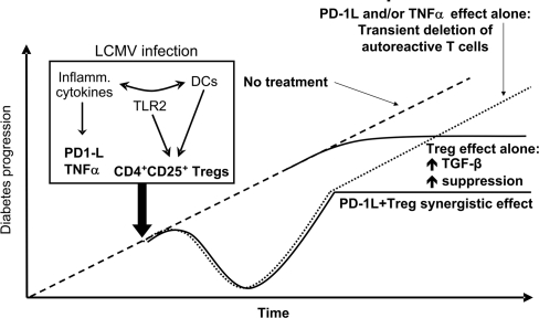
To summarize what we have learned so far regarding the mechanism by which viral infections can inhibit a diabetogenic response, please see Fig. 2. TGF-β augmentation of Tregs is not solely responsible for virally mediated prevention of type 1 diabetes. It turns out that there are important general mechanisms that occur simultaneously, while the virus infection is ongoing. Based on our findings, these are associated with the viral induction of PD-1L and tumor necrosis factor (TNF)-α (31). Both molecules have been shown to downmodulate immune responses and are important for the attrition of the antiviral immune response, when the viral infection is being cleared (with the purpose to limit excess immunopathology). Indeed, what happens after a viral infection, and this mechanism holds true for Coxsackie B viruses and other systemic infections, is that not only are the antiviral T-cells being eliminated, but PD-1L and TNF-α are mediating the bystander death of autoaggressive T-cells. This aspect of the mechanism results in a delay in the progression to type 1 diabetes but not a reduction in the overall incidence. The invigoration/activation of Treg cells is ultimately responsible for the observed reduction in incidence of type 1 diabetes in our model systems. Combined, these cytokine effects result in reduced incidence and delay of type 1 diabetes. These findings also illustrate rather elegantly that there are two components needed to prevent or cure type 1 diabetes therapeutically: 1) elimination of autoaggressive T-cells and 2) augmentation of Tregs to achieve long-term tolerance.
FIG. 2.
Conclusions and mechanistic hypothesis: How viral infections can stop type 1 diabetes. TGF-β augmentation of Tregs is not solely responsible for virally mediated prevention of type 1 diabetes. The viral induction of both PD-1L and TNF-α has been shown to downmodulate immune responses and are important for the attrition of the antiviral immune response, when the viral infection is being cleared. PD-1L and TNF-α also mediate the bystander death of autoaggressive T-cells.
Treatment of type 1 diabetes with novel combinatorial therapies.
We will now discuss potential combination therapies in recent-onset type 1 diabetes. As indicated above, there are multiple immune-modulatory steps required for the ultimate protection against type 1 diabetes as well as protection against progression of disease. Because of the multifaceted nature of this disease, a monotherapy will not be sufficient to combat type 1 diabetes. The best approach is to use a combination therapy (Table 1), with the goal being the early elimination of autoaggressive T-cells followed by inducing long-term tolerance through induction of regulatory T-cells. Combination therapies consist of a short-acting systemic immunosuppressant and an antigen-specific vaccine that synergize to enhance efficacy in preventing or delaying the onset of clinical type 1 diabetes. Long-term systemic immunosuppression is therefore not necessary, thus reducing deleterious side effects and decreasing the potential for infections and cancers (48). In contrast, direct administration of cytokines results in more side effects, and the cytokine half-life in blood is very short, necessitating repeated administration (49,50). Drugs such as anti-CD3 also have systemic side effects, and their efficacy so far has been limited to reducing the decline in C-peptide production in patients with early-onset diabetes (24,51). In turn, vaccination with β-cell auto-antigens alone has limited efficacy, albeit no side effects (52–54). However, as shown by us in animal models, efficacy can be increased through synergistic effects, which allows for a dose reduction of the systemically acting immunosuppressant (anti-CD3) and therefore increases the safety margin of the intervention (55). In summary, a potential, yet potent combinatorial therapy for type 1 diabetes, would consist of a systemically acting antibody such as anti-CD3 with an islet antigen–specific vaccine to augment Tregs. This cocktail would have limited side effects and enhance efficacy significantly.
TABLE 1.
How combination therapies will help in translating antigen-specific immunotherapy to induce Tregs
| 1. | Enhancing efficacy while reducing unwanted systemic side effects |
| 2. | Choosing the correct islet antigen |
| 3. | Choosing the right dose of antigen |
| 4. | Choosing optimal time for intervention = identifying ideal target group (pre-diabetic, recent onset) |
I will now discuss islet antigen–specific Tregs and their contribution toward establishing long-term tolerance and thus protection against the development of type 1 diabetes. To understand autoreactive islet antigen–specific Tregs, one has to consider that any adaptive immune response consists of different classes of responses such as pro-inflammatory and anti-inflammatory, and these combined responses constitute a process in equilibrium. This equilibrium can be shifted, however, if either effector or regulatory responses are increased. The result of this shift in equilibrium is either enhanced inflammation or resolution of the process. Therefore, it follows that immune regulation is context, and likely organ, dependent. Tregs will likely have as many functions and phenotypes as T effector (Teff) cells. Key effector molecules produced by Tregs are cytokines such as IL-10, TGF-β, and FoxP3, which can modulate the interaction of Teff with antigen-presenting cells (APCs) and are therefore frequently crucial in mediating tolerance. Because of this Treg/Teff balance, it is logical that any inflammatory reaction involved in to the pathogenesis of type 1 diabetes will contain some autoreactive regulatory cells that recognize islet antigens. In individuals who develop diabetes, the existing Tregs are not sufficient in numbers or activities to suppress the diabetogenic response, but modulation of Tregs causing the cells to shift to a state that produces IL-10, IL-4 or TGF-β could be a unique and powerful opportunity to augment the body's own Treg response in order to suppress islet destruction and achieve long-term tolerance. In my opinion, augmenting autologous Tregs is the only approach that could be used to treat or revert type 1 diabetes with sufficiently low side effects as well as long-term duration. Furthermore, such autologous Tregs could be induced by mucosal or DNA vaccination (56,57), which would be universally applicable and would not involve expensive or complicated techniques.
Induction of islet antigen–specific Tregs to achieve long-term tolerance.
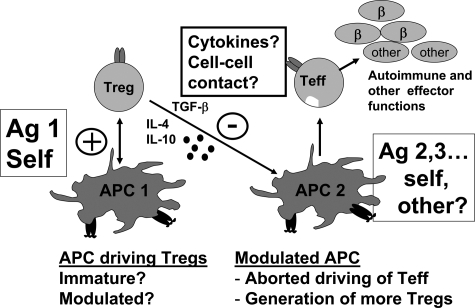
Some of the advantages of Treg activation are depicted in Fig. 3. When an APC presents β-cell proteins to an autoreactive Treg following, for example, mucosal immunization with insulin or subcutaneous immunization with GAD, autoreactive T-cells that recognize this protein, among them Tregs, become activated, proliferate, and circulate throughout the body. The activated cells will reach various organs and scan these for the antigentic protein, as shown to occur in the pancreatic draining lymph nodes and sometimes the islets in type 1 diabetes animal models (53,58). The number of cells is further augmented by proliferation within the lymph node or pancreatic islets, and soluble mediators secreted by the Tregs in these localized areas will now have multiple beneficial effects. Cytokines such as TGF-β, IL-4, and IL-10 have the potential to modulate or temper the activity of antigen-presenting and destructive islet effector cells. Seminal to this discovery is that these events occur locally in the pancreatic lymph node or islets, thus circumventing systemic immunosuppression by the host. Furthermore, regulation of the effector cells takes place, regardless of their antigentic specificity. This well-documented phenomenon was termed “bystander suppression” by Howard Weiner (54). Thus, and this is conceptually very important for therapies involving augmentation of Tregs, one does not have to know the initiating or “driving” antigens to suppress type 1 diabetes with autoreactive Tregs.
FIG. 3.
Bystander suppression by Tregs: Tolerance tag team? When an APC presents β-cell proteins to an autoreactive Treg following, for example, mucosal immunization with insulin or subcutaneous immunization with GAD, autoreactive T-cells that recognize this protein, among them Tregs, become activated, proliferate, and circulate throughout the body. The activated cells will reach various organs and scan them for the antigentic protein, as shown to occur in the pancreatic draining lymph nodes and sometimes the islets in type 1 diabetes animal models. The number of cells is further augmented by proliferation within the lymph node or pancreatic islets, and soluble mediators secreted by the Tregs in these localized areas will now have multiple beneficial effects and mediate long-term tolerance by suppressing the aggressive autoreactive effector response.
These activated Treg cells have another advantage in that they can mediate “infectious tolerance” and work as a tag team. Mechanistically, Tregs can entice APCs within the islets or pancreatic lymph node to induce and augment other types of Tregs. This phenomenon was first described by Hermann Waldman and Polly Matzinger, and Oral Alpan had a very elegant article on this topic (59–61). Thus, Tregs are a very attractive option to induce long-term tolerance while avoiding systemic side effects, and there is substantial evidence from animal models to support this concept.
Obstacles to translating antigen-specific therapies involving Tregs to the clinic.
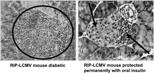
We have to ask the questions, what has hampered the translation of antigen-specific Treg therapies to the clinic and what can we do about it? You will see that combination therapies will likely provide the answers to these questions. For instance, the treatment of recent-onset diabetes can be mediated by a combinatorial therapy that will induce a protective immune response as well as provide long-term tolerogenic effects. However, there still exist several hurdles to overcome in therapeutic translation to the clinic. Figure 4 shows a representative histological picture for long-term tolerance following induction of Tregs with oral insulin in the mouse model. The left panel shows a completely destroyed islet of a diabetic RIP-LCMV mouse. The right panel shows a representative image of mild insulitis commonly found in mice protected from type 1 diabetes onset after administration of oral insulin during the pre-diabetic phase. Here, the β-cells are mostly intact, and the immune cells are located at the periphery but not within the islet. Some of the cells in the periphery do have regulatory activity, as shown by enzyme-linked immunospot analysis (62). Despite these promising results with β-cell antigen vaccinations, there are still significant questions that need to be addressed before clinical trials are initiated. Several of these issues are delineated in Table 1 and include predicting which islet antigen is optimal to use for an individual patient, defining the right dose of β-cell antigen for immunization, and choosing the proper time for intervention. However, these aspects may become less problematic as initially thought, when taken into context with a combinatorial treatment rather than as a monotherapy. The means by which combination therapy will address these concerns are described below and will be illustrated with some examples from experimental data, demonstrating how combination therapies will help.
FIG. 4.
Long-term tolerance following induction of regulatory T-cells with oral insulin: Histological representation of long-term tolerance following induction of Tregs with oral insulin in the mouse model. Shown are a completely destroyed islet of a diabetic RIP-LCMV mouse (left panel) and mild insulitis commonly found in mice protected from type 1 diabetes onset after administration of oral insulin during the pre-diabetic phase (right panel). In these images, the β-cells are mostly intact and the immune cells are located at the periphery but not within the islet.
Based on experimental evidence, not all β-cell proteins are suited to induce Treg activation, and efficacy depends on the genetic background of the host. For example, immunization with insulin, but not GAD, protects NOD mice during the pre-diabetic phase (D. Bresson and M.v.H., unpublished results). Although not shown, it is important to know that efficacy of these antigens is reversed when used in a mouse with a different genetic background, such as in B6 mice. In this case GAD immunization protects better than insulin. In the majority of genetic backgrounds, however, protection can be enhanced by co-immunization with cytokine expressing plasmids (63), indicating that combination therapy with immune modulatory compounds will not only be beneficial but also, in some cases, prevent the generation of insulin auto-antibodies (M.v.H. and G. Eisenbarth, unpublished results). Thus, efficacy of antigen-specific induction of Tregs depends on the individual genetic background, the context in which antigen presentation occurs, and the extent of activation of the preexisting Treg repertoire.
The dose of the islet antigen used for induction of Treg cells also appears to play a crucial role. For example, orally administered human or porcine insulin, or insulin conjugated to the cholera toxin B subunit to enhance efficacy, protects only at intermediate dosages, but not when low or high amounts of oral antigen are administered (64). Such bell-shaped dose-response curves are common to oral antigens and constitute a major roadblock for clinical translation because there are no suitable biomarkers to assess or predict which antigenic dose might be optimal for a given individual.
It is known from multiple animal studies that protection from diabetes by induction of Tregs is most effective when the intervention is done early, not late. Also, the time point of Treg induction in relation to the diabetogenic response is of importance. Based on our unpublished data (G. Fousteri and M.v.H., unpublished results), intranasal peptide treatment can lead in some experiments to significant acceleration of disease in older mice, whereas similar peptide treatments in younger mice were reported to have significant protection from diabetes. The occasional acceleration of the disease observed after immunotherapy in older mice is indicative of a preexisting pool of activated effector cells, which cannot be turned off by a monotherapeutic Treg-inducing vaccination process alone. This deleterious negative outcome, however, can be overcome by the use of a combination therapy or use of suitable adjuvants, for example, incomplete Freund's adjuvant. This mechanism ensures that regulatory, not islet-attacking, effector T-cells are being generated following immunization. In fact, immunization in the presence of adjuvants as a type 1 diabetes therapeutic is currently being tested in an ongoing clinical trial in Boston (65).
Thus, although promising in terms of establishing long-term tolerance, there are significant hurdles that need to be overcome before induction of islet antigen–specific Tregs can be brought to the clinic setting. These include finding the correct dose to induce and not delete Tregs, defining the proper antigen for vaccination or Treg recognition (ideally not an antigen that is already being targeted by a destructive ongoing immune response), selecting an appropriate adjuvant or immunosuppressive agent for co-vaccination, and defining the optimal time in the disease process for such an intervention. As discussed earlier, one way to circumvent some of these issues would be to combine the Treg augmentation immunization with a short-term course of a suitable, systemically acting immunosuppressant or immune modulator. Such a compound would ideally have two key abilities: 1) to dampen the destructive response attacking the islets in order to generate a sufficient window for Tregs to be augmented and 2) to immunosuppress in such a way that Tregs are not affected. An example of one such compound is anti-CD3.
Combination therapy of recent-onset type 1 diabetes using anti-CD3 and immunization with β-cell antigens.
Human clinical study results in recent-onset type 1 diabetes treated with non-FC binding anti-CD3 (24,51) have shown that a 14-day course can delay the decline of C-peptide in recently diagnosed individuals by 2–3 years. Herold and coworkers (55) have also shown augmented Treg responses after anti-CD3 treatment. These results in conjunction with the success of mouse vaccination studies using mucosal administration of β-cell antigens to induce adaptive Tregs led to the expectation that their combination (anti-CD3 and islet antigen vaccines) would result in long-lasting tolerance. We conducted a proof of principle mouse trial; we combined a 5-day course of a suboptimal anti-CD3 dose with intranasal application of a proinsulin peptide to treat recently diabetic mice using two animal models, the NOD mouse and the RIP-LCMV mouse (55). In previous studies, higher doses of anti-CD3 had reverted, thus cured, type 1 diabetes in both mouse models. With the current study, the question was whether combination therapies would result in an increased efficacy of type 1 diabetes reversion with an otherwise suboptimal dose. As expected from the earlier studies, the proinsulin peptide alone had no or very limited efficacy in recent-onset diabetes in both mouse models. Furthermore, a suboptimal anti-CD3 dose alone also exhibited very limited efficacy. In contrast, the combination of both anti-CD3 and proinsulin peptide administration exhibited significant synergy, with a 100% increase in the reversion rate for both recent-onset hyperglycemia and diabetes. Based on these encouraging results, other islet antigens were also tested for their immunomodulatory properties in conjunction with anti-CD3. Synergy with oral and nasal whole insulin immunization was observed as well as several DNA vaccines expressing insulin or GAD. Some synergy was also seen in preliminary studies using GAD-Alum administered subcutaneously. Thus, islet antigen–specific therapies in combination with anti-CD3 resulted in a synergistic effect that increased the reversion rate of recent-onset diabetes in two different animal models.
In follow-up studies, we were able to define the underlying mechanisms for the above-described synergism and therefore proceeded to test additional parameters that were clinically relevant. In Fig. 5A–C, animals that remained diabetic are shown with squares, the animals that reverted to normoglycemia are shown with diamonds, and those transiently reverted are shown with triangles. These data were correlated with age (x-axis) and blood glucose (y-axis) of the animal at the onset of hyperglycemia and the start of intervention. The upper panels show the NOD mice that underwent combination therapy, whereas the bottom panels portray the animals treated with the monotherapy anti-CD3. One striking observation was that regardless of age of diabetes onset, mice with chronically high blood glucose values were essentially refractory to the combination therapy (Fig. 5A vs. B). There was an improved response to the combination therapy when mice had a lower blood glucose value (Fig. 5B), perhaps when animals were at an earlier stage of disease progression. We would argue that this observation parallels well with data from Chatenoud and coworkers in the human anti-CD3 trial (51). If one considers blood glucose values in non–insulin-treated NOD mice as a correlate of remaining β-cell activity, individuals with high C-peptide values respond much better to anti-CD3 treatment than those with lower C-peptide values. It appears that reversion of recent-onset type 1 diabetes is not possible once β-cell mass has declined beyond a certain point.
FIG. 5.
A: Essential influence of age (reflects rapidity of diabetogenic response) and blood glucose value (BGV) (reflects remaining islet mass) at treatment onset. Forty-four percent of the mice with chronically high BGVs were essentially refractory to the combination therapy. B: Essential influence of age (reflects rapidity of diabetogenic response) and BGV (reflects remaining islet mass) at treatment onset. There is better synergy in mice with higher remaining β-cell mass (BGV <450 mg/dl): 65% of animals with consistently lower blood glucose (<450 mg/dl) achieved improved synergy with combination therapies. This lower blood glucose range is indicative of remaining islet mass. C: Essential influence of age (reflects rapidity of diabetogenic response) and BGV (reflects remaining islet mass) at treatment onset. The best synergy occurs in mice with higher β-cell mass and rapid type 1 diabetes onset. A 75% reversion rate from type 1 diabetes was observed in animals receiving the combination therapy. These animals were younger and had a more rapid type 1 diabetes onset. Only a 27% reversion rate was seen in mice treated at diabetes onset with anti-CD3 alone. The reason for this phenomenon is not known, but it is possible that younger mice have better capacity to regenerate β-cells after inception of treatment and thus reestablish euglycemia.
The other remarkable clinical correlate discovered was that younger mice with lower blood glucose levels responded very well to the combination therapy, as shown in Fig. 5C. Here, statistics revealed a 75% reversion rate from type 1 diabetes in the combination therapy–treated mice. Only a 27% reversion rate was seen in mice treated at diabetes onset with anti-CD3 alone. The reason for this phenomenon is not known, but it is possible that younger mice have better capacity to regenerate β-cells after inception of treatment and thus reestablish euglycemia.
What are the underlying mechanisms for the enhanced protection in the group that received anti-CD3 and intranasal proinsulin peptide versus monotherapy with either agent? We have observed increased induction of Tregs that react to proinsulin and a reduction in CD8+ T-cells specific for β-cell antigens in both diabetes models treated with the combination therapy (55). The ability of T-cells isolated from mice receiving the combination therapy, versus those treated with anti-CD3 alone, to produce the regulatory cytokines IL-4 or -10 in vitro in response to the proinsulin peptide was augmented. Thus, nasal peptide immunization with an insulin peptide in conjunction with anti-CD3 can lead to enhanced islet antigen–specific production of immune modulatory cytokines that are known from many earlier studies to delay or prevent type 1 diabetes. This strongly indicates that islet-adaptive Tregs were augmented by the combination therapy. In support of the clinical translation of this finding was our discovery that increased numbers of IL-10–positive Tregs at diagnosis correlated with better glycemic control 3 months later in a small clinical study (66). Thus, increasing Tregs by vaccination in conjunction with an immunomodulatory as combination therapy might improve glycemic control.
We then took a closer look at the phenotype and further function of such Tregs in the mouse models. After combinatorial treatment, Tregs were FoxP3-positive and the numbers of CD4+CD25+ Tregs following combinatorial treatment were significantly increased compared with those treated with anti-CD3 monotherapy alone. Furthermore, when the CD4+CD25+ Tregs were sorted and stimulated with the proinsulin peptide, we again found increased production of cytokines known to have immune modulatory or dampening functions (TGF-β, IL-10, and IL-4).
We next wished to understand whether this increased induction of Tregs in response to the proinsulin peptide affected the number or activity of autoreactive CD8+ lymphocytes known to attack β-cells. In both models treated with the combination therapy versus the anti-CD3 alone, or compared with pre-diabetic mice, a strong reduction in the number of autoaggressive CD8+ lymphocytes was seen. The numbers were on average reduced by threefold compared with those in untreated or pre-diabetic mice. We would argue that this loss of autoreactive CD8+ lymphocytes is the result of proinsulin-specific Treg-mediated bystander suppression. Indeed, our earlier studies published in Immunity in 1999 had first described this in mice that received Treg transfers from oral insulin–treated donors (53). The transfer of 105 CD4+ cells from an oral insulin–treated RIP-LCMV mouse that had been protected from type 1 diabetes resulted in localized reduction of autoaggressive CD8+ cells in the pancreatic lymph node but not other lymphoid sites. The site specificity of the regulatory effect is also a clinically important point and is likely due to the fact that transferred Tregs only proliferate in the pancreatic lymph node, not in other locations, as shown by CFSE (carboxyl fluorescent succinimidyl ester) labeling. We can therefore conclude from these studies that the antigen-specific component of our combination therapy, which is the intranasal administration of the proinsulin peptide, leads to an augmentation of proinsulin-specific adaptive Tregs, which are able to bystander-suppress autoaggressive CD8+ T-cells. This result was similar in two distinct diabetes models and lead to enhanced prevention of disease, when the induction of Tregs by the proinsulin peptide was combined with a short-course anti-CD3 treatment.
It is my personal opinion that only combination therapies and not monotherapies will be able to treat recent-onset diabetes, and a strong effort should be made to clinically translate such approaches. Other combinations of antigenic immunization with systemic immune modulators can be envisioned—ideally, a combination therapy will consist of an islet antigen–specific immunization, such as oral insulin, coupled with a systemic drug that dampens islet destructive immunity but does not affect Tregs. Anti-CD3, Rituxan (anti-CD20), Anakinra, and possibly ATG (antithymocyte globulin) are good candidates for this.
Future perspectives.
I would like to introduce newly emerging and exciting areas of research that could be important in translating such approaches more rapidly. One very interesting new avenue is the use of in silico modeling, or virtual NOD mice, to predict and narrow down optimal treatment protocols. We have spent the past year, thanks to an American Diabetes Association–supported grant, working with the company Entelos (67), simulating optimized treatment regimens. In this case, some interesting and surprising predictions were made, which we are currently following up to validate with wet-lab experiments in our laboratory. Entelos generated a variety of virtual NOD mice and tested under which experimental conditions most of these would revert to normoglycemia after recent-onset type 1 diabetes using anti-CD3 and oral insulin combination therapy. Less than 50% of virtual mice reverted to normoglycemia in these simulations, when anti-CD3 and oral insulin were given at the same time. This prediction is fully reflected by our current experimental data, combining anti-CD3 and oral insulin (D. Bresson and M.v.H., unpublished observations). Unexpected to us, Entelos’ simulation predicted that the therapeutic window will be much larger if anti-CD3 is given after the oral insulin. The delay in anti-CD3 administration should allow for sufficient time for Treg generation. We are currently validating this prediction. Additionally, and in relevance to the ongoing oral insulin trial DPT-1 (Diabetes Prevention Trial of Type 1 Diabetes), there is an optimal and bell-shaped dose range for oral insulin, too frequent insulin dosing is not beneficial, and few if any mice are predicted to revert from hyperglycemia. In the future, one can imagine that similar predictive simulations could guide human trial design and define optimal dosing regimens, optimal time for testing for induced T-cells (as biomarkers), and so forth.
In summary, let me share some thoughts about what I believe will solve the puzzle of type 1 diabetes in the future (Table 2). Foremost, we will need direct access to human pancreata to better understand the pathogenesis of the human disease (34). Animal studies should be used to fill crucial gaps in human pathogenesis and not vice versa. We need live imaging data from mouse and human islets to better understand the kinetics of β-cell death and regeneration. We have recently been successful at visualizing autoreactive lymphocytes entering and leaving the islets in the living animal (K. Coppieters and M.v.H., unpublished observations). We need to have diverse teams that comprise expertise from immunology, islet physiology, and complications in order to tackle this disease. We need to make a commitment to test combination therapies now, before single drugs have been approved for type 1 diabetes. Life-long immunosuppression will be difficult to justify for type 1 diabetes. We need to establish suitable biomarkers that can predict success of a given intervention.
TABLE 2.
Future directions and perspectives
| 1. | We need direct access to human pancreata to better understand the pathogenesis of the human disease. |
| 2. | We need live imaging data from mouse and human islets to better understand the kinetics of β-cell death and regeneration (2-photon and other techniques). |
| 3. | We need to have diverse teams that comprise expertise from immunology, islet physiology, and complications in order to tackle this disease. |
| 4. | We need to make a commitment to test combination therapies now, before single drugs have been approved for type 1 diabetes. |
| 5. | We need to establish suitable biomarkers that can predict success of a given intervention. Longitudinal T-cell studies will be essential, especially if the process leading to type 1 diabetes turns out to be relapsing-remitting in nature. |
| 6. | The ultimate goal should be an early childhood vaccine that redirects the immune system towards tolerance to β-cells. |
Longitudinal T-cell studies will be essential, especially if the process leading to type 1 diabetes turns out to be relapsing-remitting in nature. We presented the concept that Tregs and Teffs might undergo rapid cyclical changes during type 1 diabetes pathogenesis in a recent Nature review article (68). The ultimate goal should be an early childhood vaccine that redirects the immune system toward tolerance to β-cells.
Finally, I'd like to conclude with a more general view of an area of high importance, that is, how do we facilitate clinical translation of data relevant to type 1 diabetes, and for that matter, all clinically relevant data? We must improve the collaboration between academic researchers, industry, and funding bodies. Unfortunately, there are divergences that inhibit collaborative efforts. One major roadblock is the fact that academia is evaluated on individual achievements, not necessarily on the ability to work well in networks or in teams. In translational medicine, however, sharing of expensive resources, pooling of efforts, and involvement of larger networks is frequently unavoidable. Another example is the fact that industry is perhaps motivated too much by profit, which is not always the best driving force if one considers diseases such as type 1 diabetes, with a smaller market opportunity. Last, funding bodies are frequently driven by timeline constraints through their constituency—this can result in unrealistic timelines and, even worse, in cessation of funding for expensive endeavors after a large amount of infrastructure, as frequently needed for clinical trials, has already been created. I would like to leave you with the thought that we need to work hard and work together to change some of these paradigms. We will need to create novel coalitions and synergy and shift some of the existing paradigms. Evaluation of academic careers will have to involve factors other than the number and impact of papers. Treatment of orphaned diseases will require additional funding and incentives to attract industry. Funding bodies need to not abandon infrastructure they created based on time lines dictated by granting mechanisms. Finally, and most importantly, we all need to work together more effectively to benefit the health and overall quality of life of our patients.
Acknowledgments
The work featured prominently here is that contributed by Damien Bresson and Christophe Filippi.
The La Jolla Institute, the Brehm Coalition, the National Institutes of Health, the Juvenile Diabetes Research Foundation, and the American Diabetes Association sponsored this study.
No potential conflicts of interest relevant to this article were reported.
Many thanks to my long-term collaborators and colleagues. Most important, I would like to thank my family for their patience and support.
REFERENCES
- 1.Sutherland DE, Sibley R, Xu XZ, Michael A, Srikanta AM, Taub F, Najarian J, Goetz FC: Twin-to-twin pancreas transplantation: reversal and reenactment of the pathogenesis of type I diabetes. Trans Assoc Am Physicians 97: 80–87, 1984 [PubMed] [Google Scholar]
- 2.Eisenbarth GS: Molecular aspects of the etiology of type I diabetes mellitus. J Diabetes Complications 7: 142–150, 1993 [DOI] [PubMed] [Google Scholar]
- 3.Eisenbarth GS, Gianani R, Pugliese A, Verge CF, Pietropaolo M: Prediction and prevention of type I diabetes. Transplant Proc 26: 361–362, 1994 [PubMed] [Google Scholar]
- 4.In't Veld P, Lievens D, De Grijse J, Ling Z, Van der Auwera B, Pipeleers-Marichal M, Gorus F, Pipeleers D: Screening for insulitis in adult autoantibody-positive organ donors. Diabetes 56: 2400–2404, 2007 [DOI] [PubMed] [Google Scholar]
- 5.Cudworth AG: Type I diabetes mellitus. Diabetologia 14: 281–291, 1978 [DOI] [PubMed] [Google Scholar]
- 6.Itoh M: Immunological aspects of diabetes mellitus: prospects for pharmacological modification. Pharmacol Ther 44: 351–406, 1989 [DOI] [PubMed] [Google Scholar]
- 7.Blankenhorn EP, Descipio C, Rodemich L, Cort L, Leif JH, Greiner DL, Mordes JP: Refinement of the Iddm4 diabetes susceptibility locus reveals TCRVbeta4 as a candidate gene. Ann N Y Acad Sci 1103: 128–131, 2007 [DOI] [PubMed] [Google Scholar]
- 8.Blankenhorn EP, Rodemich L, Martin-Fernandez C, Leif J, Greiner DL, Mordes JP: The rat diabetes susceptibility locus Iddm4 and at least one additional gene are required for autoimmune diabetes induced by viral infection. Diabetes 54: 1233–1237, 2005 [DOI] [PMC free article] [PubMed] [Google Scholar]
- 9.Ellerman KE, Like AA: Susceptibility to diabetes is widely distributed in normal class IIu haplotype rats. Diabetologia 43: 890–898, 2000 [DOI] [PubMed] [Google Scholar]
- 10.Efrat S: Beta-cell replacement for insulin-dependent diabetes mellitus. Adv Drug Deliv Rev 60: 114–123, 2008 [DOI] [PubMed] [Google Scholar]
- 11.Jones PM, Courtney MJ, Burns CJ, Persaud SJ: Cell-based treatments for diabetes. Drug Discov Today 13: 888–893, 2008 [DOI] [PubMed] [Google Scholar]
- 12.Jun HS: Regeneration of pancreatic beta cells. Front Biosci 13: 6170–6182, 2008 [DOI] [PubMed] [Google Scholar]
- 13.Keymeulen B: Therapies aimed at preservation or restoration of beta cell function in type 1 diabetes. Verh K Acad Geneeskd Belg 70: 85–103, 2008 [PubMed] [Google Scholar]
- 14.Limbert C, Path G, Jakob F, Seufert J: Beta-cell replacement and regeneration: Strategies of cell-based therapy for type 1 diabetes mellitus. Diabetes Res Clin Pract 79: 389–399, 2008 [DOI] [PubMed] [Google Scholar]
- 15.Liu M, Han ZC: Mesenchymal stem cells: biology and clinical potential in type 1 diabetes therapy. J Cell Mol Med 12: 1155–1168, 2008 [DOI] [PMC free article] [PubMed] [Google Scholar]
- 16.Seissler J, Schott M: Generation of insulin-producing beta cells from stem cells: perspectives for cell therapy in type 1 diabetes. Horm Metab Res 40: 155–161, 2008 [DOI] [PubMed] [Google Scholar]
- 17.Sordi V, Bertuzzi F, Piemonti L: Diabetes mellitus: an opportunity for therapy with stem cells? Regen Med 3: 377–397, 2008 [DOI] [PubMed] [Google Scholar]
- 18.Soria B, Bedoya FJ, Tejedo JR, Hmadcha A, Ruiz-Salmeron R, Lim S, Martin F: Cell therapy for diabetes mellitus: an opportunity for stem cells? Cells Tissues Organs 188: 70–77, 2008 [DOI] [PubMed] [Google Scholar]
- 19.Soto-Gutierrez A, Navarro-Alvarez N, Caballero-Corbalan J, Tanaka N, Kobayashi N: Endoderm induction for hepatic and pancreatic differentiation of ES cells. Acta Med Okayama 62: 63–68, 2008 [DOI] [PubMed] [Google Scholar]
- 20.Stanley EG, Elefanty AG: Building better beta cells. Cell Stem Cell 2: 300–301, 2008 [DOI] [PubMed] [Google Scholar]
- 21.Urban VS, Kiss J, Kovacs J, Gocza E, Vas V, Monostori E, Uher F: Mesenchymal stem cells cooperate with bone marrow cells in therapy of diabetes. Stem Cells 26: 244–253, 2008 [DOI] [PubMed] [Google Scholar]
- 22.Monti P, Scirpoli M, Maffi P, Ghidoli N, De Taddeo F, Bertuzzi F, Piemonti L, Falcone M, Secchi A, Bonifacio E: Islet transplantation in patients with autoimmune diabetes induces homeostatic cytokines that expand autoreactive memory T cells. J Clin Invest 118: 1806–1814, 2008 [DOI] [PMC free article] [PubMed] [Google Scholar]
- 23.Roep BO: Islet autoreactive CD8 T-cells in type 1 diabetes: licensed to kill? Diabetes 57: 1156, 2008 [DOI] [PubMed] [Google Scholar]
- 24.Herold KC, Gitelman SE, Masharani U, Hagopian W, Bisikirska B, Donaldson D, Rother K, Diamond B, Harlan DM, Bluestone JA: A single course of anti-CD3 monoclonal antibody hOKT3γ1(Ala-Ala) results in improvement in C-peptide responses and clinical parameters for at least 2 years after onset of type 1 diabetes. Diabetes 54: 1763–1769, 2005 [DOI] [PMC free article] [PubMed] [Google Scholar]
- 25.Herold KC, Taylor L: Treatment of type 1 diabetes with anti-CD3 monoclonal antibody: induction of immune regulation? Immunol Res 28: 141–150, 2003 [DOI] [PubMed] [Google Scholar]
- 26.Voltarelli JC, Couri CE, Stracieri AB, Oliveira MC, Moraes DA, Pieroni F, Coutinho M, Malmegrim KC, Foss-Freitas MC, Simoes BP, Foss MC, Squiers E, Burt RK: Autologous nonmyeloablative hematopoietic stem cell transplantation in newly diagnosed type 1 diabetes mellitus. JAMA 297: 1568–1576, 2007 [DOI] [PubMed] [Google Scholar]
- 27.Ginsberg-Fellner F, Witt ME, Fedun B, Taub F, Dobersen MJ, McEvoy RC, Cooper LZ, Notkins AL, Rubinstein P: Diabetes mellitus and autoimmunity in patients with the congenital rubella syndrome. Rev Infect Dis 7 (Suppl. 1): S170–S176, 1985 [DOI] [PubMed] [Google Scholar]
- 28.Jenson AB, Rosenberg HS, Notkins AL: Pancreatic islet-cell damage in children with fatal viral infections. Lancet 2: 354–358, 1980 [PubMed] [Google Scholar]
- 29.Notkins AL: On the track of viruses. Nature 311: 209–210, 1984 [DOI] [PubMed] [Google Scholar]
- 30.Yoon JW, Morishima T, McClintock PR, Austin M, Notkins AL: Virus-induced diabetes mellitus: mengovirus infects pancreatic beta cells in strains of mice resistant to the diabetogenic effect of encephalomyocarditis virus. J Virol 50: 684–690, 1984 [DOI] [PMC free article] [PubMed] [Google Scholar]
- 31.Christen U, Benke D, Wolfe T, Rodrigo E, Rhode A, Hughes AC, Oldstone MB, von Herrath MG: Cure of pre-diabetic mice by viral infections involves lymphocyte recruitment along an IP-10 gradient. J Clin Invest 113: 74–84, 2004 [DOI] [PMC free article] [PubMed] [Google Scholar]
- 32.von Herrath MG, Fujinami RS, Whitton JL: Microorganisms and autoimmunity: making the barren field fertile? Nat Rev Microbiol 1: 151–157, 2003 [DOI] [PubMed] [Google Scholar]
- 33.Dotta F, Censini S, van Halteren AG, Marselli L, Masini M, Dionisi S, Mosca F, Boggi U, Muda AO, Prato SD, Elliott JF, Covacci A, Rappuoli R, Roep BO, Marchetti P: Coxsackie B4 virus infection of beta cells and natural killer cell insulitis in recent-onset type 1 diabetic patients. Proc Natl Acad Sci U S A 104: 5115–5120, 2007 [DOI] [PMC free article] [PubMed] [Google Scholar]
- 34.Juvenile Diabetes Research Foundation: nPod (Network for Pancreatic Organ Donors with Diabetes) helps advance study of diabetes. Available from http://www.jdrf.org/index.cfm?page_id=101980. Accessed 4 November 2008
- 35.Seewaldt S, Thomas HE, Ejrnaes M, Christen U, Wolfe T, Rodrigo E, Coon B, Michelsen B, Kay TW, von Herrath MG: Virus-induced autoimmune diabetes: most β-cells die through inflammatory cytokines and not perforin from autoreactive (anti-viral) cytotoxic T-lymphocytes. Diabetes 49: 1801–1809, 2000 [DOI] [PubMed] [Google Scholar]
- 36.von Herrath MG, Dockter J, Oldstone MB: How virus induces a rapid or slow onset insulin-dependent diabetes mellitus in a transgenic model. Immunity 1: 231–242, 1994 [DOI] [PubMed] [Google Scholar]
- 37.Smith KA, Efstathiou S, Cooke A: Murine gammaherpesvirus-68 infection alters self-antigen presentation and type 1 diabetes onset in NOD mice. J Immunol 179: 7325–7333, 2007 [DOI] [PubMed] [Google Scholar]
- 38.Bach JF: Infections and autoimmune diseases. J Autoimmun 25 (Suppl.): 74–80, 2005 [DOI] [PubMed] [Google Scholar]
- 39.Feillet H, Bach JF: On the mechanisms of the protective effect of infections on type 1 diabetes. Clin Dev Immunol 11: 191–194, 2004 [DOI] [PMC free article] [PubMed] [Google Scholar]
- 40.Horwitz MS, Bradley LM, Harbertson J, Krahl T, Lee J, Sarvetnick N: Diabetes induced by Coxsackie virus: initiation by bystander damage and not molecular mimicry. Nat Med 4: 781–785, 1998 [DOI] [PubMed] [Google Scholar]
- 41.Horwitz MS, Ilic A, Fine C, Balasa B, Sarvetnick N: Coxsackieviral-mediated diabetes: induction requires antigen-presenting cells and is accompanied by phagocytosis of beta cells. Clin Immunol 110: 134–144, 2004 [DOI] [PubMed] [Google Scholar]
- 42.Horwitz MS, Ilic A, Fine C, Rodriguez E, Sarvetnick N: Coxsackievirus-mediated hyperglycemia is enhanced by reinfection and this occurs independent of T cells. Virology 314: 510–520, 2003 [DOI] [PubMed] [Google Scholar]
- 43.Benoist C, Mathis D: Autoimmunity provoked by infection: how good is the case for T cell epitope mimicry? Nat Immunol 2: 797–801, 2001 [DOI] [PubMed] [Google Scholar]
- 44.Tang Q, Bluestone JA: Regulatory T-cell physiology and application to treat autoimmunity. Immunol Rev 212: 217–237, 2006 [DOI] [PubMed] [Google Scholar]
- 45.Bluestone JA, Thomson AW, Shevach EM, Weiner HL: What does the future hold for cell-based tolerogenic therapy? Nat Rev Immunol 7: 650–654, 2007 [DOI] [PubMed] [Google Scholar]
- 46.DiPaolo RJ, Brinster C, Davidson TS, Andersson J, Glass D, Shevach EM: Autoantigen-specific TGFbeta-induced Foxp3+ regulatory T cells prevent autoimmunity by inhibiting dendritic cells from activating autoreactive T cells. J Immunol 179: 4685–4693, 2007 [DOI] [PubMed] [Google Scholar]
- 47.Setoguchi R, Hori S, Takahashi T, Sakaguchi S: Homeostatic maintenance of natural Foxp3(+) CD25(+) CD4(+) regulatory T cells by interleukin (IL)-2 and induction of autoimmune disease by IL-2 neutralization. J Exp Med 201: 723–735, 2005 [DOI] [PMC free article] [PubMed] [Google Scholar]
- 48.Soulillou JP, Giral M: Influence of graft characteristics on the outcome of kidney transplantation. N Engl J Med 354: 2060–2062, 2006 [DOI] [PubMed] [Google Scholar]
- 49.Type 1 Diabetes: TrialNet. Available from http://www.diabetestrialnet.org/healthcare/index.htm. Accessed 4 November 2008
- 50.Benaroya Research Institute: New Diabetes Clinical Research Trials. Available from http://www.vmresearch.org/node/82. Accessed 4 November 2008
- 51.Keymeulen B, Vandemeulebroucke E, Ziegler AG, Mathieu C, Kaufman L, Hale G, Gorus F, Goldman M, Walter M, Candon S, Schandene L, Crenier L, De Block C, Seigneurin JM, De Pauw P, Pierard D, Weets I, Rebello P, Bird P, Berrie E, Frewin M, Waldmann H, Bach JF, Pipeleers D, Chatenoud L: Insulin needs after CD3-antibody therapy in new-onset type 1 diabetes. N Engl J Med 352: 2598–2608, 2005 [DOI] [PubMed] [Google Scholar]
- 52.Zhang ZJ, Davidson L, Eisenbarth G, Weiner HL: Suppression of diabetes in nonobese diabetic mice by oral administration of porcine insulin. Proc Natl Acad Sci U S A 88: 10252–10256, 1991 [DOI] [PMC free article] [PubMed] [Google Scholar]
- 53.Homann D, Holz A, Bot A, Coon B, Wolfe T, Petersen J, Dyrberg TP, Grusby MJ, von Herrath MG: Autoreactive CD4+ T cells protect from autoimmune diabetes via bystander suppression using the IL-4/Stat6 pathway. Immunity 11: 463–472, 1999 [DOI] [PubMed] [Google Scholar]
- 54.ClincialTrials.gov, a service of the U.S. National Institutes of Health: The Diabetes Prevention Trial of Type 1 Diabetes (DPT-1). Available from http://clinicaltrials.gov/ct2/show/NCT00004984?spons=%22National+Center+on+Minority+Health+and+Health+Disparities+(NCMHD)%22&spons_ex=Y&rank=7. Accessed 4 November 2008
- 55.Bresson D, Togher L, Rodrigo E, Chen Y, Bluestone JA, Herold KC, von Herrath M: Anti-CD3 and nasal proinsulin combination therapy enhances remission from recent-onset autoimmune diabetes by inducing Tregs. J Clin Invest 116: 1371–1381, 2006 [DOI] [PMC free article] [PubMed] [Google Scholar]
- 56.Therapeutics B: Randomized placebo-controlled phase I/II trial evaluating once-weekly intramuscular administration of BHT-3012 in up to 72 patients with type 1 diabetes who are on a stable dose of insulin. Available from http://www.bayhilltherapeutics.com/type1diabetes.html. Accessed 4 November 2008
- 57.Peakman M, Hussain MJ, Millward BA, Leslie RD, Vergani D: Effect of initiation of insulin therapy on T-lymphocyte activation in type 1 diabetes. Diabet Med 7: 327–330, 1990 [DOI] [PubMed] [Google Scholar]
- 58.Tang Q, Henriksen KJ, Bi M, Finger EB, Szot G, Ye J, Masteller EL, McDevitt H, Bonyhadi M, Bluestone JA: In vitro-expanded antigen-specific regulatory T cells suppress autoimmune diabetes. J Exp Med 199: 1455–1465, 2004 [DOI] [PMC free article] [PubMed] [Google Scholar]
- 59.Honey K, Cobbold SP, Waldmann H: Dominant regulation: a common mechanism of monoclonal antibody induced tolerance? Immunol Res 20: 1–14, 1999 [DOI] [PubMed] [Google Scholar]
- 60.Matzinger P: Graft tolerance: a duel of two signals. Nat Med 5: 616–617, 1999 [DOI] [PubMed] [Google Scholar]
- 61.Alpan O, Rudomen G, Matzinger P: The role of dendritic cells, B cells, and M cells in gut-oriented immune responses. J Immunol 166: 4843–4852, 2001 [DOI] [PubMed] [Google Scholar]
- 62.von Herrath MG, Dyrberg T, Oldstone MB: Oral insulin treatment suppresses virus-induced antigen-specific destruction of beta cells and prevents autoimmune diabetes in transgenic mice. J Clin Invest 98: 1324–1331, 1996 [DOI] [PMC free article] [PubMed] [Google Scholar]
- 63.Bot A, Smith D, Bot S, Hughes A, Wolfe T, Wang L, Woods C, von Herrath M: Plasmid vaccination with insulin B chain prevents autoimmune diabetes in nonobese diabetic mice. J Immunol 167: 2950–2955, 2001 [DOI] [PubMed] [Google Scholar]
- 64.Petersen JS, Bregenholt S, Apostolopolous V, Homann D, Wolfe T, Hughes A, De Jongh K, Wang M, Dyrberg T, Von Herrath MG: Coupling of oral human or porcine insulin to the B subunit of cholera toxin (CTB) overcomes critical antigenic differences for prevention of type I diabetes. Clin Exp Immunol 134: 38–45, 2003 [DOI] [PMC free article] [PubMed] [Google Scholar]
- 65.Orban T: Autoantigen vaccination in human type 1 newly diagnosed diabetes mellitus. Available from http://www.immunetolerance.org/research/autoimmune/trials/orban1.html. Accessed 4 November 2008
- 66.Sanda S, Roep BO, von Herrath M: Islet antigen specific IL-10+ immune responses but not CD4+CD25+FoxP3+ cells at diagnosis predict glycemic control in type 1 diabetes. Clin Immunol 127: 138–143, 2008 [DOI] [PubMed] [Google Scholar]
- 67.Tang Q, Adams JY, Penaranda C, Melli K, Piaggio E, Sgouroudis E, Piccirillo CA, Salomon BL, Bluestone JA: Central role of defective interleukin-2 production in the triggering of islet autoimmune destruction. Immunity 28: 687–697, 2008 [DOI] [PMC free article] [PubMed] [Google Scholar]
- 68.von Herrath M, Sanda S, Herold K: Type 1 diabetes as a relapsing-remitting disease? Nat Rev Immunol 7: 988–994, 2007 [DOI] [PubMed] [Google Scholar]


