Abstract
Digestion patterns of chromosomal DNAs of Bacillus cereus and Bacillus weihenstephanensis strains suggest that Sau3AI-type restriction modification systems are widely present among the isolates tested. In vitro methylation of plasmid DNA was used to enhance poor plasmid transfer upon electroporation to recalcitrant strains that carry Sau3AI restriction barriers.
Bacillus cereus is a gram-positive, spore-forming bacterium that can cause food spoilage and that has been associated with food poisoning outbreaks (9). B. cereus occurs ubiquitously in soil (6) and seems to be adapted to a wide range of environmental conditions, including a broad temperature range. Traditionally, B. cereus has not been considered a psychrotolerant species, but psychrotolerant strains can be isolated from the environment (11, 13). A mechanistic understanding of important traits, such as temperature survival and virulence, requires the availability of molecular tools to create knockout mutations of relevant genes. Gene knockout strategies routinely involve the introduction of plasmid or other extrachromosomal DNA into recipient strains to generate mutated derivatives of the parental strain. Successful transformation protocols have been developed for laboratory strains that may have lost important traits as a result of frequent subculturing. Diversity between pathogenicity or food spoilage properties of industrial or food isolates can be high, and consequently, traits of these isolates may be quite different from those analyzed in the (sequenced) laboratory strains. Various protocols have been developed for electroporation of gram-positive bacteria that aim at the improvement of transformation efficiency by using cell-weakening agents, various washing buffer compositions, and a variety of electric pulses. These protocols were not successful in our hands for B. weihenstephanensis DSM11821 and several other cold-tolerant food isolates, whereas when they were applied to the Bacillus cereus type strain (ATCC 14579) or Bacillus weihenstephanensis KBAB4, reasonable levels of transformants were obtained.
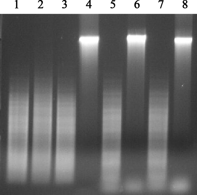
Variation in plasmid transfer frequency could reflect the presence of fortified cell walls that prevent DNA uptake and/or the presence of restriction modification (RM) systems in recalcitrant isolates (1, 8, 14). These topics were addressed in this study, and the latter possibility is suggested by the observation that genomic DNA of strain B. weihenstephanensis DSM11821 (isolated from mid-exponential cells [optical density at 600 nm, 0.5] using the GenElute kit from Sigma-Aldrich) is not digested by Sau3AI and BamHI but that DNA of B. cereus ATCC 14579 is digested by Sau3AI and BamHI, suggesting that the core recognition site (GATC) was modified in B. weihenstephanensis DSM11821. Moreover, two isoschizomers (MboI and DpnII) that recognize the same site but that are not inhibited by methylation of the cytosine residue, readily digested B. weihenstephanensis DSM11821 chromosomal DNA (Fig. 1), indicating that resistance to Sau3AI digestion has been caused by methylation of the cytosine residue of the GATC recognition site. Eighteen strains originating from various food products (Table 1) were analyzed for digestion by Sau3AI and its isoschizomers. Five out of the 18 strains tested (Table 1) were resistant to Sau3AI digestion, but this trait was not restricted to cold-tolerant strains, as two mesophilic strains (strains 1230-88 and B434) were identified as positive for methylation. Moreover, resistance to Sau3AI digestion was not shared by all cold-tolerant strains (3 out of 11 cold-tolerant strains tested were resistant to digestion). The modification of DNA could be part of a Sau3A-type RM system and appears widespread among B. cereus and B. weihenstephanensis isolates. The suggested function of RM systems in microorganisms is protection against invasion of foreign DNA, especially against phage infection (3, 7), and they are frequently associated with low transformation efficiencies in bacteria (2, 4, 5).
FIG. 1.

Digestion of chromosomal DNA of strain B. cereus ATCC 14579 (lanes 1 to 4) and strain B. weihenstephanensis DSM11821 (lanes 5 to 8). Chromosomal DNA (2 μg) was digested with either DpnII (lane 1 and 5), Sau3AI (lanes 2 and 6), or MboI (lanes 3 and 7) or left undigested (lanes 4 and 8).
TABLE 1.
Sensitivities of Bacillus cereus and Bacillus weihenstephanensis chromosomal DNA to digestion by Sau3AI, MboI, and DpnII
| Straina | Isolation source | Culture collection or referenceb | Digestion result with:
|
||
|---|---|---|---|---|---|
| Sau3AI | MboI | DpnII | |||
| B. cereus B434 | Pasteurized milk | NIZO | − | + | + |
| B. cereus B435 | Raw milk | NIZO | + | + | + |
| B. cereus B436* | Pasteurized milk | NIZO | + | + | + |
| B. cereus B437 | Pasteurized milk | NIZO | + | + | + |
| B. cereus B439 | Pasteurized milk | NIZO | + | + | + |
| B. cereus B443* | Pasteurized milk | NIZO | + | + | + |
| B. cereus 59* | Cream | 12 | + | + | + |
| B. cereus 61* | Cream | 12 | + | + | + |
| B. weihenstephanensis 401-92* | Scrambled eggs | 12 | + | + | + |
| B. weihenstephanensis 132* | Milk | 12 | + | + | + |
| B. weihenstephanensis 43-92* | Milk | 12 | − | + | + |
| B. weihenstephanensis 453-92* | Cream | 12 | − | + | + |
| B. cereus 674-98* | Scrambled eggs | 12 | + | + | + |
| B. cereus 1230-88 | Stew (food poisoning) | 12 | − | + | + |
| B. weihenstephanensis DSM11821* | Pasteurized milk | DSMZ | − | + | + |
| B. weihenstephanensis KBAB4* | Soil | 13 | + | + | + |
| B. cereus ATCC 14579 | Air | ATCC | + | + | + |
| B. cereus ATCC 10987 | Spoiled cheese | ATCC | + | + | + |
*, psychrotolerant strain (grows at temperatures of ≤7°C).
DSMZ, Deutsche Sammlung von Mikroorganismen und Zellkulturen, Braunschweig, Germany; ATCC, American Type Culture Collection; NIZO, NIZO food research, Ede, The Netherlands.
We anticipated that the presence of Sau3AI RM systems could explain at least part of the poor transfer of plasmid DNA that we experienced for some strains. To verify this hypothesis, we used an in vitro methylation procedure described previously for Helicobacter pylori isolates (4). To this end, plasmid DNA was methylated with cell extracts of B. cereus ATCC 14579, B. weihenstephanensis DSM11821, and B. cereus B434 that had been prepared from cells cultivated in LB medium at 30°C to an optical density at 600 nm of 1, and extracts were prepared as described by Donahue et al. (4) with the following modification: cells were mechanically disrupted in three runs in a minibeadbeater (BioSpec Products) in the presence of 0.1-mm Zirconia/silica beads, with cooling on ice between the runs. Strains were transformed using the protocol described by Silo-Suh et al. (10) for B. cereus with the following modifications: cells were cultivated at 30°C and the settings for electroporation were 1.2 kV, 400 Ω, and 25 μF. Transformants were selected on LB plates containing erythromycin or chloramphenicol (both at 5 μg/ml). For the two strains that harbor the Sau3AI RM system (DSM11821 and B434), in vitro methylation with cell extract of the corresponding recipient strains resulted in an improvement of transformation efficiency between approximately log 3 and log 5 transformants per μg plasmid DNA (pIL253) for these strains (Table 2). The presence of plasmid pIL253 could be confirmed by PCR using primers pIL253_fwd (TGCTCGAGTCTAGAATCGATACGA) and ery_pIl253r (TTGGCGTGTTTCATTGCTTG), which are specific to the erythromycin resistance cassette on the plasmid (data not shown). Plasmid methylated with cell extract of DSM11821 and B434 also cross-enhanced its transformation efficiency, although the number of transformants obtained was lower for strain DSM11821 than for strain B434, whose transformants were obtained with a plasmid treated with its own cell extract. It is possible that there are additional restriction enzymes in DSM11821 for which a corresponding methylase is not present in strain B434. Treatment of plasmid DNA with cell extract of strain ATCC 14579, which is negative for Sau3AI methylation, did not enhance the transformation of strains DSM11821 and B434.
TABLE 2.
Transformation efficiencies of Bacillus cereus and Bacillus weihenstephanensis strainsa
| Recipient strain | Plasmid | Transformation efficiency (no. of CFU μg−1) of:
|
|||
|---|---|---|---|---|---|
| Nontreated cells | CFEDSM11821 | CFEB434 | CFEATCC 14579 | ||
| DSM11821 | pIL253 | NT | 3.8 × 10³ | 1 × 10² | NT |
| None | 3 | 9.8 × 10⁴ | 3 × 101 | ||
| B434 | pIL253 | NT | 8.0 × 10³ | 4.8 × 10³ | NT |
| None | 2.3 × 10⁴ | ||||
| ATCC 14579 | pIL253 | 3.5 × 10⁶ | 2.2 × 10⁶ | ND | 1.5 × 10⁶ |
| None | 7.5 × 10⁵ | 2.3 × 10⁶ | |||
| KBAB4 | pIL253 | 2.5 × 10⁴ | 2.5 × 10⁴ | ND | ND |
Plasmid pIL253 was in vitro methylated with cell extracts of strain DSM11821 (CFEDSM11821), strain B434 (CFEB434), or strain ATCC 14579 (CFEATCC 14579). Each value is derived from an independent experiment. NT, no transformants were obtained; ND, not determined.
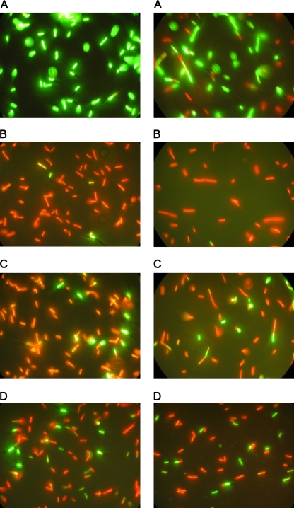
In vitro methylation resulted in a major improvement of the transformation efficiencies of strains DSM11821 and B434, but their efficiencies were still approximately 1 to 2 log units lower than that of B. cereus ATCC 14579. Electroporation requires the temporal formation of pores in the membrane and subsequently its resealing. We used the fluorescent nucleic acid dyes SYTO9 and propidium iodide (PI) of the Live/Dead BacLight bacterial viability kit (Invitrogen) to study whether pore formation and the resealing of pores upon electroporation could explain differences between strains. To this end, 100 microliters of cells immediately after electroporation was resuspended in 1.5 ml electroporation buffer (0.5 mM KH2PO4-K2HPO4, 0.5 mM MgCl2, 272 mM sucrose) containing a 5 μM concentration of the SYTO9 probe and a 30 μM concentration of the PI probe and incubated for 15 min at room temperature. Five microliters of stained cells was visualized using a Axioskop epifluorescence microscope equipped with a fluorescein isothiocyanate filter set (excitation wavelength, 450 to 490 nm; emission wavelength, >520 nm) for the detection of SYTO9- and PI-specific signals. For the time series, electroporated cells were recovered in electroporation buffer without probes and allowed to recover for 5, 10, or 30 min at room temperature prior to the addition of the probes. The SYTO9 dye can pass the bacterial membranes of intact cells, whereas the PI dye can enter only cells with a damaged or permeable membrane. Figure 2 shows that PI penetrates the cell membranes of B. cereus ATCC 14579 and B. weihenstephanensis DSM11821 upon electroporation and stains the majority of the population. When cells are allowed to recover for 5 to 30 min prior to the addition of fluorescent probes, part of the population remains unstained by PI, suggesting that pore resealing has occurred in these cells. The fractions of non-PI-stained cells were comparable for B. cereus ATCC 14579 and B. weihenstephanensis DSM11821, which indicates similar efficiencies of pore formation and recovery for the two strains. Notably, a relatively large fraction of the population loses culturability when subjected to electroporation for both B. weihenstephanensis DSM11821 and B. cereus ATCC 14579. Culturability was not improved upon extension of the recovery period (up to 3 h) (data not shown). Loss of viability after electroporation was confirmed by plate counting (there was an approximately 2-log reduction in viability [data not shown]). This suggests that the transformation protocol could be further optimized, and fluorescence staining may be a useful approach for optimizing transformation protocols without depending on extensive plate counting procedures.
FIG. 2.
Pore formation and pore resealing of the membrane of B. weihenstephanensis DSM11821 (left panels) or B. cereus ATCC 14579 (right panels) upon electroporation. Cells were labeled with fluorescent probes (PI and SYTO9) prior to electroporation (A), directly after electroporation (B), and after recovery for 5 (C) or 30 (D) minutes.
The results presented in this study show that in vitro methylation of DNA could be an approach to transfer DNA to otherwise-recalcitrant strains of B. weihenstephanensis and B. cereus. It may allow the molecular genetic analysis of undomesticated B. cereus and B. weihenstephanensis strains where RM systems prevent transformation.
Footnotes
Published ahead of print on 24 October 2008.
REFERENCES
- 1.Accetto, T., M. Peterka, and G. Avgustin. 2005. Type II restriction modification systems of Prevotella bryantii TC1-1 and Prevotella ruminicola 23 strains and their effect on the efficiency of DNA introduction via electroporation. FEMS Microbiol. Lett. 247:177-183. [DOI] [PubMed] [Google Scholar]
- 2.Alvarez, B., P. Secades, M. J. McBride, and J. A. Guijarro. 2004. Development of genetic techniques for the psychrotrophic fish pathogen Flavobacterium psychrophilum. Appl. Environ. Microbiol. 70:581-587. [DOI] [PMC free article] [PubMed] [Google Scholar]
- 3.Bickle, T. A., and D. H. Kruger. 1993. Biology of DNA restriction. Microbiol. Rev. 57:434-450. [DOI] [PMC free article] [PubMed] [Google Scholar]
- 4.Donahue, J. P., D. A. Israel, R. M. Peek, M. J. Blaser, and G. G. Miller. 2000. Overcoming the restriction barrier to plasmid transformation of Helicobacter pylori. Mol. Microbiol. 37:1066-1074. [DOI] [PubMed] [Google Scholar]
- 5.Hegna, I. K., H. Bratland, and A.-B. Kølsto. 2001. BceS1, a new addition to the type III restriction and modification family. FEMS Microbiol. Lett. 202:189-193. [DOI] [PubMed] [Google Scholar]
- 6.Hendriksen, N. B., B. M. Hansen, and J. E. Johansen. 2006. Occurrence and pathogenic potential of Bacillus cereus group bacteria in a sandy loam. Antonie van Leeuwenhoek 89:239-249. [DOI] [PubMed] [Google Scholar]
- 7.Jeltsch, A. 2002. Beyond Watson and Crick: DNA methylation and molecular enzymology of DNA methyltransferases. Chembiochem 3:274-293. [DOI] [PubMed] [Google Scholar]
- 8.Redaschi, N., and T. A. Bickle. 1996. DNA restriction and modification systems, p. 773-781. In F. C. Neidhardt et al. (ed.), Escherichia coli and Salmonella: cellular and molecular biology, 2nd ed. American Society for Microbiology, Washington, DC.
- 9.Schoeni, J. L., and A. C. Wong. 2005. Bacillus cereus food poisoning and its toxins. J. Food Prot. 68:636-648. [DOI] [PubMed] [Google Scholar]
- 10.Silo-Suh, L. A., B. J. Lethbridge, S. J. Raffel, H. He, J. Clardy, and J. Handelsman. 1994. Biological activities of two fungistatic antibiotics produced by Bacillus cereus UW85. Appl. Environ. Microbiol. 60:2023-2030. [DOI] [PMC free article] [PubMed] [Google Scholar]
- 11.Sorokin, A., B. Candelon, K. Guilloux, N. Galleron, N. Wackerow-Kouzova, S. D. Ehrlich, D. Bourguet, and V. Sanchis. 2006. Multiple-locus sequence typing analysis of Bacillus cereus and Bacillus thuringiensis reveals separate clustering and a distinct population structure of psychrotrophic strains. Appl. Environ. Microbiol. 72:1569-1578. [DOI] [PMC free article] [PubMed] [Google Scholar]
- 12.Stenfors, L. P., and P. E. Granum. 2001. Psychrotolerant species from the Bacillus cereus group are not necessarily Bacillus weihenstephanensis. FEMS Microbiol. Lett. 197:223-228. [DOI] [PubMed] [Google Scholar]
- 13.Vilas-Boas, G., V. Sanchis, D. Lereclus, M. V. Lemos, and D. Bourguet. 2002. Genetic differentiation between sympatric populations of Bacillus cereus and Bacillus thuringiensis. Appl. Environ. Microbiol. 68:1414-1424. [DOI] [PMC free article] [PubMed] [Google Scholar]
- 14.Waschkau, B., J. Waldeck, S. Wieland, R. Eichstadt, and F. Meinhardt. 2008. Generation of readily transformable Bacillus licheniformis mutants. Appl. Microbiol. Biotechnol. 78:181-188. [DOI] [PubMed] [Google Scholar]


