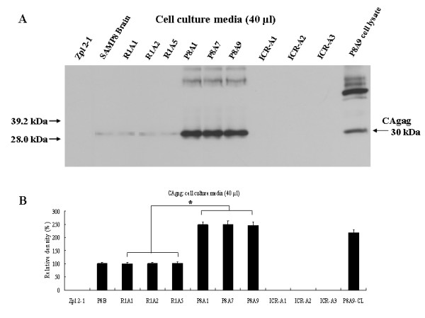
Figure 6.

Immunoblot analysis of CAgag in culture media from ICR-A, R1A and P8A cell lines. (A) Western blot analysis for expression of CAgag (40 μl of culture media) using anti-CAgag antibody. Zpl 2-1 culture media: a negative control; SAMP8 brain (12-month-old) and P8A9 cell lysate (P8A9-CL) (50 μg): positive controls. (B) Densitometry of CAgag in cell culture media. CAgag protein levels in P8A cell culture media were significantly higher than in R1A cell culture media. *statistically significant difference (p < 0.01).
