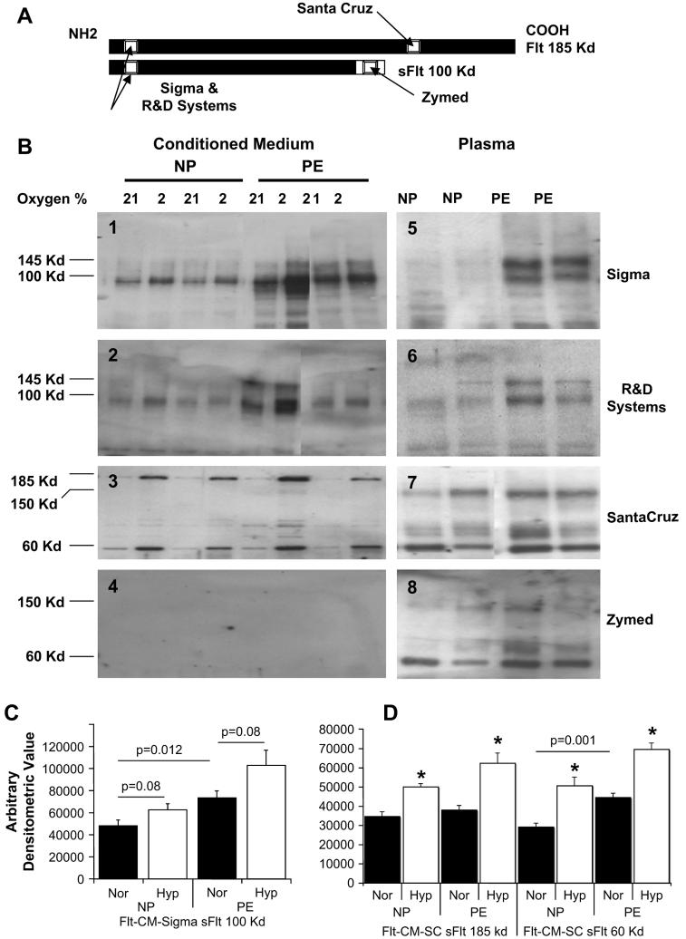
Fig. 3.
Characterization of sFlt-1 variants using different antibodies in both villous explant conditioned media and plasma from normal pregnant (NP) and preeclamptic women (PE). (A) Schematic representation of Flt (185 kDa) and sFlt (100 kDa) epitopes that are recognized by antibodies from Sigma Chemical Co., R&D Systems, Santa Cruz, and Zymed as reported by the respective manufacturers. (B) Panels 1–4 are representative Western blots using Sigma (1), R&D Systems (2), Santa Cruz (3) and Zymed (4) antibodies on villous explant conditioned media under 21% oxygen and 2% oxygen conditions. Panels 5–8 are representative Western blots using plasma samples from women with normal pregnancies and preeclampsia with the same order of antibodies. These are shown in adjacent panels to indicate the variability in sFlt-1 protein sources, molecular weight and antigenicity. (C) The 100 kDa sFlt-1 protein mass is significantly higher in preeclamptic villous explant conditioned media compared to normal pregnancy (p = 0.012) and hypoxia up regulated in response to (p = 0.08) using the Sigma antibody and densitometric quantitation. (B) Similarly, there is significant difference between normal pregnancy and preeclampsia for the 60 kDa sFlt-1 protein and significant hypoxic up regulation for both 185 and 60 kDa sFlt-1 proteins using the Santa Cruz antibody (p < 0.05 for all).

