Abstract
The activity and level of HMG-CoA reductase (HMGR) were addressed in halophilic fungi isolated from solar saltpans. Representative fungi belonging to the orders Dothideales, Eurotiales and Wallemiales have a specific pattern of HMGR regulation, which differs from salt-sensitive and moderately salt-tolerant yeasts. In all of the halophilic fungi studied, HMGR amounts and activities were the lowest at optimal growth salinity and increased under hyposaline and hypersaline conditions. This profile paralleled isoprenylation of cellular proteins in H. werneckii. Inhibition of HMGR in vivo by lovastatin impaired the halotolerant character. HMGR may thus serve as an important molecular marker of halotolerance.
Keywords: Adaptation, extremophiles, isoprenylation, lovastatin, mevalonate regulation
INTRODUCTION
Until recently, no true halophilic representatives were thought to exist within the kingdom of Fungi. Reports have, however, emerged arguing that the orders Dothideales, Eurotiales (Ascomycota) and Wallemiales (Basidiomycota) generally include genera and species adapted to growth under hypersaline conditions that represent part of the mycota of solar salterns (Gunde-Cimerman 2000, Butinar et al. 2005a,b; Zalar et al. 2005). Many unicellular eukaryotic organisms can adapt to changing environmental osmolarity mainly due to their ability to modify the sterol composition of cellular membranes in response to environmental stress (Horvath et al. 1998). 3-Hydroxy-3-methylglutaryl-CoA (HMG-CoA) reductase (HMGR, EC 1.1.1.34) is the major metabolic flux regulator of the mevalonate pathway for sterol biosynthesis, and it catalyses NADPH-dependent reductive deacylation of HMG-CoA to mevalonate. HMGR is crucial for the biosynthetic production of sterols and other isoprenoids, like protein modifying prenyl groups, in all three domains of life. Regulation of HMGR levels and activities occurs at multiple levels, including transcription, phosphorylation and protein degradation (Goldstein & Brown 1990). Little is known about regulation of HMGR activity in the new ecological group of moderately to extremely halophilic fungi that have adapted to growth in highly saline environments (Prista et al. 1997; Gunde-Cimerman 2000). We have previously reported unusual HMGR activity in the halophilic black yeast Hortaea werneckii (Petrovič et al. 1999, Vaupotič et al. 2007): while HMGR activity was highly dependent on environmental NaCl concentrations, the sterol content in H. werneckii did not change accordingly (Mejanelle et al. 2001, Turk et al. 2004), indicating that regulation of HMGR activity influences the metabolic flux of mevalonate differently to the biosynthesis of sterols, possibly at the pre-squalene level.
In this study, we have explored effects of salinity on HMGR regulation in five fungi species from solar salterns: the halotolerant Aureobasidium pullulans, and the halophilic Phaeotheca triangularis, Trimmatostroma salinum (Dothideales), Eurotium amstelodami (Eurotiales) and Wallemia ichthyophaga (Wallemiales). In particular, we have addressed the correlation between their HMGR activity and halophilic character. Two further species were included as additional references: a moderately halophilic yeast D. hansenii, and a salt-sensitive (i.e. mesophilic) yeast Saccharomyces cerevisiae. We demonstrate here a specific HMGR regulation by environmental salinity that correlates well with the halophilic character of these fungi. Focused on H. werneckii, the best characterized of the halophilic fungi from solar salterns, we also provide evidence that HMGR activity is crucial for halotolerance as well as for the changes in protein prenylation in response to changing salinity.
MATERIALS AND METHODS
Strains, media, and growth conditions
Cultures of halophilic fungi were isolated from Sečovlje salterns at the Slovenian Adriatic coast: H. werneckii (MZKI B736), P. triangularis (MZKI B741), T. salinum (MZKI B734), A. pullulans (MZKI B802), E. amstelodami (MZKI A561), W. ichthyophaga (EXF 994). These have been deposited in the culture collections of the Slovenian National Institute of Chemistry (MZKI) or of EXF at the Department of Biology, Biotechnical Faculty, University of Ljubljana. The reference strains were the salt-sensitive S. cerevisiae (MZKI K86) and the moderately halophilic D. hansenii (CBS 767), from Centraalbureau voor Schimmelcultures (CBS) Utrecht, The Netherlands. The fungi were grown at 28 °C (30 °C for S. cerevisiae) on a rotary shaker at 180 rpm in defined YNB medium adjusted to the indicated NaCl concentrations at pH 7.0. The cells were harvested in mid-exponential phase by centrifugation (4,000× g, 10 min), washed in 50 mM Tris-HCl, pH 7, and frozen in liquid nitrogen. The YNB medium agar plates were also prepared with 50 μM lovastatin (Lek). Ten μL of H. werneckii liquid culture were spotted onto agar plates and incubated for seven days prior to microscopy studies.
Measurement of HMGR activity
HMGR activity was measured as decribed previously (Petrovič, et al. 1999; Vaupotič, et al. 2007). Briefly, cell lysates were prepared from exponentially growing cells by disruption with a microdismembranator, in homogenization buffer (50 mM Tris, pH 8.5, 20 % glycerol, 0.5 % NaCl, 0.5 % Triton X-100; or at pH 7.0, without glycerol and NaCl for S. cerevisiae) containing fungal protease inhibitors (Sigma). The lysates were fractionated into soluble fraction and cellular debris by centrifugation (600× g, 15 min). After following centrifugation at 10,000× g, the supernatants were used for HMGR activity assessments. Protein concentrations were measured by spectrophotometry at 590 nm using the Bradford method with Nanoquant reagent (Roth). HMGR activity was assayed with 50 μg total protein with D-3-[3-14C]-hydroxy-3-methylglutaryl-CoA and R,S-[5-3H(N)]-mevalonolactone (NEN) as substrate and internal standard, respectively. HMGR activity was expressed as pmol HMG-CoA converted to mevalonate min-1.(mg protein)-1 and are given as means ± standard error from at least three independent experiments.
Western blotting
Cell lysates were prepared, with 20 μg protein boiled for 10 min in 5× protein-loading buffer (Fermentas), separated by SDS-PAGE on 10 % polyacrylamide gels, and transferred to PVDF membranes (Roth). Immunodetection was performed with antibodies against HMGR (Upstate) and β-actin, and secondary antibodies conjugated with HRP (Santa Cruz Biotechnology), using the ECL detection system (Amersham Bioscience).
Metabolic radiolabelling with [3H]-mevalonate
Hortea werneckii was grown in media with the indicated NaCl concentrations, without or with 50 μM lovastatin and with 0.75 μCi/mL [3H]-mevalonate ([3H]-MVA; 50 Ci/mmol) added in the early logarithmic phase. The cells were harvested during the exponential phase by centrifugation and washed several times with PBS. Total protein was isolated from 200 mg of cells using the TRIzol reagent (Invitrogen), and then solubilized in 1 % SDS. Protein concentrations were determined spectrophotometricaly using the BCA method (Pierce). A delipidation procedure was performed to release the [3H]-MVA-derived moiety from 200 μg labelled cellular protein, as described previously (Konrad & Eichler 2002). Briefly, SDS-solubilized proteins were incubated in 0.5 M HCl at 95 °C for 1 h, with vigorous shaking. Samples were extracted twice with chloroform/methanol (2:1, v/v), and radioactivity released was quantified in the organic fraction by scintillation counting. Incorporation of [3H]-MVA was expressed as pmol of incorporated [3H]-MVA per mg protein, as means ±standard error from three independent experiments.
Microscopy
For morphological analysis of cells, fungi were washed in fresh growth medium, added to glass slides and covered with a coverslip. To prevent evaporation, the coverslip was sealed with nail-polish. Cell morphology was examined under an inverted light microscope (Nikon Eclipse 300) and images taken with a digital camera (Nikon DS-5M).
RESULTS
HMGR activities and protein levels in halophilic fungi depend on environmental salinity
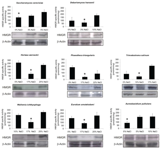
To determine enzyme activities and protein levels of HMGR, the halophilic fungi were grown at three different environmental salinities. The specific HMGR activity was responsive to changes in NaCl concentrations in all of the fungal species (Fig. 1), including S. cerevisiae and D. hansenii as reference strains. All HMGR activity profiles were similar, showing minimal activity under optimal conditions and a 2-6-fold increase in activity under hyposaline and hypersaline conditions. We also explored HMGR in these fungi at the protein level under these growth conditions. Immunoblotting with an antibody against a conserved catalytic domain of HMGR revealed that according to the enzyme activities, the HMGR protein was also lowest at optimal growth salinity and increased under hyposaline and hypersaline conditions (Fig. 1).
Fig. 1.
Regulation of fungal HMG-CoA reductase activity and protein levels by environmental salinity. Different salinities of the medium were chosen as hyposaline (0% NaCl, only for halophiles), optimal, and hypersaline conditions in different fungi. Profiles of HMGR activity and HMGR protein levels of the indicated fungi are shown. β-Actin was used as loading control. *Optimal growth salinities. R. Data represent means ±SD of three independent experiments. Significant differences (Tukey's HSD) were seen between the value marked with an arrow-head and each of the samples marked with the dot.
Inhibition of HMGR by lovastatin in vivo resulted in the salt-sensitive character of H. werneckii
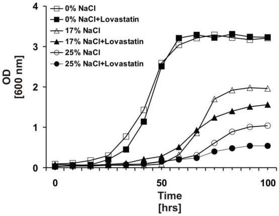
To demonstrate that HMGR activity is connected with the halotolerant character of saltern-inhabiting fungi, the growth of one of the most adaptable and halophilic yeast, H. werneckii, was monitored in the presence of sub-lethal concentrations (50 μM) of the specific HMGR inhibitor lovastatin at different NaCl concentrations. There was no effect of lovastatin on the growth curve of H. werneckii in salt-free media (Fig. 2). In contrast, lovastatin remarkably reduced growth in the otherwise physiologically optimal medium containing 17 % NaCl, an effect even more pronounced in hypersaline medium containing 25 % NaCl.
Fig. 2.
Lovastatin impaired growth ability of the halophilic H. werneckii in NaCl-containing media. Growth curves of H. werneckii in optimal (17% NaCl), hyposaline (0% NaCl) and hypersaline (25% NaCl) media without (white symbols) and with (black symbols) 50 μM lovastatin.
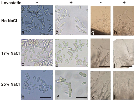
Microscopy revealed the effects of lovastatin on the morphology of H. werneckii cells (Fig. 3). In hyposaline media (Fig. 3a), the cells were significantly thinner and more elongated compared to those at optimal salinity (Fig. 3c), and most had a hardly visible septum and predominantly one unipolar bud. The only effect of lovastatin treatment was on the bipolar budding of cells (Fig. 3b). At optimal salinity (Fig. 3c), the cells grew as double-celled meristematic clusters that were slightly elongated and separated by a septum. The lovastatin treatment caused an increased number of irregularly shaped meristematic clusters of three, or even four, cells separated by a septum (Fig. 3d). In hypersaline media (Fig. 3e), the double-celled conidia were bulkier and less elongated than at optimal salinity. The effect of lovastatin was most evident under this extreme growth condition, resulting in four-celled meristematic clumps that were irregularly shaped, had a thick cell wall and well developed septa (Fig. 3f). Hyphal growth of H. werneckii was also affected by lovastatin, as seen on agar plates. No obvious growth effect of lovastatin on hyphae formation occurred in hyposaline media (Figs. 3g-h). With optimal salinity (Fig. 3i), the hyphae were highly branched and extended and had numerous buds, which were significantly reduced in number by the lovastatin treatment (Fig. 3j). In hypersaline media, the lovastatin treatment completely prevented branching of the hyphae and the formation of buds (Fig. 3k-l).
Fig. 3.
Morphological changes in cells and hyphae growth caused by salt and lovastatin in H. werneckii. Cells were grown in media or agar plates with the indicated salt concentrations without or with lovastatin. Morphology was investigated using bright field microscopy under 40x and 10x magnification for cells and hyphae, respectively. Panels a-f, bar = 60 μm; panels g-l, bar = 240 μm.
The mevalonate-derived lipid modifications of proteins correlate with HMGR activity in H. werneckii
To determine whether non-sterol mevalonate-derived lipid modifications of proteins accounted for the HMGR activity profile at these different environmental salinities, we investigated the incorporation of radioactively labelled mevalonate derivatives into proteins, as covalently linked lipid moieties. The H. werneckii cells were grown under different NaCl concentrations in the presence of [3H]-mevalonate, without or with lovastatin. After harsh acidic delipidation of the isolated proteins, the [3H]-labelled lipids released were assessed using a chloroform/methanol extraction: both the total radioactivity of the protein fractions and the lipid-derived radioactivity after protein delipidation were lowest at optimal growth salinity (17 % NaCl), and approximately 2.5-fold higher in hyposaline (0 % NaCl) and hypersaline (25 % NaCl) media (Fig. 4), following the HMGR activity profile of H. werneckii (Fig. 1). Treatment with lovastatin increased the incorporation of [3H]-mevalonate into the lipids from cellular proteins as a consequence of its the inhibitory effect on production of endogenous mevalonate. However, this was most evident in salt-free medium, where lovastatin treatment had less effect on growth.
Fig. 4.
Incorporation of [3H]-mevalonate-derived lipid moiety into cellular proteins in H. werneckii. After metabolic labelling of cellular proteins with [3H]-mevalonate, scintillation counting was carried out on total protein before delipidation (white bars) and then on the lipid fraction after protein delipidation (black bars). The data are presented as pmol incorporated radioactively labelled mevalonate per mg protein.
DISCUSSION
There have been numerous studies on stress responses of various salt-sensitive unicellular eukaryotes to saline stress. The present study of halophilic fungi represents adaptive metabolism in high-saline media rather than a stress response, due to the evolutionarily acquired halotolerance of these species. Our previous studies on ecophysiological characteristics of these fungal species have shown that H. werneckii, W. ichthyophaga, E. amstelodami, P. triangularis and T. salinum are halophiles, while A. pullulans is instead a moderately halotolerant species (Blomberg 2000, Gunde-Cimerman 2000, Butinar et al. 2005, Butinar et al. 2005, Zalar et al. 2005). Observing the changes in cellular HMGR activity we can conclude that the different HMGR activities in cells grown under different environmental salinities were the consequence of different amounts of cellular HMGR protein. The similar HMGR activity profiles in the moderately halotolerant A. pullulans and salt-sensitive S. cerevisiae, which lack the U-shaped HMGR profile, indicate that both have their growth optima in salt-free medium. This correlates well with their non-halophilic characters. Alternatively, the U-shaped HMGR profiles obtained in halophilic species corresponded well to previously proposed optimal salinities. These all showed the lowest HMGR activity at the optimal growth salinity and high HMGR activity in both salt-free medium, as hyposaline conditions for these species, and under extremely high salt concentrations, defined as hypersaline conditions, proposing the HMGR level and activity as a sensor of non-optimal growth salinity. Despite frequent reports of D. hansenii (Saccharomycetales) as a prime fungal halophile, we have shown here that according to the HMGR profile, its optimal salinity is lower than those of the halophilic fungi used in comparison. This fits well with the ecology of these species, since D. hansenii is most often isolated from sea water with only 3 % NaCl, while the other halophilic fungi were from the hypersaline waters of solar salterns, where salt concentrations can reach saturation levels.
Very little is known about the regulation of HMGR in extremophilic eukaryotes. To date, some reports have demonstrated that expression and/or activity of HMGR is regulated in response to non-optimal salinity, i.e. in the halophilic archaeon Haloferax volcanii, where authors demonstrated the HMGR at the level of protein ampunt and activity was conspicuously increased during growth at high salinity (Bidle et al. 2007). In our previous reports on halophilic black yeast, we also showed a similar response in halophilic yeast Hortaea werneckii (Petrovič et al. 1999). In our ongoing investigations into proteins linked to halophily in H. werneckii (Vaupotič et al. 2007), HMGR is one of the so-called “salt-responsive” enzymes, where proten levels and/or enzyme activities increase under both hyposaline and hypersaline conditions. We have shown that at optimal growth salinity, HMGR undergoes ubiquitination and proteasomal degradation (Vaupotič & Plemenitaŝ 2007). Here, we provide additional evidence that salinity-dependent regulation of HMGR activity in H. werneckii is linked to its halotolerant character (Figs 2, 3), as inhibition of HMGR by lovastatin resulted in a considerably more salt-sensitive phenotype. Also, the morphological changes with both salt and lovastatin clearly indicate a defect in cell proliferation (Fig. 3). These data demonstrate further that HMGR activity is required for the halotolerant character of H. werneckii. Based on the data in the present study, we can speculate that this could similarly be true for other halophilic fungi in this study, where similar HMGR enzyme activities and protein profiles were seen (Fig. 1).
As we have shown previously, the sterol composition in H. werneckii did not change significantly at different salinities (Mejanelle et al. 2001, Turk et al. 2004) as we could expect according to HMGR activity fluctuations. Therefore, we sought evidence for changes in other mevalonate-derived, but pre-squalene isoprenoid intermediates, to reflect the metabolic effects of the HMGR activity profile in H. werneckii. Derivatives of pre-squalen isoprenoids are important regulatory determinants of prenylated proteins implicated in cell-cycle progression and cell proliferation, since their attachement to proteins directes them to cellular mebranes (Brown & Goldstein 1980, Siperstein 1984). Using [3H]-mevalonate and the highly selective HMGR inhibitor lovastatin in metabolic labelling experiments, we have clearly demonstrated that modification of protein by the mevalonate-derived lipid moiety not only reflects the profile of HMGR activity, but also the responses to HMGR inhibition (Fig. 4). Combining the growth inhibitory effect of lovastatin treatment in salt containing media with the HMGR activity-dependent protein prenylation profile, we might conclude that lovastatin-mediated reduction in halotolerance was not merely the result of a non-specific inhibitory effect on overall cellular function, but rather reflects a protein-prenylation-specific event. Based on this data we can conclude, that in salterns-inhabiting fungi the regulation of HMGR activity by environmental salinity reflects more distinctively at the level of the metabolic flux through the pre-squalene part of the mevalonate pathway, rather than at the level of post-squalene regulation of sterol content. The higher activity of HMGR in hyposaline and hypersaline media could be connected with specific metabolic demands, when increased flux through the mevalonate pathway may be needed for prenylation and the subsequent membrane localisation of specific proteins that are not essential for growth in an optimal environment. In evolutionary terms, the maintenance of high levels of HMGR in hyposaline and hypersaline environments may also reflect physiological adaptation of halophilic fungi to metabolic demands under extreme conditions.
In conclusion, the key metabolic enzyme, HMGR, has been studied in a previously non-characterised group of halophilic fungi. The present study documents that both HMGR activity and protein levels in halophilic fungi depend on environmental salinity. In the extremely halotolerant H. werneckii, the biological consequence of HMGR regulation relates to posttranslational modification of proteins by prenylation. Therefore our findings provide a new insight for understanding the regulation of the mevalonate pathway as a response to changes in environmental NaCl concentrations. We propose the HMGR enzyme as an important biochemical signature of halophily in the Fungal kingdom.
Acknowledgments
Supported in part by research grant P1-0170 and in part by a Young Researcher Fellowship to T.V. from the Slovenian Research Agency. We thank Mrs. Milena Marušič for technical assistence with HMGR activity assays.
References
- Bidle KA, Hanson TE, Howell K, Nannen J (2007). HMG-CoA reductase is regulated by salinity at the level of transcription in Haloferax volcanii. Extremophiles 11: 49-55. [DOI] [PubMed] [Google Scholar]
- Blomberg A (2000). Metabolic surprises in Saccharomyces cerevisiae during adaptation to saline conditions: questions, some answers and a model. FEMS Microbiolology Letters 182: 1-8. [DOI] [PubMed] [Google Scholar]
- Brown MS, Goldstein JL (1980). Multivalent feedback regulation of HMG CoA reductase, a control mechanism coordinating isoprenoid synthesis and cell growth. Journal of Lipid Research 21: 505-517. [PubMed] [Google Scholar]
- Butinar L, Santos S, Spencer-Martins I, Oren A, Gunde-Cimerman N (2005a). Yeast diversity in hypersaline habitats. FEMS Microbiology Letters 244 229-234. [DOI] [PubMed] [Google Scholar]
- Butinar L, Zalar P, Frisvad JC, Gunde-Cimerman N (2005b). The genus Eurotium - members of indigenous fungal community in hypersaline waters of salterns. FEMS Microbiology Ecology 51 155-166. [DOI] [PubMed] [Google Scholar]
- Goldstein JL, Brown MS: Regulation of the mevalonate pathway (1990). Nature 343: 425-430. [DOI] [PubMed] [Google Scholar]
- Gunde-Cimerman N, Zalar P, Hoog GS de, Plemenitaš A (2000). Hypersaline waters in salterns: natural ecological niches for halophilic black yeasts. FEMS Microbiology Ecology 32 235-340. [DOI] [PubMed] [Google Scholar]
- Horvath I, Glatz A, Varvasovszki V, Torok Z, Pali T, Balogh G, Kovacs E, Nadasdi L, Benko S, Joo F, Vigh L: (1998). Membrane physical state controls the signaling mechanism of the heat shock response in Synechocystis PCC 6803: identification of hsp17 as a “fluidity gene”. Proceedings of the National Academy of Sciences of the United States of America 95 3513-3518. [DOI] [PMC free article] [PubMed] [Google Scholar]
- Konrad Z, Eichler J (2002). Lipid modification of proteins in Archaea: attachment of a mevalonic acid-based lipid moiety to the surface-layer glycoprotein of Haloferax volcanii follows protein translocation. Biochemical Journal, 366: 959-964. [DOI] [PMC free article] [PubMed] [Google Scholar]
- Mejanelle L, Lopez JF, Gunde-Cimerman N, Grimalt JO (2001). Ergosterol biosynthesis in novel melanized fungi from hypersaline environments. Journal of Lipid Research 42: 352-358. [PubMed] [Google Scholar]
- Petrovič U, Gunde-Cimerman N, Plemenitaš A (1999). Salt stress affects sterol biosynthesis in the halophilic black yeast Hortaea werneckii. FEMS Microbiology Letters 180 325-330. [DOI] [PubMed] [Google Scholar]
- Prista C, Almagro A, Loureiro-Dias MC, Ramos J (1997). Physiological basis for the high salt tolerance of Debaryomyces hansenii. Applied and Environmental Microbiology 63 4005-4009. [DOI] [PMC free article] [PubMed] [Google Scholar]
- Siperstein MD (1984). Role of cholesterogenesis and isoprenoid synthesis in DNA replication and cell growth. Journal of Lipid Research 25: 1462-1468. [PubMed] [Google Scholar]
- Turk M, Mejanelle L, Sentjurc M, Grimalt JO, Gunde-Cimerman N, Plemenitaš A (2004). Salt-induced changes in lipid composition and membrane fluidity of halophilic yeast-like melanized fungi. Extremophiles, 8: 53-61. [DOI] [PubMed] [Google Scholar]
- Vaupotič T, Gunde-Cimerman N, Plemenitaš A (2007). Novel 3'-phosphoadenosine-5'-phosphatases from extremely halotolerant Hortaea werneckii reveal insight into molecular determinants of salt tolerance of black yeasts. Fungal Genetetics and Biology 44: 1109-22. [DOI] [PubMed] [Google Scholar]
- Vaupotič T, Plemenitaš A (2007). Osmoadaptation-dependent activity of microsomal HMG-CoA reductase in the extremely halotolerant black yeast Hortaea werneckii is regulated by ubiquitination. FEBS Letters 581: 3391-5. [DOI] [PubMed] [Google Scholar]
- Zalar P, de Hoog GS de, Schroers HJ, Frank JM, Gunde-Cimerman N (2005). Taxonomy and phylogeny of the xerophilic genus Wallemia (Wallemiomycetes and Wallemiales, cl. et ord. nov.). Antonie van Leeuwenhoek 87: 311-328. [DOI] [PubMed] [Google Scholar]


