Abstract
Long-term potentiation (LTP), a synaptic mechanism thought to underlie memory formation, has been studied extensively at hippocampal Schaffer collateral (SC) synapses. The SC pathway transmits information to area CA1 that originates in entorhinal cortex and is processed by the dentate gyrus and area CA3. CA1 also receives direct excitatory input from entorhinal cortex via the perforant path (PP), but the role of this cortical input is less certain. Here, we report that low-frequency stimulation of PP inputs to CA1 has no lasting effect on basal SC transmission, but effectively depotentiates SC synapses that have undergone LTP in a manner that can be reversed by subsequent high-frequency stimulation of SC inputs. This depotentiation does not require NMDA receptors, group I metabotropic glutamate receptors, or L-type calcium channels, but involves adenosine acting at A1 receptors. Given the limited storage capacity of the hippocampus, these observations provide a mechanism by which input from cortex can help to reset synaptic transmission in the hippocampus and facilitate additional information processing.
Keywords: depotentiation, adenosine, hippocampus, entorhinal cortex, perforant path, temperoammonic path
Introduction
The hippocampus plays a key role in memory and is involved in several neuropsychiatric disorders (Martin et al., 2000). Hippocampal processing is required for formation of new declarative memories including the ability to place events in context and sequence (Eichenbaum, 2000). The hippocampus accomplishes these tasks using information from multiple brain regions with input from neocortex arriving via entorhinal cortex (EC) and the perforant pathway (PP) (Witter et al., 2000; van Groen et al., 2003). The hippocampus processes cortical input via a trisynaptic pathway that links layer II of entorhinal cortex to dentate gyrus, allowing coding of different components of a memory (Eichenbaum, 2000). The dentate gyrus transmits this information to area CA3 via the mossy fibers where heteroassociative memories are likely formed. CA3 subsequently sends processed information to stratum radiatum in area CA1 via the Schaffer collateral (SC) pathway. CA1 is thought to help decode memories into a form that is sent back to entorhinal cortex via the subiculum for subsequent longer-term storage in other brain regions (Lisman, 1999; Eichenbaum, 2000; Witter et al., 2000).
How memories are actually formed remains uncertain with current theories suggesting the importance of activity-dependent synaptic plasticity including long-term potentiation (LTP) and long-term depression (LTD) (Martin et al., 2000). The role of the hippocampus in memory processing is critical for initial stages of learning but less important for longer-term storage. The hippocampus is a region with limited storage capacity and there is interest in how the hippocampus resets it synapses once memories have been transmitted to cortex. Two processes are likely to be important in synaptic resetting, homosynaptic depotentiation and longer-term homeostatic changes (Kemp and Manahan-Vaughan, 2007; Turrigiano, 2007). Both mechanisms help hippocampal circuits adjust to synaptic changes, but do not take into account the potential dynamics of cortical–hippocampal interactions in regulating hippocampal function.
In addition to SC input, CA1 has direct excitatory connections with layer III of entorhinal cortex via the PP (temperoammonic path) (Colbert and Levy, 1992; Jones, 1993). These direct inputs synapse on distal pyramidal neuron dendrites in stratum lacunosum moleculare (SLM). The function of the direct PP inputs is not well understood (Buzsáki et al., 1995; Soltesz, 1995; Fernandez and Tendolkar, 2006), although increasing evidence indicates that these synapses have important modulatory effects on CA1 pyramidal neurons (Levy et al., 1998; Remondes and Schuman, 2002, 2004). PP inputs also appear to play a key role in incremental learning in a familiar environment (Nakashiba et al., 2008). In the present study, we used hippocampal slices to examine interactions between PP and SC inputs in CA1. Our results indicate that repeated low-frequency activation of the PP causes only a transient depression of SC inputs in naive slices, but results in depotentiation of SC synapses that have previously undergone LTP. This depotentiation differs mechanistically from homosynaptic SC depotentiation and provides a mechanism by which direct cortical input can direct the hippocampus to reset its function for additional processing.
Materials and Methods
Hippocampal slice preparation.
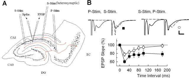
Hippocampal slices were prepared from postnatal day 30 (P30) to P32 albino rats using standard methods (Zorumski et al., 1996). Rats were anesthetized with isoflurane and decapitated. Dissected hippocampi were placed in ice-cold artificial CSF (ACSF) containing the following (in mm): 124 NaCl, 5 KCl, 2 MgSO4, 2 CaCl2, 1.25 NaH2PO4, 22 NaHCO3, 10 glucose, bubbled with 95% O2–5% CO2 at 4–6°C, and cut into 450 μm slices using a vibrotome. The slices were cut in a manner that included a significant portion of entorhinal cortex to maximize maintaining PP inputs to SLM in the CA1 region (see Fig. 1A). Acutely prepared slices were placed in an incubation chamber containing gassed ACSF for 1 h at 30°C before additional experimentation.
Figure 1.
Interactions between PP and SC inputs in the CA1 region. A, The diagram depicts the slice preparation used for these studies. The SC pathway was activated in the apical dendritic region by separate stimulating electrodes (S-Stim) placed on either side of recording electrodes in the CA1 region. The PP (P-Stim) was activated by a stimulating electrode placed near the EC where the PP enters the hippocampus. B, The panel depicts interactions between PP and SC inputs to CA1 using a paired-pulse protocol and a 20 ms interpulse interval. Traces show examples of raw data, whereas the graph shows the time course of change in EPSPs recorded in stratum radiatum after conditioning stimulation of the PP (black squares) or SC (white circles). Calibration: 1 mV, 5 ms. Error bars indicate SEM.
Hippocampal slice physiology.
At the time of study, slices were transferred individually to a submersion-recording chamber. Experiments were done at 30°C with continuous ACSF perfusion at 2 ml/min. Extracellular recordings were obtained from the apical dendritic layer (stratum radiatum) of the CA1 region for analysis of EPSPs using electrodes filled with 2 m NaCl (5–10 MΩ resistance).
EPSPs were evoked with 0.1 ms constant current pulses through a bipolar stimulating electrode in the SC pathway. A second stimulating electrode was placed in the PP to activate distal dendrites of CA1 in SLM. A control input–output curve was obtained to determine stimulus intensities for subsequent studies. Responses were monitored by applying single stimuli to the SC pathway every 60 s at half-maximal intensity. After establishing a stable baseline for at least 10 min, LTP was induced by a single 100 Hz by 1 s tetanus using the same intensity stimulus. In some studies, a 200 Hz by 1 s tetanus was administered in the presence of the NMDA receptor (NMDAR) antagonist, 2-amino-5-phosphonovalerate (APV), to induce a form of LTP not dependent on NMDARs. Input–output curves were repeated 20 and 60 min after tetanic stimulation and appear as gaps in the time course graphs shown in Figures 2–6. In some experiments, a second independent SC input to CA1 was activated using a stimulating electrode placed at a different level in stratum radiatum than the primary SC stimulating electrode and positioned on the distal (subiculum) side of the dendritic recording electrode. Again, a stimulus that evoked a half-maximal response in stratum radiatum was used for these studies.
Figure 2.
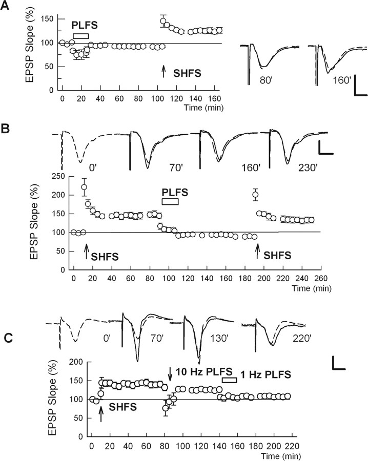
Effects of PP stimulation on synaptic plasticity in the SC pathway. A, LFS of the PP (PLFS) causes a transient depression of SC transmission that rapidly recovers after PLFS. A subsequent 100 Hz by 1 s HFS of the SC inputs (SHFS) results in LTP. B, After induction of SC LTP, PLFS results in rapid depotentiation of SC inputs. A subsequent tetanus delivered to the SC results in a return of LTP. Traces above the graph show EPSPs obtained at the times denoted, with the initial baseline response shown as a dashed trace in the last three panels. C, Depotentiation of SC LTP by PP stimulation is frequency dependent. After establishing SC LTP, 10 Hz by 900 pulse stimulation of the PP causes only a transient depression of LTP. In contrast, subsequent 1 Hz PLFS results in depotentiation. Error bars indicate SEM. Traces above the graph show representative EPSPs. Calibration: 1 mV, 5 ms.
Figure 3.
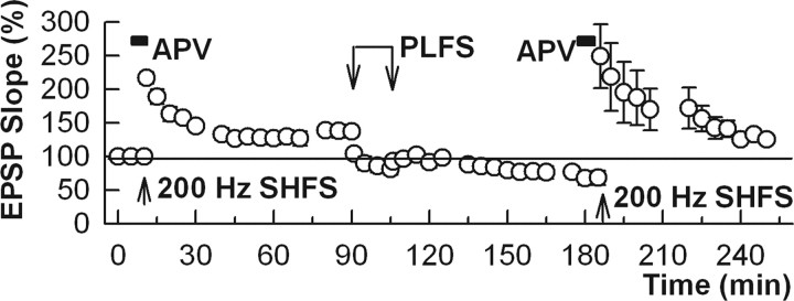
In the presence of 100 μm APV (black bar), an NMDAR antagonist, 200 Hz by 1 s high frequency stimulation of the Schaffer collateral pathway (SHFS) (arrow) induces a form of LTP that requires L-type calcium channels. After induction of this form of LTP, 1 Hz by 900 pulse stimulation of the perforant path (PLFS) depotentiates this LTP in a manner that can be reversed by a subsequent tetanus. Error bars indicate SEM.
Figure 4.
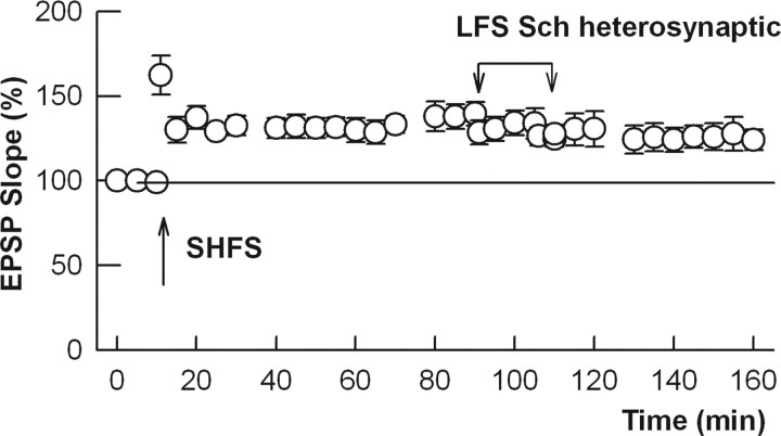
Activation of a heterosynaptic Schaffer collateral input does not produce depotentiation. The graph shows LTP after HFS (100 Hz by 1 s tetanus; arrow) of the Schaffer collateral pathway. Subsequently, a second independent Schaffer collateral input was stimulated at 1 Hz for 15 min (downward connected arrows). In contrast to PP stimulation, heterosynaptic Schaffer collateral stimulation failed to reverse LTP. Error bars indicate SEM.
Figure 5.
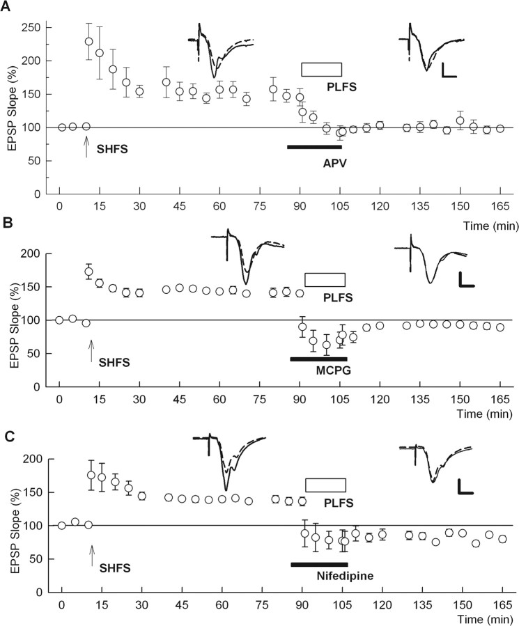
PP-induced depotentiation does not require NMDARs, mGluRs, or L-type calcium channels. A, After induction of SC LTP with a 100 Hz by 1 s HFS (SHFS), 1 Hz LFS delivered to the PP (PLFS) (open bar) depotentiates SC responses in the presence of 100 μm APV (solid bar). B, PLFS also results in depotentiation when administered in the presence of 500 μm MCPG, a broad-spectrum mGluR antagonist. C, The L-type calcium channel blocker, nifedipine (10 μm), also fails to block depotentiation. Error bars indicate SEM. Traces depict representative EPSPs. Calibration: 1 mV, 5 ms.
Figure 6.
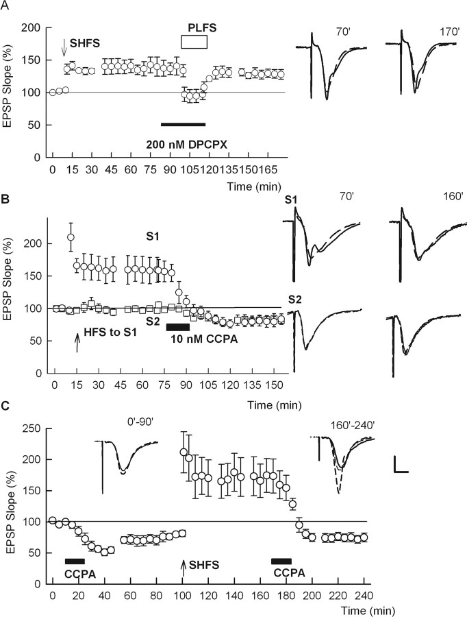
Adenosine participates in PP-induced depotentiation of SC LTP. A, When PLFS is delivered in the presence of 200 nm DPCPX, a competitive adenosine A1 receptor antagonist, depotentiation is eliminated. B, Administration of 10 nm CCPA, an adenosine A1 receptor agonist (solid bar), causes depotentiation of SC LTP (S1 pathway) and a form of LTD in a second independent SC input (S2). C, After induction of SC LTD by 10 nm CCPA, HFS of the SC input results in robust LTP. This LTP can be reversed by a second application of CCPA. Error bars indicate SEM. Traces in all panels show representative EPSPs obtained at the times specified with control responses shown as dashed lines. Calibration: 1 mV, 5 ms.
Chemicals were obtained from Sigma-Aldrich except the adenosine and metabotropic glutamate receptor (mGluR) ligands, which were from Tocris. Drugs were dissolved in ACSF at the time of experiment and administered by bath perfusion at the concentrations noted in the text. The concentrations selected for study and the durations of drug administration were based on previous studies indicating that the agents are effective at altering synaptic transmission or synaptic plasticity when administered in this manner.
Statistical analysis.
Data were collected and analyzed using PClamp software (Molecular Devices). Data in the text are expressed as mean ± SEM. A two-tailed Student t test was used for comparisons between groups. Statistical comparisons were based on input–output curves at baseline and 60 min after tetanic or 1 Hz stimulation with p < 0.05 considered significant and were done using commercial software (SigmaStat; Systat Software). Statistical analyses of data shown in Figures 2, 4, and 6 are included in supplemental Table 1 (available at www.jneurosci.org as supplemental material).
Results
Studies were conducted in the CA1 region of rat hippocampal slices. The slices included a section of EC to preserve PP (temperoammonic) inputs to SLM in distal CA1 dendrites (Fig. 1A). SC and PP inputs were activated by separate electrodes using stimuli that produced 50% maximal responses in stratum radiatum during baseline recordings. We initially examined interactions between PP and SC pathways using a paired-pulse protocol and a 20 ms interpulse interval. We found that a single conditioning PP stimulation transiently depressed SC EPSPs recorded in stratum radiatum (Fig. 1B). Similarly, previous SC stimulation transiently depressed PP evoked EPSPs. In experiments designed to examine recovery from the depression evoked by PP or SC conditioning stimuli, we lengthened the interpulse stimulation interval. We found that depression of SC responses induced by PP stimulation was longer-lived than the effects of SC stimulation on PP inputs, and took ∼1 s to decay completely. In contrast, the effects of SC stimulation on PP inputs decayed completely in ∼180 ms. The depression of SC responses produced by a single PP conditioning stimulation was not altered significantly by either the adenosine A1 receptor antagonist, 8-cyclopentyl-1,3-dipropylxanthine (DPCPX), or the GABAA receptor antagonist, picrotoxin (supplemental Fig. 1, available at www.jneurosci.org as supplemental material).
Based on the ability of PP stimulation to alter SC transmission and the potential importance of direct cortical inputs in modulating CA1 processing (Levy et al., 1998; Remondes and Schuman, 2002, 2004), we examined the effects of repeated PP stimulation on SC LTP. In naive slices, we found that repeated low-frequency PP stimulation (LFS) using 900 pulses at 1 Hz caused a transient depression of SC responses that recovered to baseline in <5 min and remained stable (Fig. 2A). A subsequent 100 Hz by 1 s high-frequency stimulation (HFS) of the SC pathway readily induced LTP, indicating no lasting effect of PP stimulation on the generation of SC long-term plasticity (supplemental Table 1, available at www.jneurosci.org as supplemental material).
In contrast to the transient effects in naive slices, we found that PP LFS rapidly depotentiated SC responses in slices in which LTP had been induced initially in the SC pathway (Fig. 2B). After PP-induced depotentiation, a subsequent SC HFS again induced LTP, indicating that PP stimulation does not have a lasting disruptive effect on the mechanisms required for SC LTP induction. The effects of 1 Hz PP LFS on SC LTP were not mimicked by 900 pulses administered at 10 Hz (Fig. 2C), indicating that PP-induced depotentiation is frequency dependent. We also examined a shorter period of PP stimulation, but found that 1 Hz by 300 pulse LFS failed to produce lasting depotentiation.
To determine whether PP-mediated depotentiation depends on the calcium source that drives LTP induction, we also examined the effects of PP stimulation on a form of LTP induced by very high-frequency stimulation (200 Hz by 1 s) in the presence of 100 μm APV, an NMDAR antagonist. Previous studies have shown that this form of LTP requires activation of L-type voltage-gated calcium channels (Grover and Teyler, 1990; Nagashima et al., 2005). Consistent with results observed with NMDAR-dependent LTP, we found that 1 Hz PP stimulation effectively depotentiated NMDAR-independent SC LTP (Fig. 3). Thus, the ability of PP stimulation to reverse SC LTP appears to be independent of the calcium source driving LTP.
To determine whether other heterosynaptic inputs also depotentiate SC LTP, we examined the effects of activating an independent SC input. For these studies, a second stimulating electrode was placed in the SC pathway at a different level in stratum radiatum than the primary SC stimulating electrode. In contrast to PP stimulation, we found that 1 Hz by 900 pulse LFS of the heterosynaptic SC input failed to persistently alter either baseline SC transmission or previously established LTP (Fig. 4).
In the SC pathway, 1 Hz by 900 pulse LFS induces either homosynaptic LTD or homosynaptic depotentiation depending on whether LFS is administered to naive slices or slices that have previously experienced LTP (Fujii et al., 1991; Izumi and Zorumski, 1993; Malenka and Bear, 2004). These forms of SC plasticity are blocked by APV, a broad-spectrum NMDAR antagonist (Malenka and Bear, 2004). In contrast, PP-induced SC depotentiation was insensitive to 100 μm APV (Fig. 5A). PP-mediated depotentiation was also insensitive to 500 μm α-methyl-4-carboxyphenylglycine (MCPG), an inhibitor of group I mGluRs that contribute to SC LTD (Bortolotto et al., 1999) (Fig. 5B), and was not altered by the L-type calcium channel blocker, nifedipine (Fig. 5C). These results indicate that PP-mediated depotentiation differs mechanistically from homosynaptic SC depotentiation or LTD.
The lack of effect of APV, MCPG, and nifedipine on PP-mediated SC depotentiation, led us to consider the role of other neuromodulators. We focused on adenosine, an agent that has been previously associated with LTD and depotentiation in the hippocampus (Abraham and Huggett, 1997; Fujii et al., 1997, 1999; Huang et al., 1999). We found that PP-mediated SC depotentiation requires activation of adenosine A1 receptors. After induction of SC LTP, delivery of PP LFS in the presence of 200 nm DPCPX, an A1 receptor antagonist (Lohse et al., 1987), blocked depotentiation (Fig. 6A). Additionally, exogenous activation of A1 receptors by 10 nm N6-chlorocyclopentyladenosine (CCPA) (Appel et al., 1995) depotentiated SC LTP and induced LTD in both the tetanized pathway and an independent SC input (Fig. 6B). After induction of LTD with CCPA, HFS delivered to the SC pathway resulted in robust LTP (Fig. 6C). This LTP was completely reversed by a second administration of the adenosine agonist.
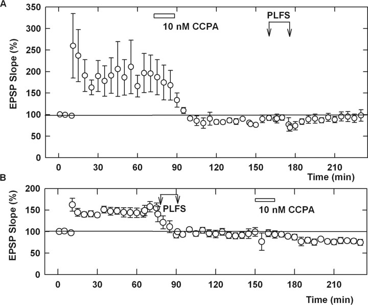
To determine whether CCPA-mediated depotentiation occludes the effects of PP stimulation, we examined PP LFS after CCPA. As described above, 10 nm CCPA depotentiated SC LTP and resulted in a persisting LTD of SC responses (Fig. 7A). Sixty minutes after CCPA-induced depotentiation, PP LFS failed to produce additional depression of SC responses. In a separate set of slices, we examined the effects of CCPA after PP-mediated depotentiation. In this case, CCPA, administered 60 min after PP-mediated depotentiation of SC LTP, produced no significant effect on SC responses during the period of acute CCPA administration, although a later developing LTD after CCPA perfusion was observed (Fig. 7B). The degree of lasting depression produced by CCPA after PP LFS was similar to that observed on basal SC responses in naive slices. A comparison of Figure 6, B and C, with Figure 7B indicates that the acute depressant effects of adenosine receptor activation on SC transmission are diminished by PP-induced depotentiation, although the longer-lasting LTD of SC transmission induced by CCPA is not altered.
Figure 7.
An adenosine agonist occludes the effects of PP LFS. A, Administration of 10 nm CCPA 60 min after induction of SC LTP results in depotentiation. Sixty minutes after CCPA-mediated depotentiation, PP LFS results in no additional depression of SC transmission. B, In a separate set of slices, LTP was induced by SC HFS. Sixty minutes after LTP induction, PP LFS resulted in SC depotentiation. Administration of 10 nm CCPA after PP-mediated depotentiation resulted in no change in SC responses during and immediately after CCPA administration, although a later developing and persistent form of LTD was induced. The degree of SC LTD induced by CCPA after PP-mediated depotentiation was similar to that observed in naive slices (Fig. 6B,C). Error bars indicate SEM. Traces show representative EPSPs. Calibration: 1 mV, 5 ms.
Discussion
Learning theories emphasize the importance of bidirectional plasticity as a mechanism by which synapses determine their operating range (Abraham and Tate, 1997; Martin et al., 2000; Govindarajan et al., 2006). In the hippocampus, repeated stimulation of a single SC input can potentiate, depress, or depotentiate its function depending on how and when the SC pathway is activated (Abraham and Tate, 1997; Malenka and Bear, 2004). Under other circumstances, SC activation itself can have no lasting effect on baseline transmission but can modulate the ability of SC synapses to undergo LTP or LTD, a form of synaptic change referred to as “metaplasticity” (Abraham and Tate, 1997). These forms of synaptic modulation are important for information processing but are limited to a single set of synaptic inputs. In the present study, we found that activation of a direct heterosynaptic input to area CA1 from entorhinal cortex depotentiates SC LTP in a manner that allows these synapses to be readily repotentiated by subsequent homosynaptic high-frequency stimulation. Entorhinal inputs to CA1 thus provide a means by which CA1 function can be directly influenced by information from cortex. This PP-mediated SC depotentiation was not mimicked by similar stimulation of a heterosynaptic SC input, suggesting unique effects of the PP input.
The ability of PP stimulation to depotentiate SC synapses creates a dynamic situation in which initial input from entorhinal cortex can be processed via the classical trisynaptic hippocampal pathway and fed back to cortex via CA1, the subiculum, and entorhinal cortex for long-term storage. In turn, entorhinal inputs directly influence the ability of the CA1 region to process new information not only by modulating the ease of SC plasticity as shown in other studies (Levy et al., 1998; Dvorak-Carbone and Schuman, 1999; Remondes and Schuman, 2002) but also by instructing SC synapses to reset their activity back to baseline for future processing. In this way, feedback from cortex may help to determine when hippocampal memory storage is no longer required. Such a function would be particularly important after memories have been transferred from hippocampus to long-term storage in various regions of cortex.
Previous studies indicate that direct PP inputs to distal dendrites in CA1 from layer III of entorhinal cortex are preferentially driven at low frequencies of layer III neuronal firing, whereas higher frequency firing of layer II entorhinal neurons is biased toward the dentate gyrus and the trisynaptic pathway (Jones, 1993; Gloveli et al., 1997; Ang et al., 2005). This frequency bias appears to result from the preferential activation of layer II and III neurons in entorhinal cortex by different frequencies of synaptic stimulation (Gloveli et al., 1997; Heinemann et al., 2000). These observations are consistent with our finding that PP-mediated SC depotentiation is clearly evident at 1 Hz, but there is little change in SC LTP with 10 Hz PP stimulation. Our results also indicate that depotentiation by low-frequency stimulation requires a significant number of synaptic activations because briefer periods of 1 Hz stimulation (3–5 min) failed to produce sustained depotentiation (data not shown). Importantly, low-frequency PP stimulation had no lasting effect on basal SC transmission and had no lasting effect on the ability of a subsequent high-frequency stimulus to induce LTP either in naive slices or slices that had previously undergone LTP.
The ability of neurons to integrate synaptic inputs and to convert those inputs into meaningful output is critical for information processing. Much of our detailed knowledge about synaptic function is based on the properties of single synapses or single sets of inputs. Recent studies examining the interactions between SC and PP excitatory synapses have begun to provide important insights into how the CA1 region integrates and uses different sets of information (Levy et al., 1998; Remondes and Schuman, 2002, 2004; Nakashiba et al., 2008). Depending on the timing and pattern of activation, PP inputs onto distal CA1 dendrites can modulate proximal SC synapses and influence the ability of SC synapses to generate LTP and LTD (Levy et al., 1998; Remondes and Schuman, 2002, 2004; Dudman et al., 2007). These direct PP inputs can also generate their own homosynaptic versions of LTP and LTD, influencing the ease with which they can alter SC function (Remondes and Schuman, 2002). These inputs are also important for modulating behavior and appear to be critical for incremental learning in a familiar environment (Nakashiba et al., 2008) and consolidation of long-term memories (Remondes and Schuman, 2004). Direct PP inputs also help to determine the precise spatial firing of CA1 place cells (Brun et al., 2008). The ability of cortical inputs to influence CA1 output may also be important for understanding defects in hippocampal processing that accompany neuropsychiatric disorders like Alzheimer's disease and major depression. In a chronic mild stress model of depression, recent studies have demonstrated a mismatch between hippocampal inputs and outputs, suggesting diminished processing through dentate gyrus but enhanced output from area CA1 (Airan et al., 2007). Interestingly, this input–output mismatch was corrected by antidepressant medications and dentate neurogenesis. In this light, the effects we have observed could provide a means by which cortical inputs can regulate overactive CA1 output. Thus, treatments that either enhance dentate processing or diminish CA1 output could be beneficial in correcting some of the features of depression. Dysregulation of the direct PP inputs also appears to contribute temporal lobe seizures and may be a principal source of hippocampal hyperexcitability in animal models of epilepsy (Ang et al., 2006).
Our results indicate that direct cortical inputs to CA1 via the PP may play a key role in depotentiating SC LTP, serving as an independent feedback mechanism for erasing previously established SC plasticity and resetting these synapses. This heterosynaptic erasure does not interfere with the subsequent ability of SC inputs to undergo LTP. Furthermore, cortically driven depotentiation acts by a mechanism that is independent of NMDARs, mGluRs, and L-type calcium channels, but involves adenosine as a diffusible messenger. Previous studies have shown that adenosine can modulate CA1 LTP and induce a form of chemical LTD (Fujii et al., 2000; Huang et al., 2001). Consistent with our experiments, previous studies have also shown that exogenous adenosine can produce a lasting depotentiation of LTP at CA1 synapses (Fujii et al., 1997, 1999; Huang et al. 1999, 2001). How adenosine produces these longer-lived effects on synaptic transmission is not entirely certain, although some evidence suggests that inhibition of adenylate cyclase (Santschi et al., 2006) or activation of phosphatases (Staubli and Chun, 1996) and mitogen-activated protein kinases (Brust et al., 2006, 2007) may be important. Our results indicate that the direct excitatory input from entorhinal cortex to area CA1 can promote adenosine release, leading to depotentiation. In some slices, we also observed that PP-mediated depotentiation or administration of an exogenous adenosine agonist resulted in not only reversal of LTP but also a degree of SC LTD in previously potentiated pathways. This may suggest some degree of basal synaptic enhancement at some SC synapses. The source of the adenosine involved in depotentiation is not certain, although recent studies indicate that release from glia may be important (Pascual et al., 2005). These studies indicate that endogenous glial-derived adenosine can modulate hippocampal synaptic plasticity, although, in our studies, adenosine derived from neurons could also contribute.
In summary, we find that direct cortical inputs to area CA1 can erase previously established LTP in the SC pathway without lasting effects on basal transmission. This form of depotentiation provides a mechanism by which the cortex can prepare the hippocampus for subsequent synaptic processing and avoid synaptic overload.
Footnotes
This work was supported by National Institutes of Health (NIH) Grant MH77791, NIH Neuroscience Blueprint Grant NS057105, and The Bantly Foundation. We thank Hiroshi Kato (Yamagata University, Yamagata, Japan) for his seminal ideas in this field and his guidance in initiating these studies.
References
- Abraham WC, Huggett A. Induction and reversal of long-term potentiation by repeated high frequency stimulation in rat hippocampal slices. Hippocampus. 1997;7:137–145. doi: 10.1002/(SICI)1098-1063(1997)7:2<137::AID-HIPO3>3.0.CO;2-K. [DOI] [PubMed] [Google Scholar]
- Abraham WC, Tate WP. Metaplasticity: a new vista across the field of synaptic plasticity. Prog Neurobiol. 1997;52:303–323. doi: 10.1016/s0301-0082(97)00018-x. [DOI] [PubMed] [Google Scholar]
- Airan RD, Meltzer LA, Roy M, Gong Y, Chen H, Deisseroth K. High-speed imaging reveals neurophysiological links to behavior in an animal model of depression. Science. 2007;317:819–823. doi: 10.1126/science.1144400. [DOI] [PubMed] [Google Scholar]
- Ang CW, Carlson GC, Coulter DA. Hippocampal CA1 circuitry dynamically gates direct cortical inputs preferentially at theta frequencies. J Neurosci. 2005;25:9567–9580. doi: 10.1523/JNEUROSCI.2992-05.2005. [DOI] [PMC free article] [PubMed] [Google Scholar]
- Ang CW, Carlson GC, Coulter DA. Massive and specific dysregulation of direct cortical input to the hippocampus in temporal lobe epilepsy. J Neurosci. 2006;26:11850–11856. doi: 10.1523/JNEUROSCI.2354-06.2006. [DOI] [PMC free article] [PubMed] [Google Scholar]
- Appel S, Mathôt RA, Langemeijer MW, IJzerman AP, Danhof M. Modeling of the pharmacodynamic interaction of an A1 adenosine receptor agonist and antagonist in vivo: N6-cyclopentyladenosine and 8-cyclopentyltheophylline. Br J Pharmacol. 1995;115:1253–1259. doi: 10.1111/j.1476-5381.1995.tb15033.x. [DOI] [PMC free article] [PubMed] [Google Scholar]
- Bortolotto ZA, Fitzjohn SM, Collingridge GL. Roles of metabotropic glutamate receptors in LTP and LTD in the hippocampus. Curr Opin Neurobiol. 1999;9:299–304. doi: 10.1016/s0959-4388(99)80044-0. [DOI] [PubMed] [Google Scholar]
- Brun VH, Leutgeb S, Wu HQ, Schwarcz R, Witter MP, Moser EI, Moser MB. Impaired spatial representation in CA1 after lesion of direct input from entorhinal cortex. Neuron. 2008;57:290–302. doi: 10.1016/j.neuron.2007.11.034. [DOI] [PubMed] [Google Scholar]
- Brust TB, Cayabyab FS, Zhou N, MacVicar BA. p38 mitogen-activated protein kinase contributes to adenosine A1 receptor-mediated synaptic depression in area CA1 of the rat hippocampus. J Neurosci. 2006;26:12427–12438. doi: 10.1523/JNEUROSCI.4052-06.2006. [DOI] [PMC free article] [PubMed] [Google Scholar]
- Brust TB, Cayabyab FS, MacVicar BA. c-Jun-N-terminal kinase regulates adenosine A1 receptor-mediated synaptic depression in the rat hippocampus. Neuropharmacology. 2007;53:906–917. doi: 10.1016/j.neuropharm.2007.09.001. [DOI] [PubMed] [Google Scholar]
- Buzsáki G, Penttonen M, Bragin A, Nádasdy Z, Chrobak JJ. Possible physiological role of the perforant path-CA1 projection. Hippocampus. 1995;5:141–146. doi: 10.1002/hipo.450050210. [DOI] [PubMed] [Google Scholar]
- Colbert CM, Levy WB. Electrophysiological and pharmacological characterization of perforant path synapses in CA1: mediation by glutamate receptors. J Neurophysiol. 1992;68:1–8. doi: 10.1152/jn.1992.68.1.1. [DOI] [PubMed] [Google Scholar]
- Dudman JT, Tsay D, Siegelbaum SA. A role for synaptic inputs at distal dendrites: instructive signals for hippocampal long-term plasticity. Neuron. 2007;56:866–879. doi: 10.1016/j.neuron.2007.10.020. [DOI] [PMC free article] [PubMed] [Google Scholar]
- Dvorak-Carbone H, Schuman EM. Patterned activity in stratum lacunosum moleculare inhibits CA1 pyramidal neuron firing. J Neurophysiol. 1999;82:3213–3222. doi: 10.1152/jn.1999.82.6.3213. [DOI] [PubMed] [Google Scholar]
- Eichenbaum H. A cortical-hippocampal system for declarative memory. Nat Rev Neurosci. 2000;1:41–50. doi: 10.1038/35036213. [DOI] [PubMed] [Google Scholar]
- Fernandez G, Tendolkar I. The rhinal cortex: “gatekeeper” of the declarative memory system. Trends Cogn Sci. 2006;10:358–362. doi: 10.1016/j.tics.2006.06.003. [DOI] [PubMed] [Google Scholar]
- Fujii S, Saito K, Miyakawa H, Ito K, Kato H. Reversal of long-term potentiation (depotentiation) induced by tetanus stimulation of the input to CA1 neurons of guinea pig hippocampal slices. Brain Res. 1991;555:112–122. doi: 10.1016/0006-8993(91)90867-u. [DOI] [PubMed] [Google Scholar]
- Fujii S, Sekino Y, Kuroda Y, Sasaki H, Ito K, Kato H. 8-Cyclopentyltheophylline, an adenosine A1 receptor antagonist, inhibits the reversal of long-term potentiation in hippocampal CA1 neurons. Eur J Pharmacol. 1997;331:9–14. doi: 10.1016/s0014-2999(97)01024-8. [DOI] [PubMed] [Google Scholar]
- Fujii S, Kuroda Y, Ito K, Kaneko K, Kato H. Effects of adenosine receptors on the synaptic and EPSP-spike components of long-term potentiation and depotentiation in the guinea-pig hippocampus. J Physiol. 1999;521:451–466. doi: 10.1111/j.1469-7793.1999.00451.x. [DOI] [PMC free article] [PubMed] [Google Scholar]
- Fujii S, Kato H, Ito K, Itoh S, Yamazaki Y, Sasaki H, Kuroda Y. Effects of A1 and A2 adenosine receptor antagonists on the induction and reversal of long-term potentiation in guinea pig hippocampal slices of CA1 neurons. Cell Mol Neurobiol. 2000;20:331–350. doi: 10.1023/A:1007014226224. [DOI] [PMC free article] [PubMed] [Google Scholar]
- Gloveli T, Schmitz D, Empson RM, Heinemann U. Frequency-dependent information flow from the entorhinal cortex to the hippocampus. J Neurophysiol. 1997;78:3444–3449. doi: 10.1152/jn.1997.78.6.3444. [DOI] [PubMed] [Google Scholar]
- Govindarajan A, Kelleher RJ, Tonegawa S. A clustered plasticity model of long-term memory engrams. Nat Rev Neurosci. 2006;7:575–583. doi: 10.1038/nrn1937. [DOI] [PubMed] [Google Scholar]
- Grover LM, Teyler TJ. Two components of long-term potentiation induced by different patterns of afferent activation. Nature. 1990;347:477–479. doi: 10.1038/347477a0. [DOI] [PubMed] [Google Scholar]
- Heinemann U, Schmitz D, Eder C, Gloveli T. Properties of entorhinal cortex projection cells to the hippocampal formation. Ann N Y Acad Sci. 2000;911:112–126. doi: 10.1111/j.1749-6632.2000.tb06722.x. [DOI] [PubMed] [Google Scholar]
- Huang CC, Liang YC, Hsu KS. A role for extracellular adenosine in time-dependent reversal of long-term potentiation by low-frequency stimulation at hippocampal CA1 synapses. J Neurosci. 1999;19:9728–9738. doi: 10.1523/JNEUROSCI.19-22-09728.1999. [DOI] [PMC free article] [PubMed] [Google Scholar]
- Huang CC, Liang YC, Hsu KS. Characterization of the mechanism underlying the reversal of long-term potentiation by low frequency stimulation at hippocampal CA1 synapses. J Biol Chem. 2001;276:48108–48117. doi: 10.1074/jbc.M106388200. [DOI] [PubMed] [Google Scholar]
- Izumi Y, Zorumski CF. Nitric oxide and long-term synaptic depression in the rat hippocampus. Neuroreport. 1993;6:1131–1134. [PubMed] [Google Scholar]
- Jones RSG. Entorhinal-hippocampal connections: a speculative view of their function. Trends Neurosci. 1993;16:58–64. doi: 10.1016/0166-2236(93)90018-h. [DOI] [PubMed] [Google Scholar]
- Kemp A, Manahan-Vaughan D. Hippocampal long-term depression: master or minion in declarative memory processes. Trends Neurosci. 2007;30:111–118. doi: 10.1016/j.tins.2007.01.002. [DOI] [PubMed] [Google Scholar]
- Levy WB, Desmond NL, Zhang DX. Perforant path activation modulates the induction of long-term potentiation of the Schaffer collateral-hippocampal CA1 response: theoretical and experimental analyses. Learn Mem. 1998;4:510–518. doi: 10.1101/lm.4.6.510. [DOI] [PubMed] [Google Scholar]
- Lisman JE. Relating hippocampal circuitry to function: recall of memory sequences by reciprocal dentate-CA3 interactions. Neuron. 1999;22:233–242. doi: 10.1016/s0896-6273(00)81085-5. [DOI] [PubMed] [Google Scholar]
- Lohse MJ, Klotz KN, Lindenborn-Fotinos J, Reddington M, Schwabe U, Olsson RA. 8-Cyclopentyl-1,3-dipropylxanthine (DPCPX)—a selective high affinity antagonist radioligand for A1 adenosine receptors. Naunyn Schmiedebergs Arch Pharmacol. 1987;336:204–210. doi: 10.1007/BF00165806. [DOI] [PubMed] [Google Scholar]
- Malenka RC, Bear MF. LTP and LTD: an embarrassment of riches. Neuron. 2004;44:5–21. doi: 10.1016/j.neuron.2004.09.012. [DOI] [PubMed] [Google Scholar]
- Martin SJ, Grimwood PD, Morris RGM. Synaptic plasticity and memory: an evaluation of the hypothesis. Annu Rev Neurosci. 2000;23:649–711. doi: 10.1146/annurev.neuro.23.1.649. [DOI] [PubMed] [Google Scholar]
- Nagashima K, Zorumski CF, Izumi Y. Propofol inhibits long-term potentiation but not long-term depression in rat hippocampal slices. Anesthesiology. 2005;103:318–326. doi: 10.1097/00000542-200508000-00015. [DOI] [PubMed] [Google Scholar]
- Nakashiba T, Young JZ, McHugh TJ, Buhl DL, Tonegawa S. Transgenic inhibition of synaptic transmission reveals role of CA3 output in hippocampal learning. Science. 2008;319:1260–1264. doi: 10.1126/science.1151120. [DOI] [PubMed] [Google Scholar]
- Pascual O, Casper KB, Kubera C, Zhang J, Revilla-Sanchez R, Sul JY, Takano H, Moss SJ, McCarthy K, Haydon PG. Astrocytic purinergic signaling coordinates synaptic networks. Science. 2005;310:113–116. doi: 10.1126/science.1116916. [DOI] [PubMed] [Google Scholar]
- Remondes M, Schuman EM. Direct cortical input modulates plasticity and spiking in CA1 pyramidal neurons. Nature. 2002;416:736–740. doi: 10.1038/416736a. [DOI] [PubMed] [Google Scholar]
- Remondes M, Schuman EM. Role for a cortical input to hippocampal area CA1 in the consolidation of long-term memory. Nature. 2004;431:699–703. doi: 10.1038/nature02965. [DOI] [PubMed] [Google Scholar]
- Santschi LA, Zhang XL, Stanton PK. Activation of receptors negatively coupled to adenylate cyclase is required for induction of long-term depression at Schaffer collateral-CA1 synapses. J Neurobiol. 2006;66:205–219. doi: 10.1002/neu.20213. [DOI] [PubMed] [Google Scholar]
- Soltesz I. Brief history of cortico-hippocampal time with a special reference to the direct entorhinal input to CA1. Hippocampus. 1995;5:120–124. doi: 10.1002/hipo.450050206. [DOI] [PubMed] [Google Scholar]
- Staubli U, Chun D. Proactive and retrograde effects on LTP produced by theta pulse stimulation: mechanisms and characteristics of LTP reversal in vitro. Learn Mem. 1996;3:96–105. doi: 10.1101/lm.3.2-3.96. [DOI] [PubMed] [Google Scholar]
- Turrigiano G. Homeostatic signaling: the positive side of negative feedback. Curr Opin Neurobiol. 2007;17:318–324. doi: 10.1016/j.conb.2007.04.004. [DOI] [PubMed] [Google Scholar]
- van Groen T, Miettinen P, Kadish I. The entorhinal cortex of the mouse: organization of the projection to the hippocampal formation. Hippocampus. 2003;13:133–149. doi: 10.1002/hipo.10037. [DOI] [PubMed] [Google Scholar]
- Witter MP, Wouterlood FG, Naber PA, Van Haeften T. Anatomical organization of the parahippocampal-hippocampal network. Ann N Y Acad Sci. 2000;911:1–24. doi: 10.1111/j.1749-6632.2000.tb06716.x. [DOI] [PubMed] [Google Scholar]
- Zorumski CF, Mennerick S, Izumi Y. Assessment of synaptic effects of nitric oxide in hippocampal neurons. Methods Neurosci. 1996;31:282–299. [Google Scholar]


