Abstract
Aim
To determine the effects of cigarette smoke (CS) exposure on the expression/activation of mitogen-activated protein kinases (MAPKs) (extracellular signal-regulated kinase [ERK1/2], p38-kinase [p38] and c-Jun NH2–terminal protein kinase [JNK]), norepinephrine (NE) levels and myocardial structure and function.
Methods
Rats were randomised to two groups: CS–exposed (n = 10) or room air (CON) (n = 12). After 5 weeks, the animals underwent echocardiography with pulse-wave Doppler flow measurements. Hearts were removed for microscopy and Western blot analysis.
Results
CS exposure was associated with significant increases in NE urinary levels and larger ventricular dimensions (mm) (CON = left ventricular end diastolic dimension [LVEDD] 7.99 ± 0.10, LV end systolic dimension [LVESD] 4.55 ± 0.20, CS = LVEDD 8.3 ± 0.10, LVESD 5.3 ± 0.09, p = 0.026, p = 0.003). There was also evidence of systolic dysfunction in the CS-exposed group compared to the CON group (fractional shortening %, CON = 43 ± 2, CS = 36 ± .09, p = 0.010). In CS-exposed hearts, significant increases in phosphorylated p38/total p38 (0.975 ± 0.05) and phosphorylated ERK1/2/totalERK1/2 (1.919 ± 0.050) were found compared to CON hearts (0.464 ± 0.008, 0.459 ± 0.050, respectively). No significant differences were found in JNK levels between the groups.
Conclusions
Increased NE levels and MAPK activation are associated with CS-related left ventricular remodelling.
Keywords: cigarette smoke, echocardiography, ventricular remodelling, mitogen-activated, protein kinases
1. Introduction
Heart disease that results from primary as well as environmental cigarette smoke (CS) exposure more than likely develops as a result of a complex interaction among the many constituents in cigarette smoke. CS contains over 4,000 different constituents, one of which is nicotine [1]. Many of the adverse cardiovascular effects of CS have been attributed to nicotine. Nicotine administration is associated with changes in cardiac output and heart rate; however, the effect of nicotine on the initiation and progression of CS-mediated cardiovascular events remains controversial [1–3]. Furthermore, small clinical trials of nicotine replacement therapies have not shown an increased cardiovascular risk, even in patients with cardiovascular disease, suggesting the need to consider CS as a whole rather than nicotine alone [4,5]. Using nicotine alone may lead to inaccurate information regarding the pathophysiological effects of CS.
Others have examined the effects of 4–5 weeks of CS exposure on echocardiographic-derived parameters and found increased left ventricular end diastolic and systolic dimensions, indicating some degree of left ventricular (LV) remodelling [6,7]. To our knowledge, no reports have examined the effects of CS on both myocardial structure and signal transduction pathways such as mitogen-activated protein kinases (MAPKs), which govern cell survival and growth in the myocardium. There is evidence from others (in humans and in animal models) that CS is associated with increased levels of norepinephrine (NE) and tumour necrosis factor α [8,9]. These latter extracellular signals activate MAPKs, such as extracellular-regulated kinase (ERK1/2), p38 kinase (p38) and c-Jun NH2–terminal protein kinase (JNK). In the myocardium, activation of MAPKs such as ERK1/2, p38 and JNK plays a key role in the pathogenesis of many processes, such as hypertrophy, heart failure and reperfusion injury [10]. Therefore, the primary objectives of this investigation were to determine in adult male rats the expression/activation of MAPKs that are involved in the regulation of cell growth, cell differentiation and cell death (ERK1/2, p38, JNK) and determine if increased expression of MAPKs parallels CS-induced histological and structural changes in the myocardium (as assessed via electron microscopy and echocardiography, respectively). Furthermore, we measured urinary levels of NE, since CS exposure in humans is associated with increased NE levels and NE is associated with the activation of MAPKs. It was hypothesized that CS exposure would significantly alter left ventricular structure and be associated with systolic and diastolic dysfunction as well as activation of MAPKs.
2. Methods
2.1. Groups and Treatment
All experiments were performed in accordance with the Guide for the Care and Use of Laboratory Animals published by the U.S. National Institutes of Health (NIH Publication No. 85-23, revised 1996) and were approved by the Institutional Animal Care and Use Committee. After arrival and a brief acclimation period, age-matched male Sprague-Dawley rats (beginning body weights 210–260 gm) were randomly divided into the following groups: control (CON) animals (n = 10) not exposed to cigarette smoke, and CS-exposed animals (n = 12) exposed to 45 mins of CS twice a day for 5 weeks.
Similar to other investigators, we used a smoke exposure system (University of Kentucky Tobacco and Health Research Institute smoking machine) which exposes rats to mainstream smoke (nose-only exposure) from a cigarette [11,12]. This system has been extensively used to study smoking-related diseases, and it allows for exposure to CS in conscious, restrained rats. As noted above, rats in the CS group received CS twice daily for 5 weeks. The University of Kentucky reference (2R1) cigarettes were used in all experiments. The 2R1 cigarette has a tar yield of 34.3 mg and nicotine yield of 2.16 mg. As a point of reference, the average yield of a cigarette marketed in the U.S. in 1994 was 12.1 mg tar and between 1.72 and 1.89 mg nicotine. Cigarettes were placed into the cigarette puffer, and a peristaltic pump was used to generate puffs at a frequency of 1/min, durations of 2.4 s and puff volumes of 38 ml [11]. Animals received two 45- to 60-min CS sessions per day. Urinary cotinine levels were measured at the end of the 5-week protocol and were quantified using an enzyme-linked radioimmunoassay (ELISA) (Cozart Bioscience, Oxfordshire, UK). A 24-hour urine creatinine measure was also taken, and cotinine levels were normalized to urinary creatinine levels. In the CON group, the cotinine level was 15 ± 5 (ng/mmol/creatinine), and it was 1102 ± 306 (ng/mmol/creatinine) in the CS group. These cotinine levels were in the range of those reported in human active smokers (i.e., those who smoked > 10 cigarettes/day) [13].
2.2. Urinary Norepinephrine
Norepinephrine (NE) levels can be influenced by physical stress, such as handling and anaesthesia. Therefore, 2 days prior to sacrifice, both groups of animals were placed into metabolic cages, and urine was collected for a 24-hour period. Urine was collected into tubes containing 6 M hydrochloric acid. The total 24-hour volume was measured, and 1-ml aliquots were stored at −20°C until assay. NE was measured using an ELISA (Rocky Mountain Diagnostics, Colorado Springs, CO). All samples were run in duplicate.
2.3. Echocardiography
All echocardiograms were performed as previously described by our laboratory and according to the American Society of Echocardiography guidelines [14,15]. Echocardiograms were performed by the same experienced sonographer using the Sequoia C256 Echocardiography System (Acuson Corporation, Mountain View, CA) and a 15.0 MHz transducer. Before the procedure, animals were anaesthetized with an initial dose of methoxyflurane, and sedation was maintained thereafter via intubation with 1% isoflurane, using a Harvard small-animal ventilator (respiratory rate 80 breaths per minute, respiratory volume 2.5 ml). Body temperature was monitored using a rectal thermistor and maintained at 37°C using a warming plate perfused with a water circulating bath. The transducer was placed on the left thorax, and M-mode and 2-dimensional echocardiography images were obtained in the parasternal long- and short-axis views by directing the ultrasound beam at the mid-papillary muscle level. The measurements listed below were obtained after well-defined, continuous interface of the anterior and posterior walls were visualized. All parameters were measured with electronic calipers, and mean calculations were obtained from three or more consecutive cardiac cycles. For all animals, three to four beats were recorded using the same transducer position. Fractional shortening (FS) and relative wall thickness (RWT) were calculated as previously described. Using pulse-wave Doppler echocardiography, we also measured signals from the left ventricular inflow and outflow track and obtained indices of diastolic function such as: isovolumic relaxation time (IVRT) (time between the closure of the aortic valve [S2] and opening of the mitral value), deceleration time (DT), and E/A ratio (the E wave represents early rapid filling of the ventricle; the A wave represents late filling of the ventricle). Mean values were used for statistical analysis.
2.4. Tissue Collection
After sacrifice, the myocardium was removed and rinsed in normal saline. A small (< 1-mm) section was removed and fixed in 2.5% glutaraldehyde-buffered (pH 7.4) solution. Two more small sections were cut, and one was flash-frozen and stored at −80°C for Western blot analysis.
2.5. Electron Microscopy
One-mm sections were subsequently postfixed in osmium, dehydrated in acetone and flat-embedded in epoxy resin. Ultrathin sections were cut and then examined with a Phillips EM-400 electron microscope (Phillips Electronic Instruments, Mount Vernon, NY). A pathologist blinded to the experiment groups examined micrographs from at least 2 blocks (10 random electron micrographs) for ultrastructural changes.
2.6. MAPK Western Blot Analysis
Using methods previously described by our laboratory, immunoblots were performed using 40-µg total protein homogenates [16]. Briefly, protein homogenates were prepared from the left ventricle, and total protein concentrations were determined by Lowry (Bio-Rad) assay. Equal amounts of protein (40 µg) were loaded onto a 10% SDS-PAGE, subjected to gel electrophoresis, and electroblotted onto nitrocellulose membranes that were blocked with milk, washed and then incubated with primary antibody. Primary antibodies employed included: rabbit polyclonal anti-phospho-p38 MAPK, anti-phospho-ERK1/2, anti-phospho-JNK anti-P38, anti-ERK1/2 and anti-JNK (Cell Signaling Technology, Inc., Boston, MA). Membranes were extensively washed and then incubated with horseradish peroxidase-conjugated anti-rabbit secondary antibody (Jackson ImmunoResearch, West Grove, PA). Bands were visualized with the ECL system (Amersham, Little Chalfont, UK) and quantified with Chemi Doc XRS (Bio-Rad) applying Quantity One software.
2.7. Statistical Analysis
All data are expressed as the mean ± SEM. Data were compared using a one-way analysis of variance (ANOVA). When a significant F ratio (p < 0.05) was found, group comparisons were made using a Fisher’s post-hoc procedure for multiple comparisons (Sigmastat v 3.5, SYSTAT Software, Richmond, CA).
3. Results
No differences were found between groups in the ending body weight (BW) or heart weight (HW)-to-tibia ratio (Table 1). The HW-to-BW ratio, however, was significantly greater in the CS group than in the CON group (Table 1). Urinary NE levels were also significantly increased in CS-exposed animals compared to CON animals (p = 0.004).
Table 1.
Body weight, heart weight and urinary norepinephrine levels. Values are mean ± SEM, BW-body weight, HW-to-BW ratio-heart weight to BW ratio.
| BW | HW-to-BW | HW-to-Tibia | Norepinephrine | |
|---|---|---|---|---|
| (gm) | (g/g) | (g/cm) | (pg/100µl) | |
| CON | 347 ± 5.8 | 3.00 ± .001 | 0.28 ± .004 | 10 ± 5 |
| CS | 365 ± 5.7 | 3.24 ± .06* | 0.31 ± .023 | 1270 ± 33* |
Indicates significantly greater than CON (p = 0.01).
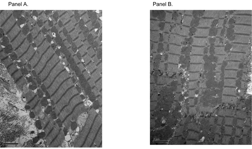
Figure 1 shows representative electron micrographs of the ultrastructure of the LV muscle from CON and CS-exposed rats. A pathologist blinded to group assignment examined and scored sections for the general appearance of the tissue, changes in intracellular organelles (such as the mitochondria and sarcoplasmic reticulum), and evidence of cell loss or myofibril disarray. No significant ultrastructural changes were found in either group.
Figure 1.
Representative electron micrographs from control (panel A) and CS-exposed animals (panel B).
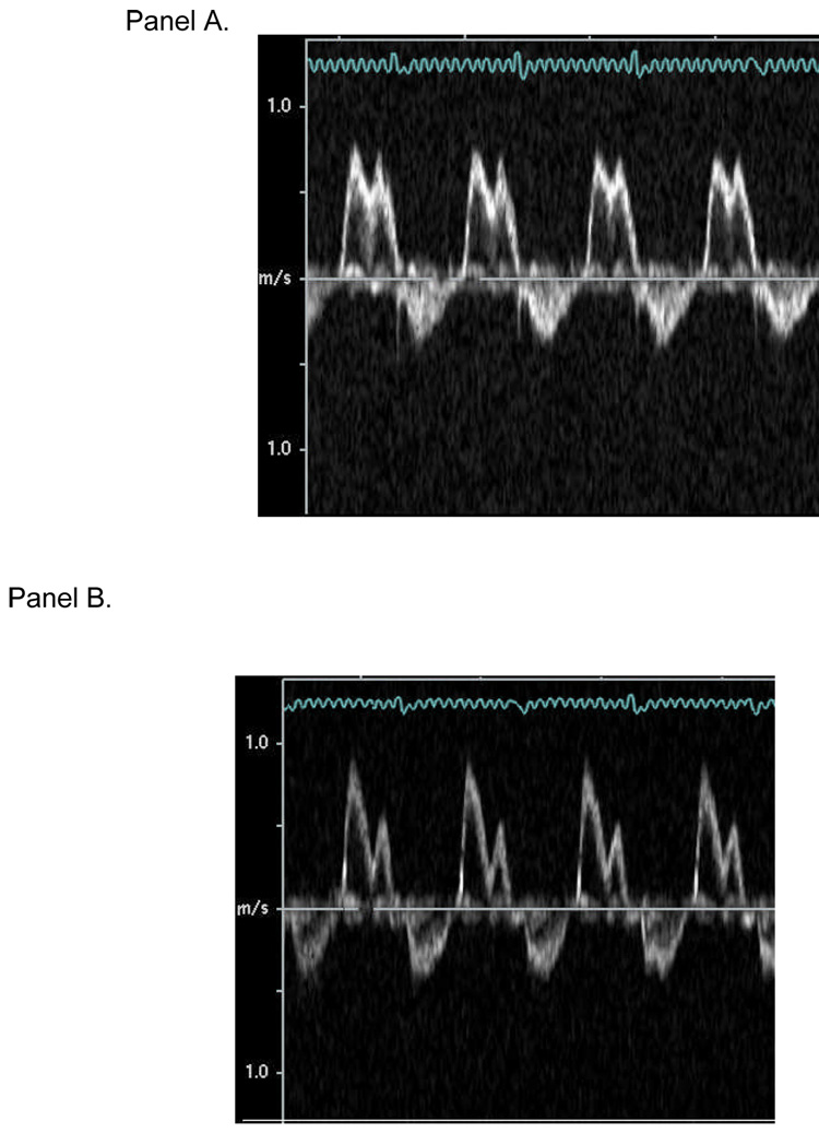
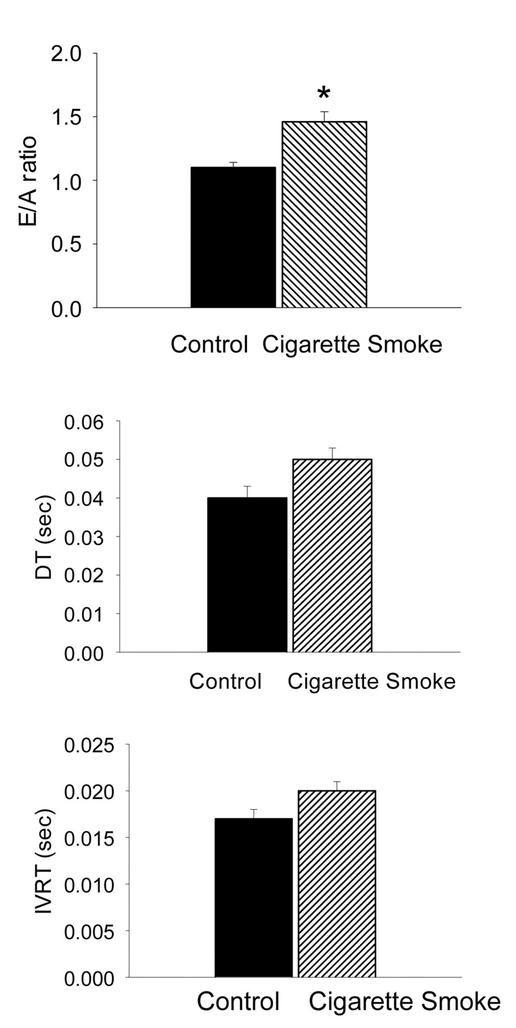
Significant increases in LVEDD and LVESD were found in the CS group compared to the CON group (Table 2). Left ventricular posterior wall thickness in diastole (LVPWD), left ventricular posterior wall thickness at systole (LVPWS) and RWT were not significantly different between the groups; however, FS was significantly lower in the CS group than in the CON group (Table 2). Using pulse-wave Doppler imaging, we found well defined signals in 94% of the animals (representative images are shown in Figure 2). We found a significant increase in the E/A ratio in the CS group compared to the CON group (Figure 3). IVRT and DT were not significantly different between groups (Figure 3).
Table 2.
Echocardiographic parameters.
| Control | CS | p | |
|---|---|---|---|
| Heart Rate (bpm) | 341 ± 11 | 316 ± 6 | 0.083 |
| LVEDD (mm) | 7.99 ± 0.1 | 8.3 ± 0.10 | 0.026 |
| LVESD (mm) | 4.55 ± 0.2 | 5.3 ± 0.09 | 0.003 |
| LVPWD (mm) | 1.40 ± 0.06 | 1.40 ± 0.07 | 0.990 |
| LVPWS (mm) | 2.40 ± 0.04 | 2.18 ± 0.14 | 0.200 |
| FS(%) | 43 ± 2.1 | 36 ± 0.9 | 0.010 |
| LV mass (gm) | 0.67 ± .03 | 0.72 ± .04 | 0.320 |
| RWT | 0.35 ± .01 | 0.33 ± .01 | 0.430 |
Abbreviations used: FS-fractional shortening, left ventricle (LV) end-diastolic dimension (LVEDD), LV end-systolic dimension (LVESD), LV posterior wall thickness in diastole (LVPWD) and LV posterior wall thickness in systole (LVPWS), relative wall thickness (RWT = 2 × LVPWD/EDD). Data are expressed as mean ± SEM.
Figure 2.
Representative pulse-wave Doppler echocardiograms from control (panel A) and CS-exposed (panel B) animals.
Figure 3.
Indices of diastolic function in control and cigarette smoke exposed animals. Data are expressed as mean ± SEM. Abbreviations used: DT-deceleration time and IVRT-isovolumic relaxation time. *Indicates significantly different from control (p = 0.01).
3.1. Western Blotting of MAPKs
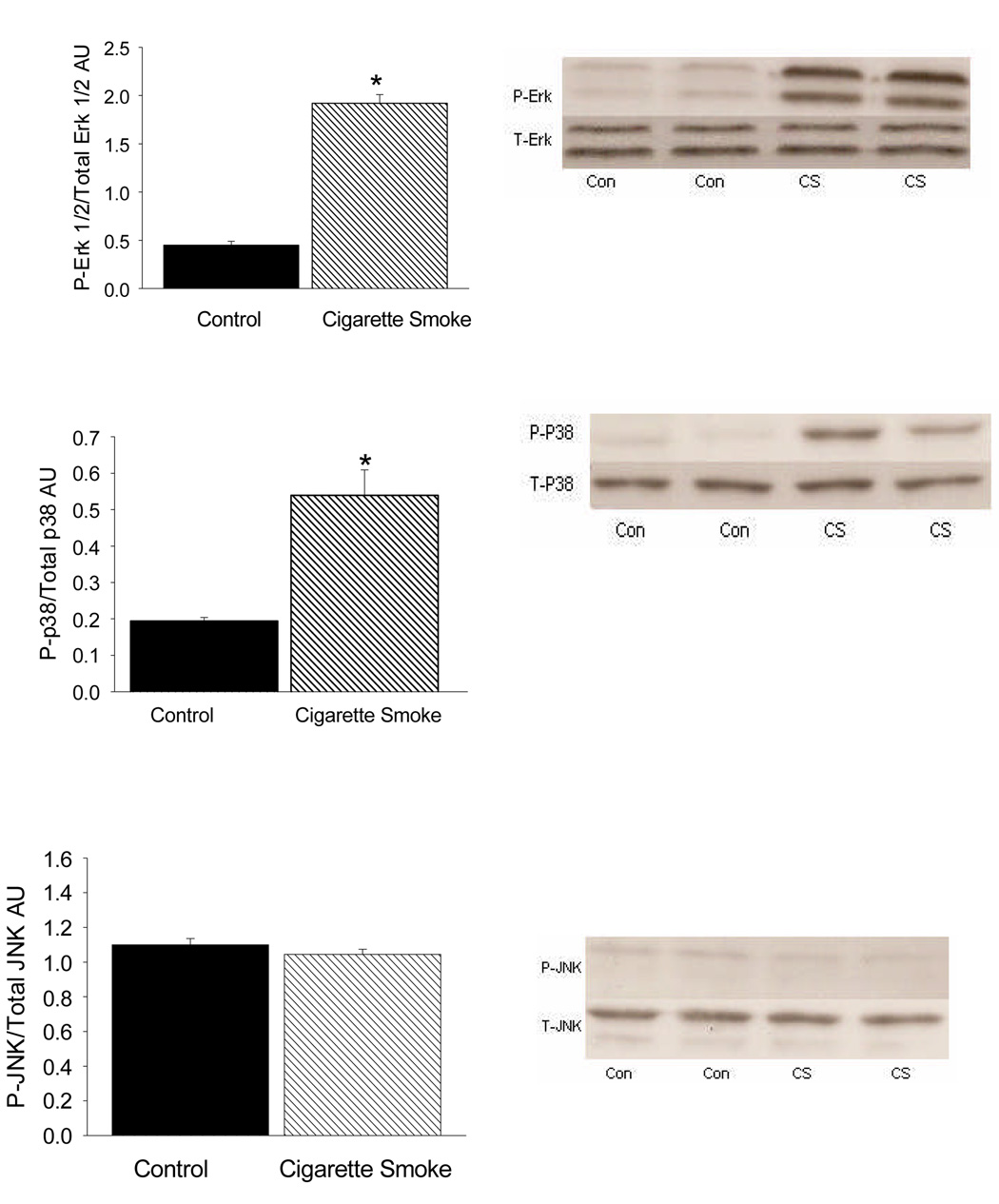
Protein levels of total p38, ERK1/2, JNK, P-p38, P-ERK1/2 and P-JNK were determined using Western blotting. Levels were expressed as the ratio of phosphorylated (P)-to-total protein levels. The levels of P-p38 and P-ERK1/2 were significantly greater in the CS-exposed hearts compared to the CON hearts (Figure 4, Panels A and B) (p < 0.01). No significant differences were found in JNK levels between the groups (Figure 4, Panel C).
Figure 4.
Activation of MAPKs. Levels were expressed as the ratio of phosphorylated (P)-to-total protein levels. Data are expressed as mean ± SEM. *Indicates significantly different from control (p < 0.01). Representative Western blots.
4. Discussion
This is the first study to demonstrate that CS-induced ventricular remodelling is linked to activation of MAPKs. Our data also suggest that CS smoke exposure is associated with systolic dysfunction and increased adrenergic drive. Collectively, these data support the involvement of MAPKs in CS-induced remodelling and cardiac dysfunction.
To date, there have been few studies examining the effects of CS on the myocardium, and to our knowledge no investigations have examined cell mechanisms that are linked to the toxic effects of CS on the myocardium. Although CS affects the entire cardiovascular system, it has been generally accepted that the most adverse effects were on the vasculature and included endothelial dysfunction and accelerated atherosclerosis [17]. However, a landmark prospective clinical study by Hartz and colleagues in 1984 established in men younger than 55 years of age (without evidence of coronary artery disease or history of myocardial infarction) that CS exposure was associated with diffuse ventricular hypokinesis [18]. The associations between CS and reduced left ventricular function and greater left ventricular mass have been re-affirmed in more recent large population-based epidemiological studies using magnetic resonance imaging to evaluate myocardial structure and function [19,20]. These data are further substantiated by our findings and those of others, which demonstrate that CS exposure is associated with ventricular remodelling [6,7].
Our findings of an increase in EDD and ESD and a decrease in FS in CS-exposed animals are similar to others, who have shown that 4 months of CS exposure is associated with cardiac remodelling and systolic dysfunction [6,7]. Changes in diastolic function after CS exposure have been equivocal in animal models and in human beings. Doppler-derived echocardiography parameters that reflect diastolic dysfunction include an increase in the E/A ratio and prolonged DT and IVRT values. In the present study, we found a significant increase in the E/A ratio in the CS group compared to CON group; however, even though DT and IVRT were increased in the CS group compared to CON, these differences were not significant. Following 4 months of CS exposure, Castardeli et al. found no change in the E/A ratio between CON and CS-exposed rats [6,7]. In human subjects, investigators have examined the acute one-time effect of CS exposure on diastolic function [21]. Alam et al. found evidence of diastolic dysfunction in healthy subjects immediately after smoking one cigarette [21]. In a similar study, 30 minutes after subjects smoked one cigarette, Karakaya et al. found evidence of diastolic dysfunction in subjects with coronary artery disease, while no significant changes were found in healthy subjects [22]. To our knowledge, there are no studies examining longer durations of CS exposure on diastolic function in human subjects. It would be interesting to determine if CS exposure alters intracellular organelles (e.g., sarcoplasmic reticulum) or molecular events (sarcoplasmic reticulum uptake of calcium) that govern diastolic function.
In our study, 5 weeks of CS exposure was associated with increased urinary NE levels. CS is associated with increased sympathetic activation and NE levels according to Cryer et al. [8] and Narkiewicz et al. [23]. Via activation of different adrenergic receptor (AR) subtypes, NE can be associated with either myocyte hypertrophy (α1A AR) or apoptosis (β1 AR) [24]. Activation of the α1A AR is also associated with increased ERK1/2 activation [25]. We did not examine cardiac tissue for evidence of apoptosis or myocyte hypertrophy (e.g., increased cell size or protein synthesis).
Our data clearly indicate that CS activates MAPKs such as p38 and ERK1/2. Our findings concur with previous studies, which have established a role for activation of MAPK signalling in the pathogenesis of CS-induced lung injury (emphysema) and inflammation [26,27]. Furthermore, others have shown that CS-induced phosphorylation of ERK1/2 precedes the activation of protooncogenes that belong to the AP-1 transcription family (e.g., fos and jun). Importantly, genes that encode for growth factors (e.g., insulin-like growth factor) and extracellular matrix metalloproteinases (MMPs) have an AP-1 site in their promoter or enhancer region [28]. MMPs are proteinases that can degrade almost all of the extracellular matrix proteins. In terms of the myocardium, increased MMP activity is associated with breakdown of the supporting fibrillar collagen network. Loss of the supporting fibrillar collagen network, along with loss of the collagen tethers, leads to myocyte slippage, ventricular wall thinning, and enlargement [29]. Mercer et al. [25] and Ning et al. [30] have demonstrated a strong causal role for ERK1/2 signalling and the activation of MMP in pulmonary epithelial cells and the consequent development of emphysema, a disease characterized by loss of alveolar attachments and parenchymal destruction.
We also found that 5 weeks of CS exposure was associated with increased p38 phosphorylation. p38 has also been referred to as a cytokine-suppressive anti-inflammatory drug-binding protein [10]. p38 is strongly activated by cytokines, such as TNFα, and other cellular stressors, such as oxidative stress and hyperosmolarity. Using an animal model, Liu et al. [31] and Li et al. [32] have shown that inhibition of p38 (in particular the α isoform) protects the heart against remodelling following myocardial infarction. In addition, Li et al. demonstrated that there were fewer infiltrating macrophages and apoptotic cells after animals were treated with the p38 inhibitor SD-282. Collectively, the latter data suggest that activation of the p38 pathway is a key mechanism in ischaemia-induced myocardial dysfunction. Considering CS exposure is associated with increased TNFα and oxidative stress [9,33], it is not surprising that we found increases in p38 phosphorylation. Using human pulmonary artery endothelial cells in culture, Low et al. [34] demonstrated that p38 mediates CS-induced endothelial permeability. It will be important to conduct future experiments to determine if there is evidence of apoptosis and inflammation and if a longer duration of CS exposure is associated with more severe changes, as well as activation of JNK.
The way in which CS activates both the p38 and ERK1/2 MAPK cascades remains unknown and could potentially occur at multiple levels: via stimulation/release hormones or by activation of upstream kinases, such as MAPK kinase kinase (MEKK) or MAPK kinases (MEK). As noted above, NE is associated with activation of the MAPK cascade (e.g., ERK1/2). CS exposure is also associated with increased cytokine levels, such as TNFα, which in turn is associated with activation of p38 MAPKs. However, it is well established that CS contains oxidants and is a potent stimulator of oxidative stress. Therefore, it is also possible that CS may (via the generation of oxidants) directly activate upstream kinases, such as MEKK or MEK [35,36]. We did not find an increase in JNK after 5 weeks of CS exposure. Perhaps the preferential activation of p38 and ERK1/2 are related to either the duration of CS exposure or overall level of oxidative stress.
We did not find any significant changes in the appearance of the sarcomeres, myofilaments, or mitochondria between groups upon ultrastructural analysis. This is in contrast to Zornoff et al. [37], who reported that 30 days of environmental tobacco smoke exposure in male Wistar rats was associated with disorganization of the myofibrils and mitochondrial swelling. In the Zornoff et al. study, animals were exposed to 10 cigarettes in the morning and then again in the afternoon. In our protocol, animals were exposed to 45 minutes of mainstream smoke (approximately 2–3 cigarettes) twice a day. The earlier investigators did not report cotinine levels; therefore, it is difficult to compare our two studies in terms of the amount of smoke exposure. However, the animals in the Zornoff protocol more than likely received more smoke exposure, perhaps explaining their morphological tissue findings. In addition, the type of cigarette (2R1) we used has a greater tar and nicotine content compared to the type of cigarette used by others [37]. Also, as noted in the methods section, the tar and nicotine content of the R21 is greater than conventional cigarettes available to the public. However, using this type of cigarette, as well as an intermittent, short-term (5-week) exposure protocol in a rodent model, our urinary cotinine levels (1102 ± 306 ng/mmol/creatinine) approximate those reported in human active smokers (i.e., those who smoked > 10 cigarettes/day) [13].
4.1 Study Limitations
There are several limitations in this study. CS induces the secretion of different types of circulating neurohumoral stimuli, such as NE, that are linked to MAPK activation. In our intermittent rodent CS model, we found large increases in urinary NE. However, we did not assay for increases in other stimuli, such as TNFα, which others have found to be increased in smoking human beings [38] and animal models exposed to CS [39]. Similar to NE, TNFα is a potent activator of MAPKs; therefore, in future studies, it would be important to determine if TNFα serum and/or myocardial tissues levels are increased after CS. Second, because we measured MAPK activation and structural and functional parameters of LV remodelling at the same time point and did not attempt to inhibit MAPK activation, we cannot conclude that CS-induced activation of MAPK leads to cardiac remodelling. Using cultured human pulmonary artery endothelial cells, Low et al. found that the addition of a p38 MPK-selective inhibitor (SB203580) prevented CS-induced p38 activation and the development of endothelial damage (i.e., increased endothelial cell permeability) [34]. p38 MAPK inhibitors are available and have been associated with positive and protective effects in animal models of inflammation [40] and myocardial injury [32]. Findings of the present study could be confirmed in future studies incorporating the use of MAPK inhibitors.
4.2 Conclusion
In conclusion, this study identified that 5 weeks of mainstream CS exposure is associated with LV remodelling, systolic dysfunction and activation of p38 and ERK1/2 MAPKs. MAPKs govern cell survival and growth in the myocardium, and others have shown that activation of MAPKs plays a key role in the pathogenesis of hypertrophy, heart failure and ischaemia/reperfusion injury. Moreover, in other tissues, CS activation of the MAPK signaling cascade is considered to be a key mechanistic event leading to cell injury, altered cell differentiation and inflammation [27]. Therefore, activation of MAPKs may represent an important event in CS-induced cardiotoxicity.
Acknowledgements
The authors thank Dr. John Van Fleet (Purdue University) for his excellent morphological analysis and Amy Tarr for her excellent laboratory assistance. This study was supported by National Institutes of Health grant AA015578. The authors thank Kevin Grandfield for editorial assistance.
Footnotes
Publisher's Disclaimer: This is a PDF file of an unedited manuscript that has been accepted for publication. As a service to our customers we are providing this early version of the manuscript. The manuscript will undergo copyediting, typesetting, and review of the resulting proof before it is published in its final citable form. Please note that during the production process errors may be discovered which could affect the content, and all legal disclaimers that apply to the journal pertain.
References
- 1.Ambrose JA, Barua RS. The pathophysiology of cigarette smoking and cardiovascular disease. JACC. 2004;43(101):1731–1737. doi: 10.1016/j.jacc.2003.12.047. [DOI] [PubMed] [Google Scholar]
- 2.Sun YP, Zhu BQ, Browne AE, et al. Nicotine does not influence arterial lipid deposits in rabbits exposed to second-hand smoke. Circulation. 2001;104:810–814. doi: 10.1161/hc3301.092788. [DOI] [PubMed] [Google Scholar]
- 3.Pellegrini MP, Newby DE, Maxwell S, Webb DJ. Short-term effects of transdermal nicotine on acute tissue plasminogen activator release in vivo on man. Cardiovasc Res. 2001;52:321–327. doi: 10.1016/s0008-6363(01)00381-9. [DOI] [PubMed] [Google Scholar]
- 4.Hubbard R, Lewis S, Smith C, et al. Use of nicotine replacement therapy and the risk of acute myocardial infarction, stroke, and death. Tob Control. 2005;14:416–421. doi: 10.1136/tc.2005.011387. [DOI] [PMC free article] [PubMed] [Google Scholar]
- 5.Benowitz NL, Gourlay SG. Cardiovascular toxicity of nicotine: Implications for nicotine replacement therapy. JACC. 1997;29(7):1422–1431. doi: 10.1016/s0735-1097(97)00079-x. [DOI] [PubMed] [Google Scholar]
- 6.Castardeli E, Duarte DR, Minicucci MF, et al. Tobacco smoke-induces left ventricular remodeling is not associated with metalloproteinase-2 or-9 activation. Eur J Heart Fail. 2007;9:1081–1085. doi: 10.1016/j.ejheart.2007.09.004. [DOI] [PubMed] [Google Scholar]
- 7.Castardeli E, Paiva SA, Matsubara BB, et al. Chronic cigarette smoke exposure results in cardiac remodeling and impaired ventricular function in rats. Arquivs Brasileiros de Cardiologia. 2005;84:320–324. doi: 10.1590/s0066-782x2005000400009. [DOI] [PubMed] [Google Scholar]
- 8.Cryer PE, Haymond MW, Santiago JV, Shah SD. Norepinephrine and epinephrine release and adrenergic mediation of smoking-associated hemodynamic and metabolic events. NEJM. 1976;295:573–577. doi: 10.1056/NEJM197609092951101. [DOI] [PubMed] [Google Scholar]
- 9.Zhang J, Liu Y, Shi J, Larson DF, Watson RR. Side-stream cigarette smoke induces dose-responses in systemic inflammatory cytokine production and oxidative stress. Exp Biol Med. 2002;227:823–829. doi: 10.1177/153537020222700916. [DOI] [PubMed] [Google Scholar]
- 10.Ravingerová T, Barancík M, Strnisková M. Mitogen-activated protein kinases: a new therapeutic target in cardiac pathology. Mol Cell Biochem. 2003 May;247(1–2):127–138. doi: 10.1023/a:1024119224033. [DOI] [PubMed] [Google Scholar]
- 11.Griffith RB, Standafer S. Simultaneous mainstream-side-stream smoke exposure systems II. The rat exposure system. Toxicology. 1985;35:13–24. doi: 10.1016/0300-483x(85)90128-3. [DOI] [PubMed] [Google Scholar]
- 12.Coggins CRE. A review of chronic inhalation studies with mainstream cigarette smoke in rats and mice. Toxicol Pathol. 1998;26:307–314. doi: 10.1177/019262339802600301. [DOI] [PubMed] [Google Scholar]
- 13.Wall MA, Johnson J, Jacob P, Benowitz NL. Cotinine in the serum, saliva, and urine of nonsmokers, passive smokers, and active smokers. Am J Public Health. 1988;78:699–701. doi: 10.2105/ajph.78.6.699. [DOI] [PMC free article] [PubMed] [Google Scholar]
- 14.Piano MR, Geenen D, Schwertz DW, Chowdhury SAK, Yuzhakova M. Cardiac remodeling secondary to long-term alcohol consumption in male and female rats. Cardiovasc Toxicol. 2007;7(4):247–254. doi: 10.1007/s12012-007-9002-y. [DOI] [PubMed] [Google Scholar]
- 15.Schiller NB, Shah PM, Crawford M, et al. Recommendations for quantitation of the left ventricle by two-dimensional echocardiography. American Society of Echocardiography Committee on Standards, Subcommittee on Quantitation of Two-Dimensional Echocardiograms. J Am Soc Echocardiogr. 1989;2(5):358–367. doi: 10.1016/s0894-7317(89)80014-8. [DOI] [PubMed] [Google Scholar]
- 16.Hu WY, Han YJ, Gu LZ, Piano MP, de Lanerolle P. Involvement of Ras-regulated myosin light chain phosphorylation in the captopril effects in spontaneously hypertensive rats. Am J Hypertens. 2007 Jan;20(1):53–61. doi: 10.1016/j.amjhyper.2006.05.024. [DOI] [PubMed] [Google Scholar]
- 17.Haustein KO. Smoking tobacco, microcirculatory changes and the role of nicotine. Int J Clin Pharmacol Ther. 1999 Feb;37(2):76–85. [PubMed] [Google Scholar]
- 18.Hartz AJ, Anderson AJ, Brooks HL, et al. The association of smoking with cardiomyopathy. New Eng J Med. 1984;311:1201–1206. doi: 10.1056/NEJM198411083111901. [DOI] [PubMed] [Google Scholar]
- 19.Heckbert SR, Post W, Pearson GDN, et al. Traditional cardiovascular risk factors in relation to left ventricular mass, volume, and systolic function by cardiac magnetic resonance imaging: the Multiethnic Study of Atherosclerosis. JACC. 2006;48(11):2285–2292. doi: 10.1016/j.jacc.2006.03.072. [DOI] [PMC free article] [PubMed] [Google Scholar]
- 20.Rosen BD, Saad MF, Shea S, et al. Hypertension and smoking are associated with reduced regional left ventricular function in asymptomatic individuals. JACC. 2006;47(6):1150–1158. doi: 10.1016/j.jacc.2005.08.078. [DOI] [PubMed] [Google Scholar]
- 21.Alam M, Samad BA, Wardell J, Andersson E, Hoglund C, Nordlander R. Acute effects of smoking on diastolic function in healthy participants : Studies by conventional Doppler echocardiography and Doppler tissue imaging. J Am Soc Echocardiogr. 2002;15(10):1232–1237. doi: 10.1067/mje.2002.124006. [DOI] [PubMed] [Google Scholar]
- 22.Karakaya O, Barutcu I, Esen AM, et al. Acute smoking-induced alterations in Doppler echocardiographic measurements in chronic smokers. Tex Heart Inst J. 2006;33(2):134–138. [PMC free article] [PubMed] [Google Scholar]
- 23.Narkiewicz K, van de Borne PJH, Hausberg M, et al. Cigarette smoking increases sympathetic outflow in humans. Circulation. 1998;98:528–534. doi: 10.1161/01.cir.98.6.528. [DOI] [PubMed] [Google Scholar]
- 24.Singh K, Xiao L, Remondino A, Sawyer DB, Colucci WS. Adrenergic regulation of cardiac myocyte apoptosis. J Cell Physiol. 2001;189:257–265. doi: 10.1002/jcp.10024. [DOI] [PubMed] [Google Scholar]
- 25.Rapacciulo A, Esposito G, Caron K, Man L, Thomas SA, Rockman HA. Important role of endogenous norepinephrine and epinephrine in the development of in vivo pressure-overload cardiac hypertrophy. JACC. 2001;38(3):876–882. doi: 10.1016/s0735-1097(01)01433-4. [DOI] [PubMed] [Google Scholar]
- 26.Mercer BA, Kolesnikova N, Sonett J, D’Armiento J. Extracellular regulated kinase/mitogen activated protein kinase is up-regulated in pulmonary emphysema and mediates matrix metalloproteinase-1 induction by cigarette smoke. J Biol Chem. 2004;279(1):19690–17696. doi: 10.1074/jbc.M313842200. [DOI] [PubMed] [Google Scholar]
- 27.Mossman BT, Lounsbury KM, Reddy SP. Oxidants and signaling by mitogen-activated protein kinases in lung epithelium. Am J Resp Cell Mol Biol. 2006;34:666–669. doi: 10.1165/rcmb.2006-0047SF. [DOI] [PMC free article] [PubMed] [Google Scholar]
- 28.Reddy SP, Mossman BT. Role and regulation of activator protein-1 in toxicant-induced responses of the lung. Am J Physiol Lung Cell Mol Physiol. 2002 Dec;283(6):L1161–L1178. doi: 10.1152/ajplung.00140.2002. [DOI] [PubMed] [Google Scholar]
- 29.Caulfield JB, Norton P, Weaver RD. Cardiac dilatation associated with collagen alterations. Mol Cell Biochem. 1992;118:171–179. doi: 10.1007/BF00299396. [DOI] [PubMed] [Google Scholar]
- 30.Ning W, Dong Y, Sun J, et al. Cigarette smoke stimulates matrix metalloproteinase-2 activity via EGR-1 in human lung fibroblasts. Am J Resp Cell Mol Biol. 2007;36:480–490. doi: 10.1165/rcmb.2006-0106OC. [DOI] [PMC free article] [PubMed] [Google Scholar]
- 31.Liu Y-H, Wang D, Rhaleb N-E, et al. Inhibition of p38 mitogen-activated protein kinase protects the heart against cardiac remodeling in mice with heart failure resulting from myocardial infarction. J Card Fail. 2005;11:74–81. doi: 10.1016/j.cardfail.2004.04.004. [DOI] [PubMed] [Google Scholar]
- 32.Li Z, Ma JY, Kerr I, et al. Selective inhibition of p38α MAPK improves cardiac function and reduces myocardial apoptosis in rat model of myocardial injury. Am J Physiol Heart Circ Physiol. 2006;291:H1972–H1977. doi: 10.1152/ajpheart.00043.2006. [DOI] [PubMed] [Google Scholar]
- 33.Barua RS, Ambrose JA, Srivastava S, DeVoe MC, Eales-Reynolds LJ. Reactive oxygen species are involved in smoking-induced dysfunction of nitric oxide biosynthesis and upregulation of endothelial nitric oxide synthase: an in vitro demonstration in human coronary artery endothelial cells. Circulation. 2003;107:2342–2347. doi: 10.1161/01.CIR.0000066691.52789.BE. [DOI] [PubMed] [Google Scholar]
- 34.Low B, Liang M, Fu J. p38 mitogen-activated protein kinase mediates sidestream cigarette smoke-induces endothelial permeability. J Pharmacol Sci. 2007;104:225–231. doi: 10.1254/jphs.fp0070385. [DOI] [PubMed] [Google Scholar]
- 35.Galan A, Garcia-Bermejo ML, Troyano A, et al. Stimulation of p38 mitogen-activated protein kinase is an early regulatory event for the cadmium-induced apoptosis in human promonocytic cells. J Biol Chem. 2000;275:11418–11424. doi: 10.1074/jbc.275.15.11418. [DOI] [PubMed] [Google Scholar]
- 36.Ueda S, Masutani H, Nakamura H, Tanaka T, Ueno M, Yodoi J. Redox control of cell death. Antioxid Redox Signal. 2002;4:405–414. doi: 10.1089/15230860260196209. [DOI] [PubMed] [Google Scholar]
- 37.Zornoff LA, Matsubara LS, Matsubara BB, et al. Beta-carotene supplementation attenuates cardiac remodeling induced by one-month tobacco-smoke exposure in rats. Toxicol Sci. 2006 Mar;90(1):259–266. doi: 10.1093/toxsci/kfj080. [DOI] [PubMed] [Google Scholar]
- 38.Takabatake N, Nakamura H, Inoue S, et al. Circulating levels of soluble Fas ligand and soluble Fas in patients with chronic obstructive pulmonary disease. Respir Med. 2000;94:1215–1220. doi: 10.1053/rmed.2000.0941. [DOI] [PubMed] [Google Scholar]
- 39.Churg A, Dai J, Tai H, Xei C, Wright JL. Tumor necrosis factor-alpha is central to acute cigarette smoke-induced inflammation and connective tissue breakdown. Am J Respir Crit Care Med. 2002;166:849–854. doi: 10.1164/rccm.200202-097OC. [DOI] [PubMed] [Google Scholar]
- 40.Jackson JR, Bolognese B, Hillegass L, Kassis S, Adams J, Griswold DE, Winkler JD. Pharmacological effects of SB 220025, a selective inhibitor of P38 mitogen-activated protein kinase, in angiogenesis and chronic inflammatory disease models. J Pharmacol Exp Ther. 1998;284(2):687–692. [PubMed] [Google Scholar]


