Abstract
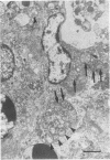
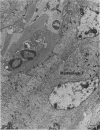
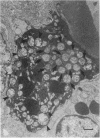
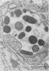
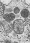
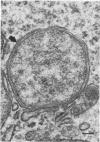
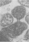
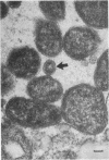
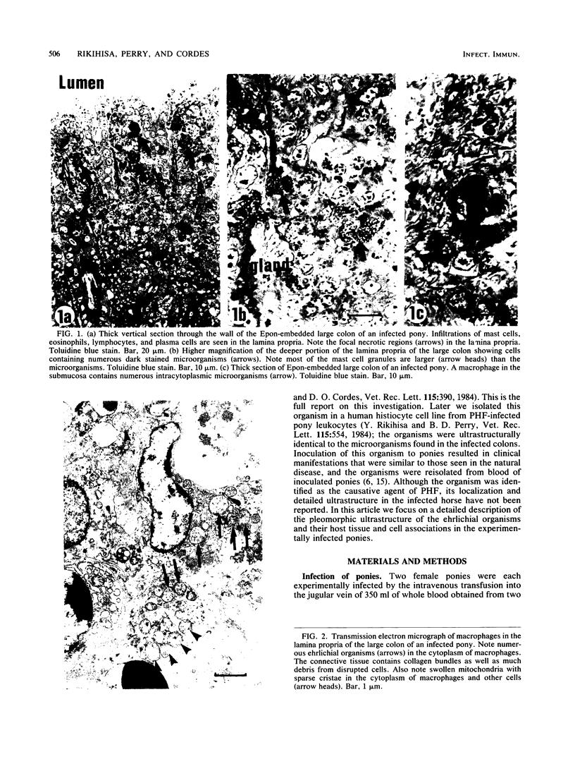
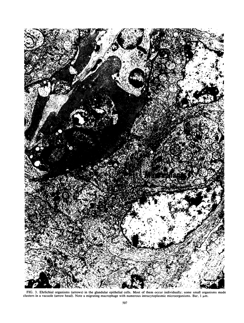
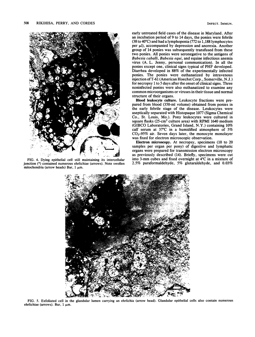
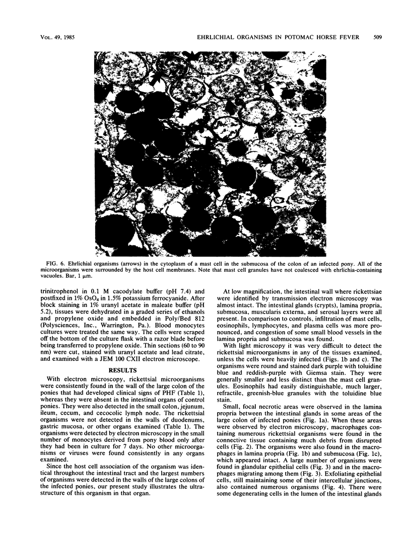
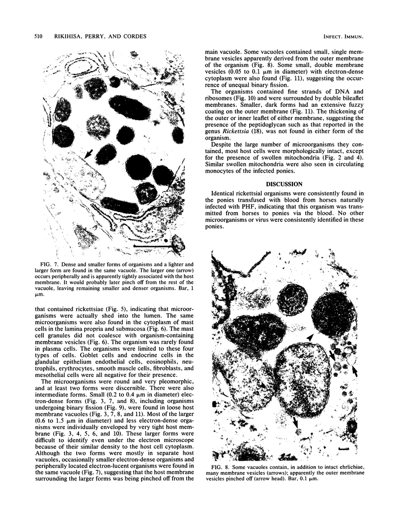
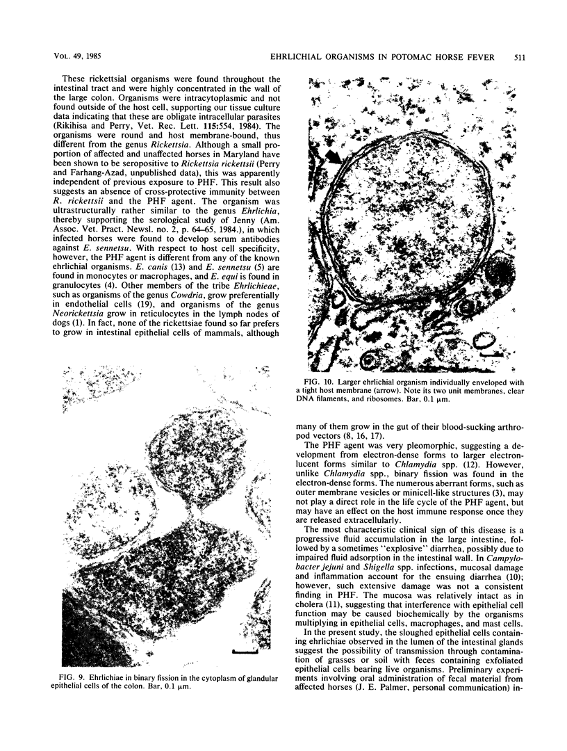
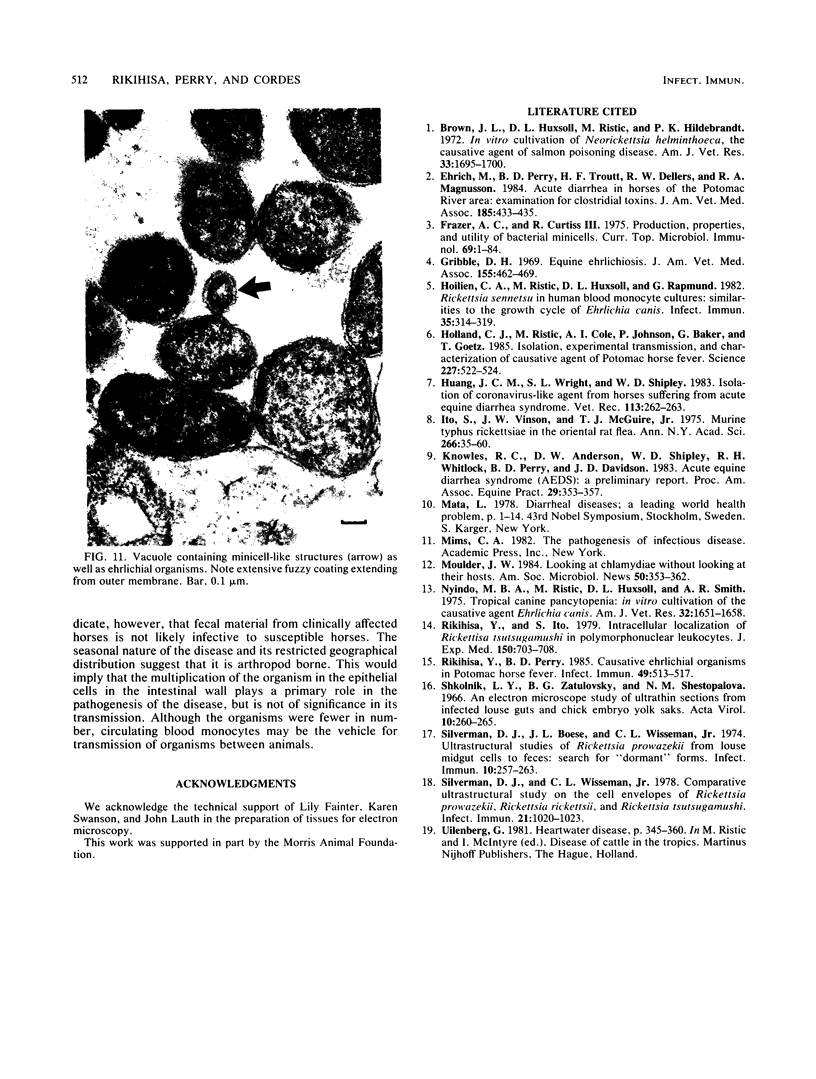
Potomac horse fever is characterized by fever, anorexia, leukopenia, profuse watery diarrhea, dehydration, and high mortality. An ultrastructural investigation was made to search for any unusual microorganisms in the digestive system, lymphatic organs, and blood cells of ponies that had developed clinical signs after transfusion with whole blood from horses naturally infected with Potomac horse fever. A consistent finding was the presence of rickettsial organisms in the wall of the intestinal tract of these ponies. The organisms were found mostly in the wall of the large colon, but fewer organisms were found in the small colon, jejunum, and cecum. The organisms were also detected in cultured blood monocytes. In the intestinal wall, many microorganisms were intracytoplasmic in deep glandular epithelial cells and mast cells. Microorganisms were also found in macrophages migrating between glandular epithelial cells in the lamina propria and submucosa. The microorganisms were round, very pleomorphic, and surrounded by a host membrane. They contained fine strands of DNA and ribosomes and were surrounded by double bileaflet membranes. Their ultrastructure was very similar to that of the genus Ehrlichia, a member of the family Rickettsiaceae. The high frequency of detection of the organism in the wall of the intestinal tract, especially in the large colon, indicates the presence of organotrophism in this organism. Infected blood monocytes may be the vehicle for transmission between organs and between animals. The characteristic severe diarrhea may be induced by the organism directly by impairing epithelial cell functions or indirectly by perturbing infected macrophages and mast cells in the intestinal wall or by both.
Full text
PDF
Images in this article
Selected References
These references are in PubMed. This may not be the complete list of references from this article.
- Brown J. L., Huxsoll D. L., Ristic M., Hildebrandt P. K. In vitro cultivation of Neorickettsia helminthoeca, the causative agent of salmon poisoning disease. Am J Vet Res. 1972 Aug;33(8):1695–1700. [PubMed] [Google Scholar]
- Ehrich M., Perry B. D., Troutt H. F., Dellers R. W., Magnusson R. A. Acute diarrhea in horses of the Potomac River area: examination for clostridial toxins. J Am Vet Med Assoc. 1984 Aug 15;185(4):433–435. [PubMed] [Google Scholar]
- Frazer A. C., Curtiss R., 3rd Production, properties and utility of bacterial minicells. Curr Top Microbiol Immunol. 1975;69:1–84. doi: 10.1007/978-3-642-50112-8_1. [DOI] [PubMed] [Google Scholar]
- Gribble D. H. Equine ehrlichiosis. J Am Vet Med Assoc. 1969 Jul 15;155(2):462–469. [PubMed] [Google Scholar]
- Hoilien C. A., Ristic M., Huxsoll D. L., Rapmund G. Rickettsia sennetsu in human blood monocyte cultures: similarities to the growth cycle of Ehrlichia canis. Infect Immun. 1982 Jan;35(1):314–319. doi: 10.1128/iai.35.1.314-319.1982. [DOI] [PMC free article] [PubMed] [Google Scholar]
- Holland C. J., Ristic M., Cole A. I., Johnson P., Baker G., Goetz T. Isolation, experimental transmission, and characterization of causative agent of Potomac horse fever. Science. 1985 Feb 1;227(4686):522–524. doi: 10.1126/science.3880925. [DOI] [PubMed] [Google Scholar]
- Huang J. C., Wright S. L., Shipley W. D. Isolation of coronavirus-like agent from horses suffering from acute equine diarrhoea syndrome. Vet Rec. 1983 Sep 17;113(12):262–263. doi: 10.1136/vr.113.12.262. [DOI] [PubMed] [Google Scholar]
- Ito S., Vinson J. W., McGuire T. J., Jr Murine typhus Rickettsiae in the Oriental rat flea. Ann N Y Acad Sci. 1975;266:35–60. doi: 10.1111/j.1749-6632.1975.tb35087.x. [DOI] [PubMed] [Google Scholar]
- Nyindo M. B., Ristic M., Huxsoll D. L., Smith A. R. Tropical canine pancytopenia: in vitro cultivation of the causative agent--Ehrlichia canis. Am J Vet Res. 1971 Nov;32(11):1651–1658. [PubMed] [Google Scholar]
- Rikihisa Y., Ito S. Intracellular localization of Rickettsia tsutsugamushi in polymorphonuclear leukocytes. J Exp Med. 1979 Sep 19;150(3):703–708. doi: 10.1084/jem.150.3.703. [DOI] [PMC free article] [PubMed] [Google Scholar]
- Rikihisa Y., Perry B. D. Causative ehrlichial organisms in Potomac horse fever. Infect Immun. 1985 Sep;49(3):513–517. doi: 10.1128/iai.49.3.513-517.1985. [DOI] [PMC free article] [PubMed] [Google Scholar]
- Shkolnik L. Y., Zatulovsky B. G., Shestopalova N. M. Ultrastructure of Rickettsia prow azeki. An electron microscope study of ultrathin sections from infected louse guts and chick embryo yolk sacs. Acta Virol. 1966 May;10(3):260–265. [PubMed] [Google Scholar]
- Silverman D. J., Boese J. L., Wisseman C. L., Jr Ultrastructural studies of Rickettsia prowazeki from louse midgut cells to feces: search for "dormant" forms. Infect Immun. 1974 Jul;10(1):257–263. doi: 10.1128/iai.10.1.257-263.1974. [DOI] [PMC free article] [PubMed] [Google Scholar]
- Silverman D. J., Wisseman C. L., Jr Comparative ultrastructural study on the cell envelopes of Rickettsia prowazekii, Rickettsia rickettsii, and Rickettsia tsutsugamushi. Infect Immun. 1978 Sep;21(3):1020–1023. doi: 10.1128/iai.21.3.1020-1023.1978. [DOI] [PMC free article] [PubMed] [Google Scholar]


