Abstract
The transcription factor Runx1 is a key regulator of definitive hematopoiesis in the embryo and the adult. Lineage-specific expression of Runx1 involves transcription and post-transcription control through usage of alternative promoters and diverse 3′UTR isoforms, respectively. We identified and mapped microRNA (miR) binding sites on Runx1 3′UTR and show that miR-27a, miR-9, miR-18a, miR-30c, and miR-199a* bind and post-transcriptionally attenuate expression of Runx1. miR-27a impacts on both the shortest (0.15 kb) and longest (3.8 kb) 3′UTRs and, along with additional miRs, might contribute to translation attenuation of Runx1 mRNA in the myeloid cell line 416B. Whereas levels of Runx1 mRNA in 416B and the B cell line 70Z were similar, the protein levels were not. Large amounts of Runx1 protein were found in 70Z cells, whereas only minute amounts of Runx1 protein were made in 416B cells and overexpression of Runx1 in 416B induced terminal differentiation associated with megakaryocytic markers. Induction of megakaryocytic differentiation in K562 cells by 12-o-tetradecanoylphorbol-13-acetate markedly increased miR-27a expression, concomitantly with binding of Runx1 to miR-27a regulatory region. The data indicate that miR-27a plays a regulatory role in megakaryocytic differentiation by attenuating Runx1 expression, and that, during megakaryopoiesis, Runx1 and miR-27a are engaged in a feedback loop involving positive regulation of miR-27a expression by Runx1.
Keywords: megakaryocyte differentiation, micro RNA, Runx transcription factor
The mammalian RUNX1 belongs to the runt domain family of transcription factors. The three family members, RUNX1, RUNX2, and RUNX3, are lineage-specific gene expression regulators in major developmental pathways (1–3). Although all three RUNX proteins recognize the same DNA motif, the functional overlaps are minor and each has a distinct subset of biological functions. This lack of functional redundancy results from a tightly regulated spatio-temporal expression of the RUNX genes by transcriptional and post-transcriptional control mechanisms (3–7).
We have previously shown that transcription of RUNX1/Runx1 is regulated by two distantly located promoters designated P1 and P2 for the distal and proximal, respectively (4). P1 or P2 primary transcripts are processed into a diverse repertoire of alternatively spliced mRNAs that are differentially expressed in various cell types and at different developmental stages (3). These alternatively spliced transcripts differ in their coding regions and in their 5′ and 3′UTRs (8–10). The large repertoire of Runx1 3′UTRs, ranging in size between 150 and 4,000 bp is generated by alternative cleavage and polyadenylation (11). These various 3′UTR isoforms could play role in translation efficiency and stability of Runx1 mRNA through interactions with regulatory proteins and microRNAs (miRs) (12). miRs are a class of regulatory, single-stranded RNAs of ≈22 nucleotides that attenuate gene expression post-transcriptionally through base pairing with the 3′UTR (13–16), thereby controlling cell proliferation and differentiation (17–19). For most miR-target interactions, miRs seem to affect gene expression as rheostats that make fine-scale adjustments to protein output (20).
In the mouse embryo, expression of Runx1 is first detected in the emerging hematopoietic system, including hematopoietic stem cells (21–23), a finding that correlates well with earlier reports that homozygous disruption of Runx1 results in a complete absence of fetal liver hematopoiesis (24, 25). Post-natally, Runx1 is highly expressed in several hematopoietic lineages including myeloid and B- and T-lymphoid cells (2, 3, 6, 26, 27). Mx1-Cre-mediated excision of Runx1 in adult mice caused inhibition of megakaryocytic maturation, an increase in hematopoietic progenitor cells, defects in T- and B-lymphocyte development (28–30), as well as progressive splenomegaly with an expansion of myeloid compartment resulting from increased self-renewal of myeloid progenitors (28). Of note, as RUNX1 resides on chromosome 21, an increased gene dosage occurs in Down syndrome, the phenotypic manifestation of trisomy 21, and patients with Down syndrome have an increased risk of developing acute megakaryoblastic leukemia (31, 32).
As a key regulator of hematopoiesis, Runx1 expression is subjected to lineage-specific regulation by miRs (33). Fontana et al. (34) have reported that, during monocytopoiesis, Runx1 expression is attenuated by the 17–5p-20a-106a miR cluster. Here we identified potential miR binding sites within the longest (3.8 kb) 3′UTR of Runx1 and show that miR-27a, miR-9, miR-18a, miR-30c, and miR-199a* bind and post-transcriptionally attenuate Runx1 expression. The impact of miR-27a on the shortest (0.15 Kb) 3′UTR is more pronounced than on the longest (3.8 kb) 3′UTR. miR-27a might contribute to translation attenuation of Runx1 mRNA in the 416B myeloid cell line, as reflected in the high ratio of Runx1 mRNA/protein level in these cells. Moreover, enforced expression of Runx1 induced 416B cells to terminally differentiate and acquire megakaryocytic features. Conversely, in the 70Z cell line, which expresses similar levels of Runx1 mRNA as 416B cells, miR-27a expression is lower than in 416B cells and Runx1 is readily translated. In the K562 cell line, induction of megakaryocytic differentiation markedly increases expression of miR-27a concomitantly with the binding of Runx1 to a putative miR-27a regulatory region. Using chromatin immunoprecipitation (ChIP)-Solexa sequencing (ChIP-Seq) we pinpointed Runx1 binding to this region. The data suggest that miR-27a plays a role in megakaryocytic differentiation by attenuating Runx1 expression and that, during megakaryopoiesis, Runx1 and miR-27a are engaged in a regulatory feedback loop. Runx1, which, in early hematopoiesis, is activated by upstream regulators such as the Gata2/Scl/Lmo2 complex (7, 35), positively regulates miR-27a expression. In turn, miR-27a modulates the level of Runx1 during megakaryopoiesis.
Results
Post-Transcriptional Regulation of Runx1 in the Myeloid Progenitor Cell Line 416B.
To assess post-transcriptional regulation of Runx1 expression we compared mRNA and protein levels in two different hematopoietic cell lines, the murine myeloid progenitor cell line 416B (36) and the pre-B lymphoblast cell line 70Z/3 (37). Quantitative RT-PCR (qRT-PCR) and Western blots indicated that, although similar levels of Runx1 mRNA were present in the two cell lines (Fig. 1A), large amounts of Runx1 protein were found in 70Z cells whereas only minute amounts of Runx1 protein were made in 416B cells (Fig. 1B). Furthermore, when Runx1 was overexpressed in 416B cells, it induced terminal differentiation associated with occasional appearance of binucleated cells and cells that were positively stained for the megakaryocytic marker acetylcholine esterase [supporting information (SI) Fig. S1]. Incubation in the presence of the proteasome inhibitor MG-132 indicated that the low level of Runx1 protein in 416B cells was not a result of proteasome-mediated degradation of Runx1 (data not shown). These data raised the possibility that Runx1 expression in 416B cells is regulated post-transcriptionally by miRs and led us to investigate this possibility.
Fig. 1.
Post-transcriptional attenuation of Runx1 in 416B cells. (A) qRT-PCR analysis of Runx1 mRNA in 416B and 70Z cells. HPRT1 was used as an internal control. (B) Western blot analysis of nuclear lysates of 416B and 70Z cells reacted with anti-Runx1 polyclonal antibody. LaminB1 was used as protein loading control. (C) Northern blot analysis of miRs expressed in HEK293 (lane 2) and 416B (lane 4) cells. 5S rRNA bands were used as reference for RNA loading.
Validation of miR Binding Sites Within Runx1 3′UTRs.
By using the miR target prediction algorithms TargetScan (38–40) and PicTar (41), we identified putative miR binding sites along the ≈3.8 kb 3′UTR of Runx1 (Fig. S2). Several of these potential regulators of Runx1 mRNA translation could be detected in 416B cells, including miRs 17–5p, 27a, 106a and b, 30c, and 20a and b (Fig. 1C). miR-27a has two binding sites within the 3′UTR, located 99 and 126 bp downstream of the stop codon and upstream of the most 5′ polyA signal (Fig. S3). This miR is therefore capable of targeting all known Runx1 mRNA isoforms (Fig. S3). To directly assess the impact of miR-27a on Runx1 3′UTRs, we cloned the 155 bp shortest 3‘UTR (S3′UTR; GenBank accession no. D13802) into Renilla (RL) luciferase vector (pRL-S3′UTR). Co-transfection of pRL-S3′UTR and miR-27a into HEK293 cells resulted in a pronounced (≈50%) reduction of luciferase activity compared with miR-negative control (miR-NC)-transfected cells (Fig. 2A). Mutations introduced in the 3′UTR miR-27a binding sites, either the first (Mut1; also see Fig. S2) or the second (Mut2), significantly reduced the inhibitory effect of miR-27a, and mutation in both sites [Mut (1 + 2)] completely abolished it (Fig. 2A). Furthermore, transfection of increasing amounts of anti-miR-27a inhibitor oligonucleotide (antago-miR-27a) resulted in a dose-dependent impairment of miR-27a activity (Fig. 2B). These data indicate that miR-27a binds to Runx1 3′UTR and thereby attenuates its expression. This conclusion is supported by the data presented in Fig. 2C, which show that miR-27a inhibits the production of Runx1 protein made in HEK293 by a genuine Runx1 cDNA bearing the S3′UTR. Moreover, miR-27a inhibited the translation of endogenous Runx1 mRNA in 70Z cells without affecting mRNA levels (Fig. 2D), underscoring the importance of miR-27a in regulation of Runx1 expression. Of note, miR-9 impact on endogenous Runx1 was lower than that of miR-27a (Fig. 2D). This occurrence could have resulted from the lower “grade” (39) of miR-9 binding sites on Runx1 3′UTR compared with miR-27a binding sites, and to their more distal location on the L3′UTR (see Fig. S3). By using transfection assays, we have similarly demonstrated that miRs 9, 18a, 30c, and 199a* attenuate Runx1 3′UTR-mediated expression (Fig. S4). Interestingly, the major Runx1 isoform expressed in 416B cells seems to have a relatively long (≈3 kb) 3′UTR (Fig. S5A) that, in addition to miR-27a, also includes binding sites for additional miRs (Figs. S2 and S3), several of which were detected in 416B cells (Fig. 1C and data not shown).
Fig. 2.
MiR-27a inhibits Runx1 3′UTR-dependent expression. (A) Inhibition of luciferase 3′UTR reporter. Relative expression of luciferase activity in HEK293 cells co-transfected with pRL-S3′UTR, pRL-Mut1-S3′UTR, pRL-Mut2-S3′UTR, or pRL-Mut(1 + 2)-S3′UTR and miR-27a (0.1 nM). Cells co-transfected with scrambled miR oligo (miR-NC 0.1 nM) served as controls. Relative to miR-NC, the effect of miR-27a was significant [P = 0.007, P = 0.002, P = 0.01, and P = 0.02 for S3′UTR, Mut1, Mut2, and Mut(1 + 2), respectively]. Data shown are representative of at least two independent experiments done in triplicate. Error bars indicate SD. (B) Antago-miR27a diminished miR-27a activity. Relative expression of luciferase activity in HEK293 cells co-transfected with miR-27a, pRL-S3′UTR, and increasing amounts of antago-miR-27a. Cells co-transfected with scrambled antago-miR-NC served as controls. Data shown are representative of at least two independent experiments done in triplicate. Error bars indicate SD. Firefly luciferase was used as an internal control in A and B. (C) Inhibition of Runx1 cDNA expression by miR-27a. Western blot analysis of nuclear lysates of HEK293 cells co-transfected with pcRunx1, pEGFP-3xNLS, and miR-27a and either miR-NC or miR-9 as controls. miR-9 was used as a negative control along with miR-NC, because pcRunx1 contained the S3′UTR lacking miR-9 binding site. EGFP-3xNLS was used to monitor transfection efficiency and protein loading. Control of non-transfected cells is also indicated (Non). (D) Inhibition of endogenous Runx1 mRNA translation by miR-27a. 70Z cells were transfected with miR-27a, miR-9, and miR-NC and endogenous Runx1 mRNA and protein were monitored by qRT-PCR (Lower) and Western blots (Upper). HPRT1 was used as qRT-PCR internal control and Emerin as a control for protein loading. Ratios of Runx1/Emerin were derived by densitometry analysis using ImageJ (55). Note that miR-27a mediates stronger effect on the protein compared with miR-9. This may be related to the proximal positioning and higher “quality” of miR-27a binding sites within the 3′UTR of Runx1 (Figs. S2 and S3). Neither miR-9 nor miR-27a affected the level of Runx1 mRNA, but mediated translation inhibition leading to an approximate two- to threefold decrease in the steady-state level of Runx1 protein.
The Size of Runx1–3′UTR Affects the Impact of miR-27a.
As mentioned earlier, in vivo expression of Runx1 gives rise to a repertoire of mRNAs with 3′UTRs that differ in length as a result of alternative usage of polyadenylation signals. The type of 3′UTR may depend on developmental stage, tissue, and/or cell type. It was recently shown that alterations in the profile of expressed 3′UTR isoforms in proliferating cells largely affected miR-mediated gene expression regulation (42). We addressed whether the size of Runx1 3′UTR affects the impact of miR-27a on Runx1 expression.
First, we cloned a ≈3.8 kb fragment, corresponding to the longest Runx1 3‘UTR (L3′UTR; GenBank accession no. AK145098), into a Firefly (FF) luciferase expression vector (pFF-L3′UTR). Second, we used site-directed mutagenesis to mutate the first three polyA signals [at positions (nt) 137, 388, and 732 downstream of the stop codon] of L3′UTR (11), to facilitate the production of longer 3′UTR (pFF-L*3′UTR). Northern blot of polyA+-RNA from pFF-L*3′UTR-transfected HEK293 confirmed the significant size increase in the 3′UTR made by pFF-L*3′UTR (Fig. S5B).
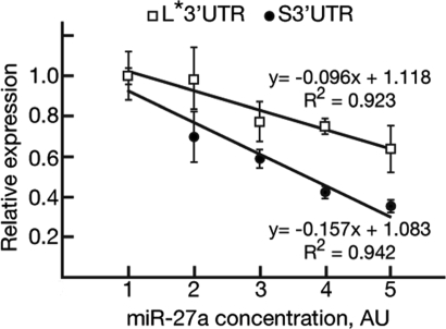
By using a dual luciferase assay, we compared the inhibitory effect of miR-27a on pRL-S3′UTR and pFF-L*3′UTR expression. As indicated earlier, miR-27a has two binding sites, both on the shortest (i.e., 155 bp) 3′UTR, and it can thus target all known Runx1 mRNA isoforms. miR-27a was approximately twofold more effective on pRL-S3′UTR than on pFF-L*3′UTR (Fig. 3), indicating that location and “context” of the miR-27a site within Runx1 3′UTR play a role in its efficacy. miR-27a efficacy could be influenced by differences in accessibility of the binding site imposed by the secondary structure of the 3′UTR (43), a hypothesis supported by “RNAfold” analysis (43) (data not shown). We conclude that, as a result of the location/context effect, miR-27a binding sites are less accessible on L3′UTR compared with the S3′UTR, rendering the mRNAs bearing L3′UTR less sensitive to inhibition by miR-27a.
Fig. 3.
The size of the 3′UTR affects the ability of miR-27a to attenuate Runx1 expression. HEK293 cells were co-transfected with pRL-S3′UTR or pFF-L*3′UTR and increasing amounts of miR-27a or miR-NC (transfection control). Numbers at the x axis [arbitrary units (AU) 1–5] correspond to increasing concentrations of miR-27a as follows: 0 nM, 0.1 nM, 0.3 nM, 1 nM, and 3 nM. The effect of miR-27a on reporter activity was calculated relative to the corresponding miR-NC transfected cells (defined as 1.0). Reporter expression was normalized to internal control luciferase activity. The slope values (−0.096 and −0.157 for L*3′UTR and S3′UTR, respectively) indicate that the S3′UTR-dependent translation is more sensitive than L*3′UTR-dependent translation to inhibition by miR-27a. Of note, a square r of ≈0.93 indicates the close relationships between the values comprising the slopes. Data shown are representative of at least two independent experiments done in triplicate. Error bars indicate SD.
Systematic bias of alternative polyadenylation, regulated by both cis-elements and transacting factors, occurs in several human tissues (44), and a shift toward mRNAs with shortened 3′UTR was observed after activation of primary murine CD4+ T lymphocytes (45). Similarly, the profiles of Runx1 3′UTR during megakaryopoiesis may shift by alternative usage of polyadenylation sites and thereby affect the ability of miR-27a to attenuate Runx1 expression.
Runx1 Regulates miR-27a During Megakaryocytic Differentiation of K562 Cells.
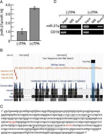
We next addressed the possibility that Runx1 and miR-27a are engaged in a mutual regulatory loop using the K562 leukemia cell line (46), which expresses both Runx1 and miR-27a. K562 cells are a common progenitor of megakaryocytic and erythroid lineage that can be induced to differentiate into erythrocytes or megakaryocytes by treatment with hemin or phorbol 12-o-tetradecanoylphorbol-13-acetate (TPA), respectively (47). Consistent with the observation that forced expression of Runx1 in 416B led to their terminal differentiation into the megakaryocytic lineage (Fig. S1), TPA-mediated differentiation of K562 cells was associated with increased expression of RUNX1 (5, 48). Significantly, a marked induction of miR-27a expression was observed following TPA treatment (Fig. 4A), raising the possibility that RUNX1 directly regulates miR-27a transcription. Compatible with this hypothesis, ChIP using the human acute megakaryocytic leukemia cell line CMK with anti-RUNX1 Ab followed by Solexa sequencing (i.e., ChIP-Seq) revealed a highly conserved putative regulatory region containing two consensus RUNX binding sites ≈10 kb upstream of the miR-27a locus (Fig. 4 B and C). To confirm the biological significance of RUNX1 binding to this region, we next performed ChIP-PCR analysis of TPA-treated and untreated K562 cells. TPA treatment markedly enhanced the binding of RUNX1 to this region (Fig. 4D). The results indicate that, upon induction of megakaryocytic differentiation, RUNX1 binds to a putative miR-27a regulatory region and up-regulates its expression. Collectively, the data presented here support the notion that, during megakaryopoiesis, RUNX1 activity is modulated by miR-27a and that Runx1 and miR-27a are engaged in a mutual regulatory loop.
Fig. 4.
Regulation of miR-27a expression by Runx1 during megakaryocytic differentiation. (A) qRT-PCR analysis of miR-27a levels in K562 cells untreated or treated with TPA (30 nM, 96 h). miR-16 served as an endogenous control. (B) Conserved regions of the ≈0.5 kb genomic element (denoted by the “YourSeq” black rectangle) identified by an anti-Runx1-ChIP-Seq of CMK cells. This putative regulatory element is located on chr19: 13,818,269–13,818,800 of the March 2006 Human Genome Assembly [http://genome.ucsc.edu/ (56)], ≈10 kb upstream of the miR-27a locus. (C) DNA sequence of the ≈0.5 kb genomic element (denoted by the “YourSeq” black rectangle in B). RUNX binding sites (shown in red) are located at nucleotide positions 238 and 361. The proximal Runx site is conserved down to Opossum. (D) ChIP-PCR assay demonstrates TPA-induced binding of Runx1 to miR-27a regulatory element. ChIP was performed on K562 cells incubated with or without TPA (30 nM, 8 h). Primers used for PCR (italicized and underlined by arrows in C) spanned the more proximal RUNX binding site on DNA derived from ChIP before (input) and after IP with the Runx1 antibody (Runx1) or with control non-immune serum (NIS). PCR of CD19 promoter region lacking RUNX binding sites served as an internal negative control.
Discussion
Runx1 is an important cell lineage-specific regulator of hematopoiesis. Its expression is tightly controlled by a combination of transcriptional and post-transcriptional mechanisms, including long range enhancer-mediated transcription and alternative usage of the P1/P2 promoters, which generate multiple mRNA isoforms differing in their 5′- and 3′-UTRs (3, 6, 7).
Runx1 expression in the early myeloid progenitor cell line 416B is largely regulated post-transcriptionally (Fig. 1). Given the diverse repertoire of 3′UTRs and distinct spatial/temporal expression of Runx1 (3), the involvement of miR-mediated post-transcriptional regulation was investigated. Analysis revealed that five known miRs interact with Runx1 short and long 3′UTRs and affect 3′UTR-dependent translation. Of these miRs, miR-27a has two closely clustered binding sites located on the short 3′UTR (Figs. S2 and S3), rendering all Runx1 mRNA isoforms targets to miR-27a-mediated attenuation. Furthermore, the two miR-27a binding sites have features characteristic of high-efficacy miR-binding sites (39) (see Fig. S3). Of note, miR binding sites for the 17–5p-20a-106a cluster regulating RUNX1 expression during monocytopoiesis (34) are confined to Runx1 L3′UTR (Fig. S2), rendering the S3′UTR isoforms refractory to translation inhibition by the 17–5p-20a-106a cluster. Significantly, Runx1 L3′UTR is less sensitive to inhibition by miR-27a than the S3′UTR, conceivably because of differences in accessibility imposed by the secondary structure of the L3′UTR. Thus, differences in the profile of Runx1 mRNA isoforms in various cell lineages and during cell differentiation may affect the ability of miR-27a to attenuate Runx1 expression. We have previously reported that TPA induction of megakaryocytic differentiation in K562 cells was associated with P1/P2 promoter shift and preferential translation of the P2 internal ribosome entry site containing RUNX1 mRNA isoforms (5). Whether these internal ribosome entry sites containing mRNAs are relatively immune to miR-mediated translation attenuation (48), or whether TPA affects the 3′UTRs profile thereby influencing miR-27a-mediated attenuation, are interesting issues to address in the future.
The myeloid progenitor cell line 416B could form both granulocyte- and megakaryocyte- containing spleen colonies upon transplantation into irradiated mice, indicating that these cells maintain a bi-potential granulocyte/megakaryocyte differentiation capability (36). However, 416B cells could not be induced to differentiate in vitro by a variety of known differentiation-inducing agents (36), but forced expression of GATA1, GATA2 (49, 50), or Runx1 (Fig. S1) induced 416B cells to express megakaryocytic markers. This occurrence is in line with the notion that RUNX1 plays a role in megakaryocytic lineage commitment through functional and physical interactions with GATA1 (48). Of potential relation, GATA2 is a potential target of miR-27a (38–41). The common progenitor megakaryocytic/erythroid cell line K562, to the contrary, can be induced to differentiate in vitro into erythrocytes or megakaryocytes by treatment with hemin or TPA, respectively (47). We therefore used K562 cells to demonstrate that, upon induction of megakaryocytic differentiation, expression of miR-27a markedly increased, concomitantly with the binding of Runx1 to conserved RUNX binding sites upstream of miR-27a locus, indicating that, during megakaryocytic differentiation, RUNX1 acts to positively regulate transcription of miR-27a. Furthermore, by using ChIP-Seq, we showed that RUNX1 binds to a putative miR-27a regulatory region. Of note, down-regulation of miR-27a expression occurs upon induction of K562 cells toward erythroid differentiation (51), raising the possibility that Runx1-mediated regulation of miR-27a plays role in the erythroid/megakaryocytic lineage determination.
Taken together, the data indicate a role for miR-27a in attenuating Runx1 expression during megakaryocytic differentiation and support the possibility that, during megakaryopoiesis, Runx1 and miR-27a are engaged in a regulatory feedback loop. Runx1, which in early hematopoiesis is up-regulated by the Gata2/Scl/Lmo2 complex (7), positively regulates miR-27a expression. miR-27a then modulates the steady-state levels of Runx1 during megakaryopoiesis.
Materials and Methods
Cell Culture.
The murine myeloid progenitor cell line 416B (36) was obtained from Marella de Bruijn (Oxford, UK) and maintained in Fischer medium (Gibco), 20% heat-inactivated horse serum (Sigma), 2 mM L-glutamine, and penicillin/streptomycin (Gibco) at 37 °C with 5% CO2. The murine pre-B lymphoblast cell line 70Z was maintained in RPMI medium 1640 (Gibco), 10% FBS (Gibco), 0.05 mM 2-mercaptoethanol, 2 mM L-glutamine (Gibco), and 1% penicillin/streptomycin (Gibco) at 37 °C with 5% CO2. The HEK293 cell line was maintained in DMEM (Gibco) supplemented with 10% FBS (Gibco), 2 mM L-glutamine (Gibco), and penicillin/streptomycin (Gibco) at 37 °C with 5% CO2. CMK and K562 cells were maintained in RPMI medium 1640 (Gibco), 10% FBS (Gibco), 2 mM L-glutamine (Gibco), and 1% penicillin/streptomycin (Gibco) at 37 °C with 5% CO2.
Protein and RNA Analysis.
Cells were collected and washed once in PBS solution, and proteins or RNA were extracted and analyzed by Western or Northern blotting as previously described (52).
qRT-PCR.
Total RNA was obtained using an EZ-RNA Kit (Biological Industries) and reverse transcribed with random hexamers using SuperScript III (Invitrogen). TaqMan quantitative PCR was performed using the LightCycler 480 System (Roche) according to standard procedures. HPRT1 was used as endogenous control. Commercial ready-to-use primers/probe mixes were used (Applied Biosystems).
miR qRT-PCR.
miR-specific RT reactions were performed by using stem-loop RT primers and a TaqMan miRNA reverse transcription kit (Applied Biosystems). Relative quantification of miR-27a was assessed by qPCR using a TaqMan miRNA assay (Applied Biosystems). MiR-16 was used as an endogenous control (Applied Biosystems). Northern blot analysis of miR was performed as described elsewhere (53). Using qRT-PCR data we also derived the endogenous copy number of miR-27a in the hematopoietic cell lines used in this study. 70Z and K562 cell lines contained between 500 and 1,000 copies per cell whereas 416B contained between 1,500 and 3,000 copies per cell. These miR-27a concentrations were obtained through an accurate qRT-PCR determination of the known amount of exogenously transfected miR-27a into 70Z cells, and comparisons of qRT-PCR cycle threshold differences of transfected and non-transfected 70Z cells. The endogenous concentrations of miR-27a in 416B and K562 were then determined and deduced by qRT-PCR cycle threshold in these cell lines.
Transfections and Dual-Reporter Assays.
For dual luciferase assay, 3′UTR segments corresponding to the two Runx1 isoforms (accession nos. D13802 and AK145098; designated S3′UTR and L3′UTR, respectively) were amplified by PCR from genomic DNA and inserted into the XbaI and FseI sites of pGL4 vector (Promega), immediately downstream of the luciferase stop codon. The following primer sets were used to generate specific fragments: 3′UTR-F, 5′-ATGACATCTAGAGCTGAGCGCCATCGCCATCG-3′; S3′UTR-R, 5′-AGTTCAGGCCGGCCAAGGGTATAAAATCTTTCTTTTATTCACAGCATTG-3′; and L3′UTR-R, 5′-GAATATGGCCGGCCACCCCCAATACATGTGTTTTACTGCTATTTC-3′. HEK293 cells were co-transfected in 24-well plates with 0.1 μg of the reporter vector, 0.035 μg of control vector (Promega), and the relevant concentration of miRNA precursor (Ambion) or anti-miR miRNA inhibitor (Ambion) using Lipofectamine 2000 according to the manufacturer protocol (Invitrogen). Firefly and Renilla luciferase activities were measured 24 h after transfection using a dual luciferase assay kit (Promega). To mutate/delete 3–5 bp of miR seed-match sequence, we used site-directed mutagenesis by whole plasmid synthesis with PfuUltra (Stratagene). Mutations were confirmed by plasmid DNA sequencing. To assess miR-27a effect on Runx1 ectopic expression, HEK293 cells were co-transfected in 9-cm plates with 0.5 μg pcRunx1, 1.5 μg pcEGFP-3xNLS and miR-27a, miR-9, or miR-NC (30 nM; Dharmacon). pcRunx1 vector contains CMV promoter-driven Runx1 cDNA bearing the S3′UTR. Cells were harvested 48 h after transfection for protein measurement by Western blot. For transfections of mature miRs into 70Z cells, cells were co-transfected in six-well plates with the appropriate miR (150 nM; Dharmacon) and Cy3-Anti-miR-NC (15 nM; Ambion). Transfection was monitored by qRT-PCR as indicated earlier and by Cy3 fluorescence.
ChIP.
ChIP assays were performed essentially as described (54). Chromatin from ≈107 cells was fragmented to an average size of 500 bp using a Microson XL ultrasonic cell disruptor (Misonix). For immunoprecipitation, 4 μl of anti-RUNX1 antibody (52) was added to 1.5 mL of soluble chromatin. Rabbit pre-immune serum was used as a control. ChIP-Seq was performed according to Solexa protocols. For ChIP-PCR analysis, an aliquot of precipitated DNA was analyzed by PCR using primers specific for the human miR-23a-27a-24–2 upstream promoter region identified by ChIP-Seq analysis (forward primer, 5′- CCCAGCCGGCCCACAGGTCA; reverse primer, 5′-GATCCCGGCTCAGTCCATAACACA). Primers for human CD19 promoter region (forward primer, 5′-AGAAGGAGTCTATGTGCCCA; reverse primer, 5′-CCTCCAGACTCACAACACTT) served as an internal negative control.
Supplementary Material
Acknowledgments.
We thank Dr. Daniela Amann-Zalcenstein, Dr. Edna Ben-Asher, Dr. Shirley Horn-Saban, and Dr. Hershel Safer for help with Solexa sequencing data acquisition and analysis; and Dr. Marella de Bruijn for the 416B cells and the information about Runx1 expression in these cells. This work was supported by the European Union AnEUploidy project, Israel Science Foundation, and Shapell Family Biomedical Research Foundation of the Weizmann Institute of Science.
Footnotes
The authors declare no conflict of interest.
This article contains supporting information online at www.pnas.org/cgi/content/full/0811466106/DCSupplemental.
References
- 1.Cameron ER, Neil JC. The Runx genes: lineage-specific oncogenes and tumor suppressors. Oncogene. 2004;23:4308–4314. doi: 10.1038/sj.onc.1207130. [DOI] [PubMed] [Google Scholar]
- 2.De Brujin MF, Speck NA. Core-binding factors in hematopoiesis and immune function. Oncogene. 2004;23:4238–4248. doi: 10.1038/sj.onc.1207763. [DOI] [PubMed] [Google Scholar]
- 3.Levanon D, Groner Y. Structure and regulated expression of mammalian RUNX genes. Oncogene. 2004;23:4211–4219. doi: 10.1038/sj.onc.1207670. [DOI] [PubMed] [Google Scholar]
- 4.Ghozi MC, Bernstein Y, Negreanu V, Levanon D, Groner Y. Expression of the human acute myeloid leukemia gene AML1 is regulated by two promoter regions. Proc Natl Acad Sci USA. 1996;93:1935–1940. doi: 10.1073/pnas.93.5.1935. [DOI] [PMC free article] [PubMed] [Google Scholar]
- 5.Pozner A, et al. Transcription-coupled translation control of AML1/RUNX1 is mediated by cap- and internal ribosome entry site dependent mechanisms. Mol Cell Biol. 2000;20:2297–2307. doi: 10.1128/mcb.20.7.2297-2307.2000. [DOI] [PMC free article] [PubMed] [Google Scholar]
- 6.Pozner A, et al. Developmentally regulated promoter-switch transcriptionally controls Runx1 function during embryonic hematopoiesis. BMC Dev Biol. 2007;7:84. doi: 10.1186/1471-213X-7-84. [DOI] [PMC free article] [PubMed] [Google Scholar]
- 7.Nottingham WT, et al. Runx1-mediated hematopoietic stem-cell emergence is controlled by a Gata/Ets/SCL-regulated enhancer. Blood. 2007;110:4188–4197. doi: 10.1182/blood-2007-07-100883. [DOI] [PMC free article] [PubMed] [Google Scholar]
- 8.Miyoshi H, et al. Alternative splicing and genomic structure of the AML1 gene involved in acute myeloid leukemia. Nucleic Acids Res. 1995;23:2762–2769. doi: 10.1093/nar/23.14.2762. [DOI] [PMC free article] [PubMed] [Google Scholar]
- 9.Levanon D, et al. A large variety of alternatively spliced and differentially expressed mRNAs are encoded by the human acute myeloid leukemia gene AML1. DNA Cell Biol. 1996;15:175–185. doi: 10.1089/dna.1996.15.175. [DOI] [PubMed] [Google Scholar]
- 10.Zhang YW, et al. A novel transcript encoding an N-terminally truncated AML1/PEBP2aB protein interferes with transactivation and blocks granulocytic differentiation of 32Dc13 myeloid cell. EMBO J. 1997;14:341–350. doi: 10.1128/mcb.17.7.4133. [DOI] [PMC free article] [PubMed] [Google Scholar]
- 11.Levanon D, et al. Architecture and anatomy of the genomic locus encoding the human leukemia-associated transcription factor RUNX1/AML1. Gene. 2001;262:23–33. doi: 10.1016/s0378-1119(00)00532-1. [DOI] [PubMed] [Google Scholar]
- 12.Bartel DP, Chen CZ. Micromanagers of gene expression: the potentially widespread influence of metazoan microRNAs. Nat Rev Genet. 2004;5:396–400. doi: 10.1038/nrg1328. [DOI] [PubMed] [Google Scholar]
- 13.Ambros V. The functions of animal microRNAs. Nature. 2004;431:350–355. doi: 10.1038/nature02871. [DOI] [PubMed] [Google Scholar]
- 14.He L, Hannon GJ. MicroRNAs: small RNAs with a big role in gene regulation. Nat Rev Genet. 2004;5:522–531. doi: 10.1038/nrg1379. [DOI] [PubMed] [Google Scholar]
- 15.Bartel DP. MicroRNAs: genomics, biogenesis, mechanism, and function. Cell. 2004;116:281–297. doi: 10.1016/s0092-8674(04)00045-5. [DOI] [PubMed] [Google Scholar]
- 16.Valencia-Sanchez MA, Liu J, Hannon GJ, Parker R. Control of translation and mRNA degradation by miRNAs and siRNAs. Genes Dev. 2006;20:515–524. doi: 10.1101/gad.1399806. [DOI] [PubMed] [Google Scholar]
- 17.Naguibneva I, et al. The microRNA miR-181 targets the homeobox protein Hox-A11 during mammalian myoblast differentiation. Nat Cell Biol. 2006;8:278–284. doi: 10.1038/ncb1373. [DOI] [PubMed] [Google Scholar]
- 18.Chen JF, et al. The role of microRNA-1 and microRNA-133 in skeletal muscle proliferation and differentiation. Nat Genet. 2006;38:228–233. doi: 10.1038/ng1725. [DOI] [PMC free article] [PubMed] [Google Scholar]
- 19.Zhao Y, Samal E, Srivastava D. Serum response factor regulates a muscle-specific microRNA that targets Hand2 during cardiogenesis. Nature. 2005;436:214–220. doi: 10.1038/nature03817. [DOI] [PubMed] [Google Scholar]
- 20.Baek D, et al. The impact of microRNAs on protein output. Nature. 2008;455:64–71. doi: 10.1038/nature07242. [DOI] [PMC free article] [PubMed] [Google Scholar]
- 21.North T, et al. Cbfa2 is required for the formation of intra-aortic hematopoietic clusters. Development. 1999;126:2563–2575. doi: 10.1242/dev.126.11.2563. [DOI] [PubMed] [Google Scholar]
- 22.North TE, et al. Runx1 expression marks long-term repopulating hematopoietic stem cells in the midgestation mouse embryo. Immunity. 2002;16:661–672. doi: 10.1016/s1074-7613(02)00296-0. [DOI] [PubMed] [Google Scholar]
- 23.Cai Z, et al. Haploinsufficiency of AML1 affects the temporal and spatial generation of hematopoietic stem cells in the mouse embryo. Immunity. 2000;13:423–431. doi: 10.1016/s1074-7613(00)00042-x. [DOI] [PubMed] [Google Scholar]
- 24.Wang O, et al. Disruption of the Cbfa2 gene causes necrosis and hemorrhaging in the central nervous system and blocks definitive hematopoiesis. Proc Natl Acad Sci USA. 1996;93:3444–3449. doi: 10.1073/pnas.93.8.3444. [DOI] [PMC free article] [PubMed] [Google Scholar]
- 25.Okuda T, van Deursen J, Hiebert SW, Grosveld G, Downing JR. AML1, the target of multiple chromosomal translocations in human leukemia, is essential for normal fetal liver hematopoiesis. Cell. 1996;84:321–330. doi: 10.1016/s0092-8674(00)80986-1. [DOI] [PubMed] [Google Scholar]
- 26.North TE, Stacy T, Matheny CJ, Speck NA, de-Bruijn MF. Runx1 is expressed in adult hematopoiesis stem cells and differentiating myeloid and lymphoid cells, but not in maturing erythroid cells. Stem Cells. 2004;22:158–168. doi: 10.1634/stemcells.22-2-158. [DOI] [PubMed] [Google Scholar]
- 27.Lorsbach RB, et al. Role of RUNX1 in adult hematopoiesis: analysis of RUNX1-IRES-GFP knock-in mice reveals differential lineage expression. Blood. 2004;103:2522–2529. doi: 10.1182/blood-2003-07-2439. [DOI] [PubMed] [Google Scholar]
- 28.Growney JD, et al. Loss of Runx1 perturbs adult hematopoiesis and is associated with a myeloproliferative phenotype. Blood. 2005;106:494–504. doi: 10.1182/blood-2004-08-3280. [DOI] [PMC free article] [PubMed] [Google Scholar]
- 29.Ichikawa M, et al. AML-1 is required for megakaryocytic maturation and lymphocytic differentiation, but not for maintenance of hematopoietic stem cells in adult hematopoiesis. Nat Med. 2004;10:299–304. doi: 10.1038/nm997. [DOI] [PubMed] [Google Scholar]
- 30.Putz G, Rosner A, Nuesslein I, Schmitz N, Buchholz F. AML1 deletion in adult mice causes splenomegaly and lymphomas. Oncogene. 2006;25:929–939. doi: 10.1038/sj.onc.1209136. [DOI] [PubMed] [Google Scholar]
- 31.Izraeli S, Rainis L, Hertzberg L, Smooha G, Birger Y. Trisomy of chromosome 21 in leukemogenesis. Blood Cells Mol Dis. 2007;39:156–159. doi: 10.1016/j.bcmd.2007.04.004. [DOI] [PubMed] [Google Scholar]
- 32.Zipursky A, Peeters M, Poon A. Megakaryoblastic leukemia and Down's syndrome: a review. Pediatr Hematol Oncol. 1987;4:211–230. doi: 10.3109/08880018709141272. [DOI] [PubMed] [Google Scholar]
- 33.Georgantas RW, 3rd, et al. CD34+ hematopoietic stem-progenitor cell microRNA expression and function: a circuit diagram of differentiation control. Proc Natl Acad Sci USA. 2007;104:2750–2755. doi: 10.1073/pnas.0610983104. [DOI] [PMC free article] [PubMed] [Google Scholar]
- 34.Fontana L, et al. MicroRNAs 17–5p-20a–106a control monocytopoiesis through AML1 targeting and M-CSF receptor upregulation. Nat Cell Biol. 2007;9:775–787. doi: 10.1038/ncb1613. [DOI] [PubMed] [Google Scholar]
- 35.Landry JR, et al. Runx genes are direct targets of Scl/Tal1 in the yolk sac and fetal liver. Blood. 2008;111:3005–3014. doi: 10.1182/blood-2007-07-098830. [DOI] [PubMed] [Google Scholar]
- 36.Dexter TM, Allen TD, Scott D, Teich NM. Isolation and characterisation of a bipotential haematopoietic cell line. Nature. 1979;277:471–474. doi: 10.1038/277471a0. [DOI] [PubMed] [Google Scholar]
- 37.Paige CJ, Kincade PW, Ralph P. Murine B cell leukemia cell line with inducible surface immunoglobulin expression. J Immunol. 1978;121:641–647. [PubMed] [Google Scholar]
- 38.Lewis BP, Burge CB, Bartel DP. Conserved seed pairing, often flanked by adenosines, indicates that thousands of human genes are microRNA targets. Cell. 2005;120:15–20. doi: 10.1016/j.cell.2004.12.035. [DOI] [PubMed] [Google Scholar]
- 39.Grimson A, et al. MicroRNA targeting specificity in mammals: determinants beyond seed pairing. Mol Cell. 2007;27:91–105. doi: 10.1016/j.molcel.2007.06.017. [DOI] [PMC free article] [PubMed] [Google Scholar]
- 40.Lewis BP, Shih IH, Jones-Rhoades MW, Bartel DP, Burge CB. Prediction of mammalian microRNA targets. Cell. 2003;115:787–798. doi: 10.1016/s0092-8674(03)01018-3. [DOI] [PubMed] [Google Scholar]
- 41.Krek A, et al. Combinatorial microRNA target predictions. Nat Genet. 2005;37:495–500. doi: 10.1038/ng1536. [DOI] [PubMed] [Google Scholar]
- 42.Majoros WH, Ohler U. Spatial preferences of microRNA targets in 3′ untranslated regions. BMC Genomics. 2007;8:152. doi: 10.1186/1471-2164-8-152. [DOI] [PMC free article] [PubMed] [Google Scholar]
- 43.Hofacker IL, Stadler PF. Memory efficient folding algorithms for circular RNA secondary structures. Bioinformatics. 2006;22:1172–1176. doi: 10.1093/bioinformatics/btl023. [DOI] [PubMed] [Google Scholar]
- 44.Zhang H, Lee JY, Tian B. Biased alternative polyadenylation in human tissues. Genome Biol. 2005;6:R100. doi: 10.1186/gb-2005-6-12-r100. [DOI] [PMC free article] [PubMed] [Google Scholar]
- 45.Sandberg R, Neilson JR, Sarma A, Sharp PA, Burge CB. Proliferating cells express mRNAs with shortened 3′ untranslated regions and fewer microRNA target sites. Science. 2008;320:1643–1647. doi: 10.1126/science.1155390. [DOI] [PMC free article] [PubMed] [Google Scholar]
- 46.Lozzio CB, Lozzio BB. Human chronic myelogenous leukemia cell-line with positive Philadelphia chromosome. Blood. 1975;45:321–334. [PubMed] [Google Scholar]
- 47.Huo XF, et al. Differential expression changes in K562 cells during the hemin-induced erythroid differentiation and the phorbol myristate acetate (PMA)-induced megakaryocytic differentiation. Mol Cell Biochem. 2006;292:155–167. doi: 10.1007/s11010-006-9229-0. [DOI] [PubMed] [Google Scholar]
- 48.Elagib KE, et al. RUNX1 and GATA-1 coexpression and cooperation in megakaryocytic differentiation. Blood. 2003;101:4333–4341. doi: 10.1182/blood-2002-09-2708. [DOI] [PubMed] [Google Scholar]
- 49.Visvader JE, Elefanty AG, Strasser A, Adams JM. GATA-I but not SCL induces megakaryocytic differentiation in an early myeloid line. EMBO J. 1992;11:4557–4564. doi: 10.1002/j.1460-2075.1992.tb05557.x. [DOI] [PMC free article] [PubMed] [Google Scholar]
- 50.Visvader J, Adams JM. Megakaryocytic differentiation induced in 416B myeloid cells by GATA-2 and GATA-3 transgenes or 5-azacytidine is tightly coupled to GATA-1 expression. Blood. 1993;82:1493–1501. [PubMed] [Google Scholar]
- 51.Choong ML, Yang HH, McNiece I. MicroRNA expression profiling during human cord blood-derived CD34 cell erythropoiesis. Exp Hematol. 2007;35:551–564. doi: 10.1016/j.exphem.2006.12.002. [DOI] [PubMed] [Google Scholar]
- 52.Ben Aziz-Aloya R, et al. Expression of AML1-d, a short human AML1 isoform, in embryonic stem cells suppresses in vivo tumor growth and differentiation. Cell Death Differ. 1998;5:765–773. doi: 10.1038/sj.cdd.4400415. [DOI] [PubMed] [Google Scholar]
- 53.Lau NC, Lim LP, Weinstein EG, Bartel DP. An abundant class of tiny RNAs with probable regulatory roles in Caenorhabditis elegans. Science. 2001;294:858–862. doi: 10.1126/science.1065062. [DOI] [PubMed] [Google Scholar]
- 54.Ainbinder E, et al. Mechanism of rapid transcriptional induction of tumor necrosis factor alpha-responsive genes by NF-kappaB. Mol Cell Biol. 2002;22:6354–6362. doi: 10.1128/MCB.22.18.6354-6362.2002. [DOI] [PMC free article] [PubMed] [Google Scholar]
- 55.Abramoff MD, Magelhaes PJ, Ram SJ. Image processing with ImageJ. Biophotonics Int. 2004;11:36–42. [Google Scholar]
- 56.Kent W, et al. The human genome browser at UCSC. Genome Res. 2002;12:996–1006. doi: 10.1101/gr.229102. [DOI] [PMC free article] [PubMed] [Google Scholar]
- 57.Zajicek J. Studies on the histogenesis of blood platelets. Acta Haematol. 1954;12:238–244. doi: 10.1159/000204625. [DOI] [PubMed] [Google Scholar]
- 58.Krüger J, Rehmsmeier M. RNAhybrid: microRNA target prediction easy, fast and flexible. Nucleic Acids Res. 2006;34:W451–W454. doi: 10.1093/nar/gkl243. [DOI] [PMC free article] [PubMed] [Google Scholar]
Associated Data
This section collects any data citations, data availability statements, or supplementary materials included in this article.


