Abstract
The relative fitness of arthropod-borne pathogens within the vector can be a major determinant of pathogen prevalence within the mammalian host population. Strains of the tick-borne rickettsia Anaplasma marginale differ markedly in transmission efficiency, with a consequent impact on pathogen strain structure. We have identified two A. marginale strains with significant differences in the transmission phenotype that is effected following infection of the salivary gland. We have proposed competing hypotheses to explain the phenotypes: (i) both strains are secreted equally, but there is an intrinsic difference in infectivity for the mammalian host, or (ii) one strain is secreted at a significantly higher level and thus represents delivery of a greater pathogen dose. Quantitative analysis of pathogen replication and secretion revealed that the high-efficiency St. Maries strain replicated to a 10-fold-higher titer and that a significantly greater percentage of infected ticks secreted A. marginale into the saliva and did so at a significantly higher level than for the low-efficiency Israel vaccine strain. Furthermore, the transmission phenotype of the vaccine strain could be restored to that of the St. Maries strain simply by increasing the delivered pathogen dose, either by direct inoculation of salivary gland organisms or by increasing the number of ticks during transmission feeding. We identified morphological differences in the colonization of each strain within the salivary glands and propose that these reflect strain-specific differences in replication and secretion pathways linked to the vector-pathogen interaction.
The predominance of a specific pathogen strain in the host population reflects its overall fitness advantage and is a major determinant of the consequent disease pattern (7, 17). We have investigated the strain structure of the tick-borne bacterium Anaplasma marginale in its natural reservoir hosts, domestic and wild ruminants, and identified a predominance of specific strains within spatially and temporally defined reservoir host populations (4, 12, 17, 19). We hypothesize that strain predominance is determined by the strain-specific transmission fitness within the tick vector. This overall hypothesis is supported by identification of genetically distinct A. marginale strains with marked differences in transmissibility (24). However, the basis for these strain-specific differences in transmissibility is poorly understood.
During tick acquisition feeding on an infected reservoir host, A. marginale enters the midgut epithelium and undergoes initial replication before transiting to tick salivary glands and invading the epithelial cells (6, 24). Within the salivary gland epithelial cells, A. marginale undergoes a second round of replication, and it is secreted into the saliva concomitant with tick transmission feeding on a new mammalian host (6, 12, 24). Accordingly, both the midgut and salivary gland have been identified as sites where transmission differences among A. marginale strains are manifested (3, 21, 24). At the level of the midgut, it is unclear whether specific strains differ in their ability to enter the midgut epithelial cells or whether the difference is in subsequent replication. In contrast, within the salivary gland epithelium, there is a specific transmission phenotype that occurs postinvasion (12, 24). Highly efficiently transmitted strains replicate to 106 to 107 organisms per salivary glands and, as shown using the St. Maries strain of A. marginale, can be consistently transmitted to naïve animals by feeding fewer than 10 infected Dermacentor andersoni ticks (5, 18, 20). Interestingly, the Israel vaccine strain (A. marginale subsp. centrale) also invades the salivary glands but is not transmitted using cohorts of 100 ticks (24).
We propose two alternative hypotheses to explain the different transmission phenotypes represented by the A. marginale St. Maries and the Israel vaccine strains. The first is that there is a decrease in replication of the vaccine strain within the tick vector and/or a reduced secretion into the saliva and thus insufficient organisms to exceed the minimal infective dose for transmission. If this is correct, then the infection threshold could be reached by simply increasing the number of transmission-feeding ticks to achieve the same level of organisms secreted by the highly efficient St. Maries strain of A. marginale. Alternatively, the second hypothesis is that there is an intrinsic decreased infectivity of the vaccine strain during a passage within the tick vector, and thus reaching the threshold would require secretion of a significantly greater number of organisms of the vaccine strain than of the St. Maries strain. Here we describe the testing of these hypotheses and present the results in context of vector-pathogen interactions that affect the pathogen strain structure in the mammalian reservoir host population.
MATERIALS AND METHODS
Infection of ticks and pathogen replication within tick salivary glands.
The specific-pathogen-free Reynolds Creek colony of Dermacentor andersoni and the St. Maries and the Israel vaccine strains used in these experiments have been described in detail previously (6, 21, 24). Adult male D. andersoni ticks were allowed to acquisition feed for 7 days on calves infected with either the St. Maries or the Israel vaccine strain. Following an additional 7 days of incubation at 26°C to allow complete digestion of the blood meal and eliminate any possibility of mechanical transmission, ticks were then transmission fed on naïve (competitive enzyme-linked immunosorbent assay-seronegative and msp5 PCR-negative) age- and gender-matched Holstein calves (12, 24). Cohorts of both acquisition-fed and transmission-fed ticks were dissected and midgut and salivary glands isolated from individual ticks for determination of infection rate (percentage of fed ticks that acquired infection) and infection level (bacterial numbers in each tissue). The infection rate was determined by msp5 PCR amplification, and organisms were quantified to determine infection level using real-time PCR as previously described in detail for both strains (6, 24).
Pathogen localization within salivary glands.
The presence of each strain in the granular acinar cells of the salivary glands was examined using immunohistochemistry, followed by subcellular localization using transmission electron microscopy. For immunohistochemistry, transmission-fed ticks were fixed in 10% formalin and embedded in paraffin, and sequential 4-μm sections were deparaffinized in Clear-Rite and then hydrated in an ethanol gradient. Sections were treated with citrate solution (pH 6) (Zymed, Carlsbad, CA) for antigen retrieval and steamed for 20 min as previously described (6, 21, 23). The sections were stained using 2 μg/ml of monoclonal antibodies specific for each strain. Antibody ANAR49 binds the St. Maries strain, and its use in immunohistochemical detection of this strain has been previously reported (21). To develop a monoclonal antibody reactive with the Israel vaccine strain, mice were immunized with organisms isolated from infected erythrocytes and hybridomas generated using standard procedures as previously described for A. marginale (14, 16). Hybridoma supernatants were screened for reactivity with the vaccine strain by immunoblotting and by immunohistochemistry. Monoclonal antibody 132/704.13 was identified as reactive with the vaccine strain in both assays, and supernatants from twice-cloned hybridomas were used for immunohistochemistry on infected ticks (21). Following the primary antibodies, ANAR49 for the St. Maries strain and 132/704.13 for the vaccine strain, horseradish peroxidase-labeled anti-mouse immunoglobulin (Dako Corp., Carpinteria, CA) and 3-amino-9-ethylcarbazole containing hydrogen peroxide were used to detect binding. Sections were counterstained with Mayer's hematoxylin (6, 21). Monoclonal antibodies against Trypanosoma brucei (2 μg/ml) were used as a negative control on sequential sections of the same ticks exposed either to the St. Maries strain or the Israel vaccine strain.
For subcellular localization of bacteria, salivary glands were dissected and fixed in 2% paraformaldehyde and 2% glutaraldehyde in 0.1 M cacodylate buffer at 4°C (10, 11). Tissues were rinsed in 0.1 M cacodylate buffer, postfixed in 2% OsO4 for 2 h at room temperature, and then rinsed in cacodylate buffer. Following dehydration in an ethanol gradient, samples were infiltrated with acetone and embedded in Spurr's resin. Thin sections (90 nm) were placed on nickel grids and stained in 4% uranyl acetate for 10 min and in Reynolds lead for 3 min. Sections were examined on a JEOL JEM 1200 EX transmission electron microscope.
Pathogen secretion.
To quantify the salivary secretion of A. marginale, salivation was induced in a cohort of infected, transmission-fed ticks. Briefly, approximately 10 μl of dopamine hydrochloride (100 mg/ml in a 1.2% saline solution) was inoculated into the membrane surrounding the base of the coxa of the fourth leg of individual ticks using a 12.7- by 0.21-mm needle (8, 22). Saliva was collected directly from the mouth parts during a 20-min period and the total collected volume immediately placed into 50 μl cell lysis solution (Qiagen Inc., Valencia, CA) with proteinase K (2 μg/ml) and incubated at 56°C overnight. Following incubation, dilution with 450 μl of cell lysis solution with glycogen (70 μg/ml), and removal of proteins, genomic DNA was precipitated in 100% isopropanol, washed in 70% ethanol, and resuspended in 30 μl of hydration solution (Qiagen Inc., Valencia, CA). A. marginale-positive saliva samples were identified by using msp5 PCR and the bacteria quantified in positive samples using real-time PCR (24).
Strain-specific quantitative transmission.
Based on the observed levels of replication and salivary secretion for each strain, two approaches were used to determine if these quantitative differences between strains accounted for the phenotypic differences in transmissibility. For the first, salivary glands were dissected from transmission-fed ticks, and homogenates were prepared in RPMI 1640 medium and inoculated intravenously into splenectomized, naïve calves (9). The number of salivary glands used was determined by the differences in organism levels in the salivary glands between the two strains as quantified by real-time PCR. For the second approach, the number of transmission-feeding ticks infected with the vaccine strain was increased, based on the tick infection rate and levels in saliva, to approximate a similar delivery inoculum represented by feeding 10 ticks infected with the St. Maries strain. The ability of ≤10 Reynolds Creek colony D. andersoni adult males to transmit the St. Maries strain has been replicated and reported previously (5, 18, 20, 24). Following either salivary gland homogenate inoculation or tick feeding, calves were monitored by microscopic examination of Giemsa-stained blood smears and infection was confirmed by msp5 PCR (24). The strain identity was confirmed by msp5 amplicon sequencing and alignment with the strain-specific sequences previously reported for these two strains (1, 15).
RESULTS
Infection of ticks and pathogen replication within tick salivary glands.
Ticks were acquisition fed on calves infected with either the St. Maries or the Israel vaccine strain. During the 7-day acquisition-feeding period, the mean bacteremia levels were 108 organisms per ml of blood for each strain, as determined by msp5 real-time PCR (data not shown). The tick infection rate for the St. Maries strain following acquisition feeding was 100% (Table 1), with mean levels of 106.4 ± 0.45 per midgut and 105.3 ± 0.76 per salivary gland pair. During transmission feeding, further replication within the salivary glands increased the levels of the St. Maries strain 100 times (Table 1). The tick infection rate for the Israel vaccine strain was similar at approximately 90%; however, the levels in both midgut and salivary glands were decreased 10-fold compared to those of the St. Maries strain (Table 1). Similar to the case for the St. Maries strain, the levels of the vaccine strain increased 100 times during transmission feeding but remained at reduced levels compared to those of the St. Maries strain (Table 1).
TABLE 1.
Anaplasma strain-specific infection rates and levels in Dermacentor andersoni
| Strain and feeding | Midgut
|
Salivary glands
|
||
|---|---|---|---|---|
| Infection ratea (no. positive/no. tested) | Infection levelb (mean ± SD) | Infection rate (no. positive/no. tested) | Infection level (mean ± SD) | |
| St. Maries | ||||
| Acquisitionc | 100 (25/25) | 106.4 ± 0.45 | 100 (25/25) | 105.3 ± 0.76 |
| Transmissiond | 100 (25/25) | 106.7 ± 0.61 | 100 (25/25) | 107.4 ± 0.68 |
| Israel vaccine | ||||
| Acquisitionc | 92 (23/25) | 105.3 ± 0.59 | 88 (22/25) | 104.5 ± 0.77 |
| Transmissiond | 92 (23/25) | 105.2 ± 0.87 | 88 (22/25) | 106.1 ± 0.89 |
Percentage of fed ticks that acquired infection.
Number of bacteria per midgut or salivary gland pair.
Ticks were acquisition fed on calves infected with each specific strain.
Ticks were transmission fed on naïve (competitive enzyme-linked immunosorbent assay-seronegative, msp5 PCR-negative) calves.
Pathogen localization within salivary glands.
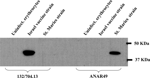
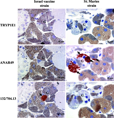
The specificity of the monoclonal antibodies was confirmed by immunoblotting using both strains (Fig. 1). These antibodies were then used to localize bacteria within the salivary glands of transmission-fed ticks using immunohistochemistry. Both strains colonized the granular acini of the salivary gland (Fig. 2). However, the St. Maries strain consistently developed multiple colonies within the acini, while the vaccine strain formed only large single colonies (Fig. 2). There was no reactivity of either strain with the negative control monoclonal antibody against Trypanosoma brucei (Tryp1E1) on sequential sections of the same infected ticks (Fig. 2).
FIG. 1.
Reactivity of strain-specific monoclonal antibodies. 132/704.13, antibody reactive with the Israel vaccine strain; ANAR49, antibody reactive with the St. Maries strain. Uninfect. erythrocytes, uninfected bovine erythrocytes as negative control. The positions of the molecular size markers are indicated on the right.
FIG. 2.
Localization of Anaplasma colonies (arrows) within the granular acinar cells (G) of Dermacentor andersoni salivary glands. TRYP1E1, isotype matched control monoclonal antibody reactive with Trypanosoma brucei; ANAR49, antibody binding the St. Maries strain; 132/704.13, antibody binding the Israel vaccine strain.
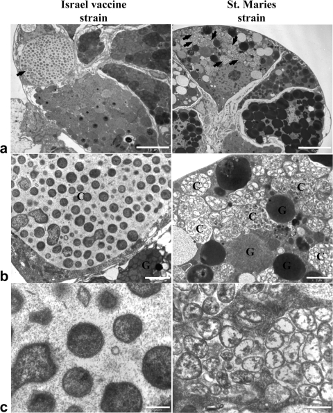
This same pattern of infection within granular acinar cells was confirmed by transmission electron microscopy. The Israel vaccine strain formed large single colonies with relatively low numbers of electron-dense organisms, characteristic of the infectious state, within the colonies, while the St. Maries strain developed multiple colonies composed of closely packed electron-lucent organisms, characteristic of the replicative state (11), and were consistently located adjacent to acinar granules (Fig. 3). Salivary glands from ticks of the same colony fed identically but on uninfected animals did not, as expected, contain bacterial colonies (data not shown).
FIG. 3.
Transmission electron micrographs of Anaplasma colonies within tick salivary glands. (a) Granular acinar cells of salivary glands containing Anaplasma colonies. Bar, 10 μm; magnification, ×2,000. Arrows indicate the colonies. (b) Single vaccine strain colony and multiple St. Maries strain colonies within the salivary glands. Bar, 2 μm; magnification, ×10,000. C, Anaplasma colonies; G, granules of the acinar cells. (c) Individual organisms within Anaplasma colonies. Bar, 0.5 μm; magnification, ×40,000.
Pathogen secretion.
Saliva from transmission-fed ticks infected with the St. Maries strain contained a mean level of 104.1 A. marginale organisms per μl, and >90% of the individual ticks were positive (Table 2). In contrast, only 30% of the individual ticks were saliva positive for the vaccine strain, and the levels were 10-fold lower than those of the St. Maries strain (Table 2).
TABLE 2.
Quantification of Anaplasma in saliva of transmission-fed Dermacentor andersonia
| Strain | Salivary glands
|
Saliva
|
||
|---|---|---|---|---|
| Infection rateb (no. positive/no. tested) | Infection levelc (mean ± SD) | Infection rated (no. positive/no. tested) | Infection levele (mean ± SD) | |
| St. Maries | 100 (25/25) | 107.5 ± 0.62 | 96 (24/25) | 104.1 ± 0.86 |
| Israel vaccine | 92 (46/50) | 106.5 ± 0.69 | 30 (15/50) | 103.1 ± 0.49 |
The salivary glands and saliva represent the same cohort of ticks.
Percentage of fed ticks that were infected.
Number of bacteria per salivary gland pair.
Percentage of fed ticks that secreted bacteria in the saliva.
Number of bacteria per μl of saliva.
Strain-specific quantitative transmission.
Calves (n = 2) were inoculated with salivary gland homogenates prepared from 10 ticks infected with the St. Maries strain. This inoculum contained 108.4 organisms of the St. Maries strain of A. marginale. Both calves were infected and progressed to develop acute high-level bacteremia (≥108 Anaplasma organisms/ml of blood) as determined by microscopic examination of Giemsa-stained blood smears. The identity of the St. Maries strain was confirmed by amplification of msp5 (Fig. 4) followed by sequencing the amplicon to identify the strain-specific sequence. Calves were identically inoculated with salivary gland homogenates of ticks infected with the Israel vaccine strain using two different inoculum sizes: inoculation with homogenates of 15 ticks representing 107.2 bacteria (n = 2 calves) and inoculation with homogenates of 150 ticks representing 108.2 bacteria (n = 2 calves). Both sets of calves became infected and progressed to develop acute high-level bacteremia (≥108 Anaplasma organisms/ml of blood). The identity of the vaccine strain was confirmed by amplification of msp5 (Fig. 4) followed by sequencing the amplicon to identify the strain-specific sequence. Having demonstrated that the vaccine strain organisms in the salivary gland were infectious, we tested whether increasing the number of ticks infected with the vaccine strain to approximate the saliva levels represented by 10 St. Maries strain-infected ticks would result in vaccine strain transmission. As both the percentage of ticks with organisms in saliva and the number of organisms per μl of saliva were decreased for the vaccine strain compared to the St. Maries strain, a >35-fold increase in the number of vaccine strain-infected ticks was predicted to be sufficient for successful transmission. A total of 425 adult male D. andersoni ticks were transmission fed for 7 days on a naïve calf. Bacteremia was detected microscopically, and the identity of the vaccine strain was confirmed by sequencing the msp5 PCR amplicon (data not shown). As a positive control, the St. Maries strain was transmitted to a separate naïve calf at the same time.
FIG. 4.
Transmission by inoculation of salivary gland Anaplasma homogenates. Calves were inoculated with homogenates containing 108.4 St. Maries strain organisms (C31861 and C32003), 107.2 vaccine strain organisms (C1201 and C1205), or 108.2 vaccine strain organisms (C1210 and C1213). PCR amplification of msp5 from preinoculation blood (lane 1) or during acute bacteremia (lane 2) is shown. The positions of the molecular size markers are indicated on the right.
DISCUSSION
The St. Maries strain is a prototypically high-transmission-efficiency strain with consistent transmission in multiple replicate trials to naïve calves using 10 infected adult male D. andersoni ticks (5, 24). Consistent with this phenotype, trials using one and three infected ticks have also resulted in transmission of the St. Maries strain (18, 20). In contrast, replicate trials using 100 adult male D. andersoni ticks, which were acquisition fed during either the acute (24) or persistent (M. F. B. M. Galletti, unpublished data) phase of infection, have failed to transmit the Israel vaccine strain. The present study indicates that the low-transmission-efficiency phenotype of the Israel vaccine strain in D. andersoni largely reflects delivery of a diminished dose during tick feeding compared to the St. Maries strain. This diminished dose reflects a significantly lower level within the tick vector, fewer ticks secreting organisms in the saliva, and a lower number of organisms secreted into the saliva. Although we have shown that both strains are infectious when directly inoculated into susceptible animals, in the absence of 50% infectious dose determination for both strains, we cannot conclude that the two strains have equal intrinsic infectivity. Thus, diminished intrinsic infectivity may also contribute to the low-efficiency transmission phenotype of the vaccine strain. Nonetheless, the primary determinant of transmission efficiency appears to be the pathogen dose in the saliva.
A quantitative basis for transmission efficiency phenotypes has consequences for our understanding of transmission both epidemiologically and mechanistically at the level of the pathogen-vector interaction. A. marginale strains have previously been reported as “non-tick transmissible,” consistently raising the question as to how these strains were propagated in the field, given the very low efficiency of mechanical transmission (3, 24). A quantitative basis for transmission efficiency phenotype rather than a binary function (a strain is or is not tick transmissible) provides an explanation of how low-efficiency strains can be transmitted but only under conditions of very high tick burden. The D. andersoni tick burden on cattle under natural conditions is normally low and thus would favor strains with high transmission efficiencies (26). This is supported by the predominance of the EMΦ strain, a strain consistently transmitted using ≤10 ticks, within a host reservoir population under conditions of natural transmission (5). However, tick burden can increase dramatically based on shifts in climate and land use, resulting in episodic high tick burdens that could allow transmission of low-efficiency strains.
Mechanistically, the strain-specific quantitative differences in replication and secretion expand the search for the pathogen determinants of transmission efficiency. Previous investigation has focused primarily, if not solely, on A. marginale surface molecules, with the presumption that successful infection of either midgut epithelial cells or salivary gland epithelial cells was the primary determinant of transmissibility (2, 3, 13). Our findings indicate that infection of the salivary gland epithelium is not the key determinant. While both strains invade and colonize granular acinar cells within the tick salivary gland, there are clear morphological differences in colony structure. The high-transmission-efficiency St. Maries strain formed multiple colonies positioned close to host cell granules and containing densely packed bacteria with the bacterial cell morphology associated with the replicative state (11). In contrast, the Israel vaccine strain formed predominately single colonies containing relatively fewer organisms exhibiting the replicative-state morphology. While these observations are, at present, limited to morphology, they support an expanded investigation of strain-specific differences in metabolic and replicative pathways in addition to surface proteins. Furthermore, identifying pathways that lead to salivary secretion may uncover key vector-pathogen interactions underlying transmission phenotypes, with potential for blocking transmission.
Whether the Israel vaccine strain, which is presently classified as Anaplasma marginale subsp. centrale, is representative of currently circulating low-transmission-efficiency A. marginale strains is unknown. However, characterization of multiple A. marginale strains has revealed a broad range of transmission phenotypes, including that of the vaccine strain (3, 5, 12, 24, 25). Expanding the investigation to additional wild-type strains is a clear next step to better understanding the basis for strain-specific variations in transmission efficiency and the resulting patterns of strain predominance in the mammalian reservoir host populations.
Acknowledgments
We express our gratitude to Ralph Horn, Sara Davis, Kathy Mason, Nancy Kumpula-McWhirter, James Allison, and Melissa Flatt for their excellent assistance.
This work was supported by NIH grant AI44005, BARD grant US-3315-02C, USDA grant ARS-CRIS 5348-32000-027-00D, and The Welcome Trust grant GR075800M. Massaro Ueti was supported by NIH grant T32 AI007025.
Editor: R. P. Morrison
Footnotes
Published ahead of print on 27 October 2008.
REFERENCES
- 1.Brayton, K. A., L. S. Kappmeyer, D. R. Herndon, M. J. Dark, D. L. Tibbals, G. H. Palmer, T. C. McGuire, and D. P. Knowles, Jr. 2005. Complete genome sequencing of Anaplasma marginale reveals that the surface is skewed to two superfamilies of outer membrane proteins. Proc. Natl. Acad. Sci. USA 102844-849. [DOI] [PMC free article] [PubMed] [Google Scholar]
- 2.de la Fuente, J., J. C. Garcia-Garcia, E. F. Blouin, and K. M. Kocan. 2003. Characterization of the functional domain of major surface protein 1a involved in adhesion of the rickettsia Anaplasma marginale to host cells. Vet. Microbiol. 91265-283. [DOI] [PubMed] [Google Scholar]
- 3.de la Fuente, J., J. C. Garcia-Garcia, E. F. Blouin, B. R. McEwen, D. Clawson, and K. M. Kocan. 2001. Major surface protein 1a effects tick infection and transmission of Anaplasma marginale. Int. J. Parasitol. 311705-1714. [DOI] [PubMed] [Google Scholar]
- 4.de la Fuente, J., E. J. Golsteyn Thomas, R. A. Van Den Bussche, R. G. Hamilton, E. E. Tanaka, S. E. Druhan, and K. M. Kocan. 2003. Characterization of Anaplasma marginale isolated from North American bison. Appl. Environ. Microbiol. 695001-5005. [DOI] [PMC free article] [PubMed] [Google Scholar]
- 5.Futse, J. E., K. A. Brayton, M. J. Dark, D. P. Knowles, Jr., and G. H. Palmer. 2008. Superinfection as a driver of genomic diversification in antigenically variant pathogens. Proc. Natl. Acad. Sci. USA 1052123-2127. [DOI] [PMC free article] [PubMed] [Google Scholar]
- 6.Futse, J. E., M. W. Ueti, D. P. Knowles, Jr., and G. H. Palmer. 2003. Transmission of Anaplasma marginale by Boophilus microplus: retention of vector competence in the absence of vector-pathogen interaction. J. Clin. Microbiol. 413829-3834. [DOI] [PMC free article] [PubMed] [Google Scholar]
- 7.Hanley, K. A., J. T. Nelson, E. E. Schirtzinger, S. S. Whitehead, and C. T. Hanson. 2008. Superior infectivity for mosquito vectors contributes to competitive displacement among strains of dengue virus. BMC Ecol. 81. doi: 10.1186/1472-6785-8-1. [DOI] [PMC free article] [PubMed] [Google Scholar]
- 8.Kaufman, W. R., and P. A. Nuttall. 1996. Amblyomma variegatum (Acari: Ixodidae): mechanism and control of arbovirus secretion in tick saliva. Exp. Parasitol. 82316-323. [DOI] [PubMed] [Google Scholar]
- 9.Kocan, K. M., W. L. Goff, D. Stiller, W. Edwards, S. A. Ewing, P. L. Claypool, T. C. McGuire, J. A. Hair, and S. J. Barron. 1993. Development of Anaplasma marginale in salivary glands of male Dermacentor andersoni. Am. J. Vet. Res. 54107-112. [PubMed] [Google Scholar]
- 10.Kocan, K. M., J. A. Hair, and S. A. Ewing. 1980. Ultrastructure of Anaplasma marginale Theiler in Dermacentor andersoni Stiles and Dermacentor variabilis (Say). Am. J. Vet. Res. 411966-1976. [PubMed] [Google Scholar]
- 11.Kocan, K. M., D. Stiller, W. L. Goff, P. L. Claypool, W. Edwards, S. A. Ewing, T. C. McGuire, J. A. Hair, and S. J. Barron. 1992. Development of Anaplasma marginale in male Dermacentor andersoni transferred from parasitemic to susceptible cattle. Am. J. Vet. Res. 53499-507. [PubMed] [Google Scholar]
- 12.Leverich, C. K., G. H. Palmer, D. P. Knowles, Jr., and K. A. Brayton. 2008. Tick-borne transmission of two genetically distinct Anaplasma marginale strains following superinfection of the mammalian reservoir host. Infect. Immun. 764066-4070. [DOI] [PMC free article] [PubMed] [Google Scholar]
- 13.Lohr, C. V., F. R. Rurangirwa, T. F. McElwain, D. Stiller, and G. H. Palmer. 2002. Specific expression of Anaplasma marginale major surface protein 2 salivary gland variants occurs in the midgut and is an early event during tick transmission. Infect. Immun. 70114-120. [DOI] [PMC free article] [PubMed] [Google Scholar]
- 14.McGuire, T. C., G. H. Palmer, W. L. Goff, M. I. Johnson, and W. C. Davis. 1984. Common and isolate-restricted antigens of Anaplasma marginale detected with monoclonal antibodies. Infect. Immun. 45697-700. [DOI] [PMC free article] [PubMed] [Google Scholar]
- 15.Molad, T., K. A. Brayton, G. H. Palmer, S. Michaeli, and V. Shkap. 2004. Molecular conservation of MSP4 and MSP5 in Anaplasma marginale and A. centrale vaccine strain. Vet. Microbiol. 10055-64. [DOI] [PubMed] [Google Scholar]
- 16.Palmer, G. H., G. Eid, A. F. Barbet, T. C. McGuire, and T. F. McElwain. 1994. The immunoprotective Anaplasma marginale major surface protein 2 is encoded by a polymorphic multigene family. Infect. Immun. 623808-3816. [DOI] [PMC free article] [PubMed] [Google Scholar]
- 17.Palmer, G. H., D. P. Knowles, Jr., J. L. Rodriguez, D. P. Gnad, L. C. Hollis, T. Marston, and K. A. Brayton. 2004. Stochastic transmission of multiple genotypically distinct Anaplasma marginale strains in a herd with high prevalence of Anaplasma infection. J. Clin. Microbiol. 425381-5384. [DOI] [PMC free article] [PubMed] [Google Scholar]
- 18.Scoles, G. A., A. B. Broce, T. J. Lysyk, and G. H. Palmer. 2005. Relative efficiency of biological transmission of Anaplasma marginale (Rickettsiales: Anaplasmataceae) by Dermacentor andersoni (Acari: Ixodidae) compared with mechanical transmission by Stomoxys calcitrans (Diptera: Muscidae). J. Med. Entomol. 42668-675. [DOI] [PubMed] [Google Scholar]
- 19.Scoles, G. A., T. F. McElwain, F. R. Rurangirwa, D. P. Knowles, and T. J. Lysyk. 2006. A Canadian bison isolate of Anaplasma marginale (Rickettsiales: Anaplasmataceae) is not transmissible by Dermacentor andersoni (Acari: Ixodidae), whereas ticks from two Canadian D. andersoni populations are competent vectors of a U.S. strain. J. Med. Entomol. 43971-975. [DOI] [PubMed] [Google Scholar]
- 20.Scoles, G. A., J. A. Miller, and L. D. Foil. 2008. Comparison of the efficiency of biological transmission of Anaplasma marginale (Rickettsiales: Anaplasmataceae) by Dermacentor andersoni Stiles (Acari: Ixodidae) with mechanical transmission by the horse fly, Tabanus fuscicostatus Hine (Diptera: Muscidae). J. Med. Entomol. 45109-114. [DOI] [PubMed] [Google Scholar]
- 21.Scoles, G. A., M. W. Ueti, S. M. Noh, D. P. Knowles, and G. H. Palmer. 2007. Conservation of transmission phenotype of Anaplasma marginale (Rickettsiales: Anaplasmataceae) strains among Dermacentor and Rhipicephalus ticks (Acari: Ixodidae). J. Med. Entomol. 44484-491. [DOI] [PubMed] [Google Scholar]
- 22.Stich, R. W., J. R. Sauer, J. A. Bantle, and K. M. Kocan. 1993. Detection of Anaplasma marginale (Rickettsiales: Anaplasmataceae) in secretagogue-induced oral secretions of Dermacentor andersoni (Acari: Ixodidae) with the polymerase chain reaction. J. Med. Entomol. 30789-794. [DOI] [PubMed] [Google Scholar]
- 23.Ueti, M. W., G. H. Palmer, G. A. Scoles, L. S. Kappmeyer, and D. P. Knowles. 2008. Persistently infected horses are reservoirs for intrastadial tick-borne transmission of the apicomplexan parasite Babesia equi. Infect. Immun. 763525-3529. [DOI] [PMC free article] [PubMed] [Google Scholar]
- 24.Ueti, M. W., J. O. Reagan, Jr., D. P. Knowles, Jr., G. A. Scoles, V. Shkap, and G. H. Palmer. 2007. Identification of midgut and salivary glands as specific and distinct barriers to efficient tick-borne transmission of Anaplasma marginale. Infect. Immun. 752959-2964. [DOI] [PMC free article] [PubMed] [Google Scholar]
- 25.Wickwire, K. B., K. M. Kocan, S. J. Barron, S. A. Ewing, R. D. Smith, and J. A. Hair. 1987. Infectivity of three Anaplasma marginale isolates for Dermacentor andersoni. Am. J. Vet. Res. 4896-99. [PubMed] [Google Scholar]
- 26.Wilkinson, P. R., and J. E. Lawson. 1965. Difference of sites of attachment of Dermacentor andersoni stiles to cattle in southeastern Alberta and in south central British Columbia, in relation to possible existence of genetically different strains of ticks. Can. J. Zool. 43408-411. [DOI] [PubMed] [Google Scholar]


