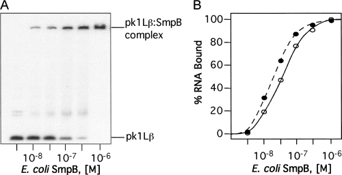
FIGURE 8.
Gel-shift analysis of binding the SmpB protein to tmRNAs. The 3′-[32P]-labeled tmRNAs (10−9 M) were titrated with the E. coli SmpB protein. Aliquots of binding mixture were analyzed by electrophoresis on a 5% polyacrylamide gel in TGE buffer. Binding of SmpB to tmRNA derivatives was quantified using a Typhoon PhosphorImager. (A) Gel-shift analysis of pk1Lβ binding to SmpB. (B) Graphical representation of PhosphorImager-derived data illustrating the binding of pkL1β (open circles) and tmRNA(H8hp) (solid circles) to SmpB. Binding curves for other mutant tmRNAs were very similar to the tmRNA(H8hp) curve and therefore are not shown.

