Abstract
Odontomas generally appear as small, solitary or multiple radio-opaque lesions found on routine radiographic examinations. Traditionally, odontomas have been classified as benign odontogenic tumors and are subdivided into complex or compound odontomas morphologically. Compound odontomas commonly occur in the incisor-canine region of the maxilla and complex odontomas are frequently located in the premolar and molar region of both jaws. Occasionally, odontoma may cause disturbances in the eruption of teeth such as impaction, delay eruption or retention of primary teeth. In general, odontomas occur more often in the permanent dentition and are very rarely associated with the primary teeth. In this report; two cases of compound odontoma associated with primary teeth is presented. (Eur J Dent 2007;1:45–49)
Keywords: Odontoma, Primary teeth
INTRODUCTION
Odontomas are developmental anomalies resulting from the growth of completely differentiated epithelial and mesenchymal cells that give rise to functional ameloblast and odontoblast.1 Odontomas have been classified as benign odontogenic tumours and are subdivided into complex or compound odontomas morphologically.2 Compound odontomas commonly occur in the incisor-canine region of the maxilla and complex odontomas are frequently located in the premolar and molar region of both jaws.3
Odontomas generally appear as small, solitary or multiple radio-opaque lesions found on routine radiographic examination. Odontoma may cause disturbances in the eruption of teeth such as impaction, delayed eruption or retention of primary teeth.4 There are very few reports of odontomas associated with primary teeth in the literature. In general, odontomas occur more often in the permanent dentition and are very rarely associated with the primary teeth.3,5,6
In this case report; odontomas in two child, which are very rarely diagnosed associated with primary teeth, are presented.
CASE REPORTS
CASE 1
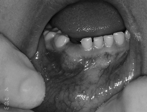
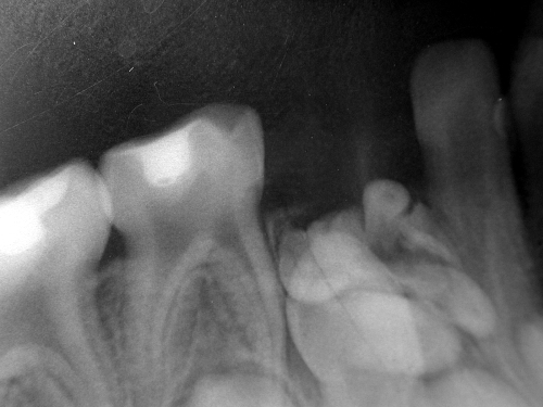
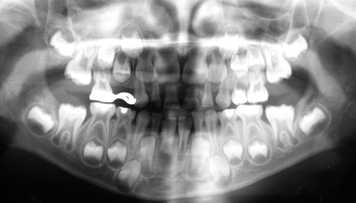
A girl aged 4 years presented with an unerupted right mandibular primary canine tooth and swelling in that region (Figure 1). Her medical story was clear. There was no history of trauma to her oro-facial region. There was no family history of unerupted teeth or hypodontia. Periapical radiograph of the lower canine region showed that multiple radio-opaque structures were present around the crown of the an unerupted canine (Figure 2). A provisional diagnosis of an odontoma was made, and the patient was scheduled for surgical removal of the lesion.
Figure 1.
Intraoral view of Case 1.
Figure 2.
Radiographic view of the odontomas.
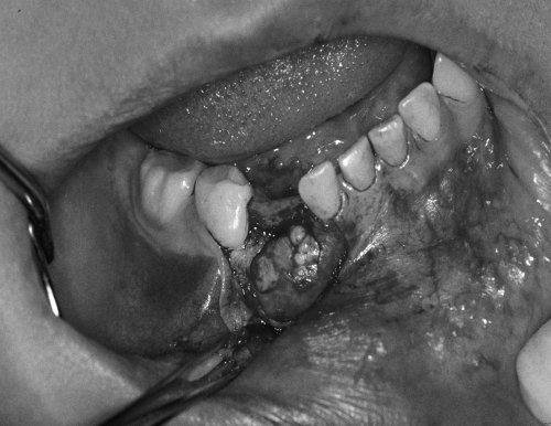
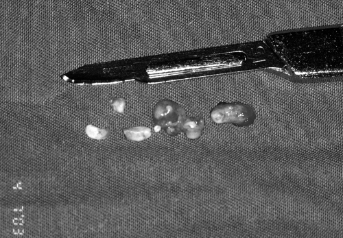
The operation was performed under local anaesthesia. Buccal mucoperiosteal flap was raised in the lower canine region. Bone was removed with a bur (Figure 3) and then multiple odontomas were removed (Figure 4). Right primary canine was unerupted. The chance of re-eruption of impacted primary canine was not slim, but it was not extracted during the operation. The surgical wound was closed primarily with 3/0 Vicryl sutures. Histologically a diagnosis of compound odontoma was made. Post-operative recovery was uneventful. The patient was followed up regularly to see eruption status of the tooth. At the end of the 2-year follow up visit, the primary canine tooth was in the dental arch (Figure 5).
Figure 3.
View of the odontomas after a buccal mucoperiosteal flap was raised.
Figure 4.
Odontomas after surgical removal.
Figure 5.
Panoramic radiograph taken two years after the operation.
CASE 2
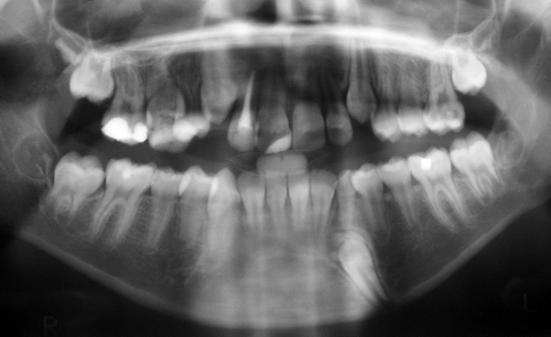
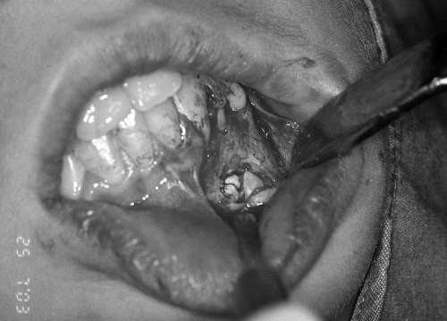
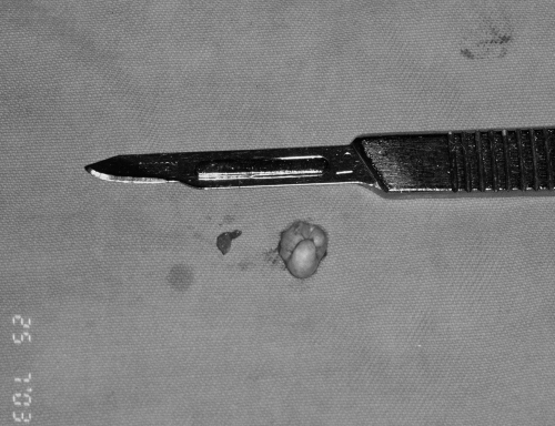
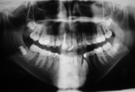
A girl aged 13 years, was referred for management of swelling in the left mandibular primary canine tooth region. Her medical history was clear. Intraorally all the primary teeth were exfoliated except mandibular left primary canine and maxillary right primary second molar tooth. There were restorations in the permanent teeth. A panoramic radiograph was taken. Radio-opaque structure was seen overlapping the crown of the left permanent canine tooth (Figure 6). The provisional diagnosis was odontoma impeding the eruption of the left permanent canine tooth. Under local anaesthesia, odontoma was removed surgically (Figures 7, 8). Surgical procedure was the same as first patient. Histologic examination revealed a diagnosis of compound odontoma. The child’s recovery was normal and intraoral healing was satisfactory. At the end of the 2-year follow-up visit, although the radiographic examination revealed that the left permanent canine’s eruption level in the alveolar bone was higher than before in the following two years, it still was unerupted (Figure 9). In addition, there was inadequate space for the canine to erupt in the lower arch and she had some other orthodontic problems. Thus the patient was referred to department of orthodontics. She is still having an orthodontic treatment.
Figure 6.
Panoramic radiograph of Case 2.
Figure 7.
View of the lesion after flap was raised.
Figure 8.
Removed odontoma.
Figure 9.
View of the canine tooth two years after the operation.
DISCUSSION
Pediatric dentists often encounter the problem of impacted teeth. However, these are mainly permanent teeth and rarely primary teeth. “Tooth impaction” refers to situations where failure to erupt appears to be due to a mechanical blocking and the tooth remains unerupted beyond the normal time of eruption. The condition is caused by systemic or local aetiologic factors.7 Factors contributing to impaction include developmental anomalies such as malposition, dilaceration, ankylosis, tumours, odontoma, dentigerous cysts, presence of supernumerary teeth and systemic-genetic interrelation such as clediocranial dysostosis and hypopituitarism.4,8 Impaction of an anterior primary tooth is very rare. When it occurs it is most often associoted with the presence of a supernumerary tooth or odontoma.9 Most cases of impacted primary teeth reported in the past were found to be caused by odontomas.7 In our first case, odontoma was the cause of the impaction of the primary canine tooth. Occasionally, odontoma may cause disturbances in the eruption of teeth such as impaction, delayed eruption or retention of primary teeth.3,4 In our second case, odontoma was the cause of retention of the primary canine tooth. Diagnosis of odontomas associated with primary teeth, as in the present cases, is unusual. A summary of cases diagnosed in the primary dentition is shown in Table 1.3,8–22
Table 1.
Odontomas associated with the primary dentition.
| Age of patients | Location | Type of odontoma | Publication |
|---|---|---|---|
| 4-year-old | Maxillary | Compound | Axel10 |
| 4-year-old | Maxillary | Compound | Aimes11 |
| 3-year, 6 month-old | Maxillary | Compound | Aimes12 |
| 4-year-old | Mandibular | Complex | Hitchin and White13 |
| 4 year, 11 month-old | Maxillary | Compound | Hitchin and Dekonor14 |
| 8 year, 7 month-old | Maxillary | Compound | Hitchin and Dekonor14 |
| 5-year-old | Maxillary | Compound | Noonan15 |
| 6-year-old | Maxillary | Compound | Stajcic3 |
| 2-year-old | Maxillary | Not stated | Brunetto et al9 |
| 1 year, 2 month-old | Maxillary | Compound | Haishima et al16 |
| 1 year, 8 month-old | Maxillary | Compound | Haishima et al16 |
| 3 year, 6 month-old | Maxillary | Compound | Bacetti17 |
| 3-year-old | Maxillary | Compound | Olivero et al18 |
| 30 month-old | Maxillary | Compound | Long et al19 |
| 3-year-old | Maxillary | Complex | Motokawa et al8 |
| 4-year-old | Maxillary | Compound | Yassin20 |
| 2 year, 5 month-old | Maxillary | Compound | Yeung et al21 |
| 4 year, 8 month-old | Maxillary | Complex | Sheehy et al22 |
When impacted primary teeth have enough space to erupt in the dental arch, surgical exposure with removal of the overlying gingiva or any overlying odontoma should be performed and the impacted teeth kept under observation for three months. When the tooth fails to erupt, orthodontic traction should be applied. When there is insufficent space for the tooth to erupt, it may necessary to increase the space by uprighting inclined neighbouring teeth. If there is no expectation of eruption, the teeth should be extracted.7 In our first case impacted primary canine tooth normally started re-erupt spontaneously one month after the operation but long-term observation is necessary until the tooth erupt. At the end of the 2-year follow up, primary canine tooth was seen in its normal position in the dental arch. In our second case, permanent canine tooth was impacted because of inadequate space for eruption. For that reason and the other orthodontic problems; she was referred to department of orthodontics.
The degree of calcification of odontoma in the primary dentition is sometimes less than is seen in relation to permanent teeth and radiographic features are therefore more weakly radio-opaque. It is important therefore, to examine the radiographs carefully.16
Paediatric dentists and oral and maxillofacial surgeons often encounter the problem of impacted teeth. However, these are mainly permanent teeth and rarely primary teeth. In this case report, odontoma was the cause of impaction and retention of primary tooth. If there is an impacted or retantive primary teeth, odontoma can be the cause. If the cause is odontoma, detailed radiographic examination and the treatment must be done.
CONCLUSIONS
Early diagnosis and management of odontomas in the primary dentition are essential in order to prevent later complications, such as failure of eruption of the primary and permanent teeth.
REFERENCES
- 1.Shafer WG, Hine MK, Levy BM. A textbook of oral pathology. 4. Philadelphia: WB. Saunders Company; 1983. p. 308. [Google Scholar]
- 2.Kramar IRH, Pindporg JJ, Shear M. World health organization international histological classification of tumours histological typing of odontogenic tumours. 2. Berlin Heidelberg: Springer-Verlag; 1992. p. 11. [Google Scholar]
- 3.Stajcic ZZ. Odontoma associated with a primary tooth. J Pedod. 1988;12:415–420. [PubMed] [Google Scholar]
- 4.Snawder KD. Delayed eruption of the anterior primary teeth and their management: report of a case. ASDC J Dent Child. 1974;41:382–384. [PubMed] [Google Scholar]
- 5.de Oliveira BH, Campos V, Marcal S. Compound odontoma-diagnosis and treatment: three case reports. Pediatr Dent. 2001;23:151–157. [PubMed] [Google Scholar]
- 6.Noonan RG. A compound odontoma associated with a deciduous tooth. Oral Surg, Oral Med, Oral Pathol. 1971;32:740–742. doi: 10.1016/0030-4220(71)90298-2. [DOI] [PubMed] [Google Scholar]
- 7.Otsuka Y, Mitomi T, Tomizawa M, Noda T. A review of clinical features in 13 cases of impacted primary teeth. Int J Paediatr Dent. 2001;11:57–63. doi: 10.1046/j.1365-263x.2001.00236.x. [DOI] [PubMed] [Google Scholar]
- 8.Motokawa W, Braham RL, Morris ME, Tanaka M. Surgical exposure and orthodontic alignment of an unerupted primary maxillary second molar impacted by an odontoma and a dentigerous cyst: a case report. Quintessence Int. 1990;21:159–162. [PubMed] [Google Scholar]
- 9.Brunetto AR, Turley PK, Brunetto AP, Regattieri LR, Nicolau GV. Impaction of a primary maxillary canine by an odontoma: surgical and orthodontic management. Pediatr Dent. 1991;13:301–302. [PubMed] [Google Scholar]
- 10.Axel AL. Supernumerary teeth in cyst: Report of case. JADA. 1937;24:457–459. [Google Scholar]
- 11.Aimes ABP. Compound composite odontoma in a child aged 4 years. Australian Dent J. 1947;51:160–161. [PubMed] [Google Scholar]
- 12.Aimes ABP. Compound composite odontoma in a child aged 3 1/2 years. Australian Dent J. 1952;56:239–240. [PubMed] [Google Scholar]
- 13.Hitchin AD, White JW. A dentinoma related to the deciduous dentition. British Dent J. 1955;98:163–165. [Google Scholar]
- 14.Hitchin AD, Dekonor E. Two cases of compound composite odontomas associated with deciduous teeth. British Dent J. 1963;114:26–28. [Google Scholar]
- 15.Noonan RG. A compound odontoma associated with a deciduous tooth. Oral Surg Oral Med Oral Pathol. 1971;32:740–742. doi: 10.1016/0030-4220(71)90298-2. [DOI] [PubMed] [Google Scholar]
- 16.Haishima K, Haishima H, Yamada Y, Tomizawa M, Noda T, Suzuki M. Compound odontomes associated with impacted maxillary primary central incisors: report of two cases. Int J Paediatr Dent. 1994;4:251–256. doi: 10.1111/j.1365-263x.1994.tb00143.x. [DOI] [PubMed] [Google Scholar]
- 17.Bacetti T. Interceptive approach to tooth eruption abnormalities: 10 year follow-up of a case. J Clin Pediatr Dent. 1995;19:297–300. [PubMed] [Google Scholar]
- 18.Oliveira JA, da Silva CJ, Costa IM, Loyola AM. Calcifying odontogenic cyst in infancy: Report of case associated with compound odontoma. ASDC J Dent Child. 1995;62:70–73. [PubMed] [Google Scholar]
- 19.Long WR, Curbox SC, Cowan JE. Arch-length asymmetry related to an odontoma in a three-year-old. ASDC J Dent Child. 1998;65:212–213. [PubMed] [Google Scholar]
- 20.Yassin OM. Delayed eruption of maxillary primary cuspid associated with compound odontoma. J Clin Pediatr Dent. 1999;23:147–149. [PubMed] [Google Scholar]
- 21.Yeung KH, Cheung CT, Tsang MMH. Compound odontoma associated with an unerupted and dilacerated maxillary primary central incisor in a young patient. Int J Paediatr Dent. 2003;13:208–212. doi: 10.1046/j.1365-263x.2003.00456.x. [DOI] [PubMed] [Google Scholar]
- 22.Sheehy EC, Odell EW, Al-Jaddir G. Odontomas in the primary dentition: Literature review and case report. J Dent Child. 2004;71:73–76. [PubMed] [Google Scholar]


