

Abstract
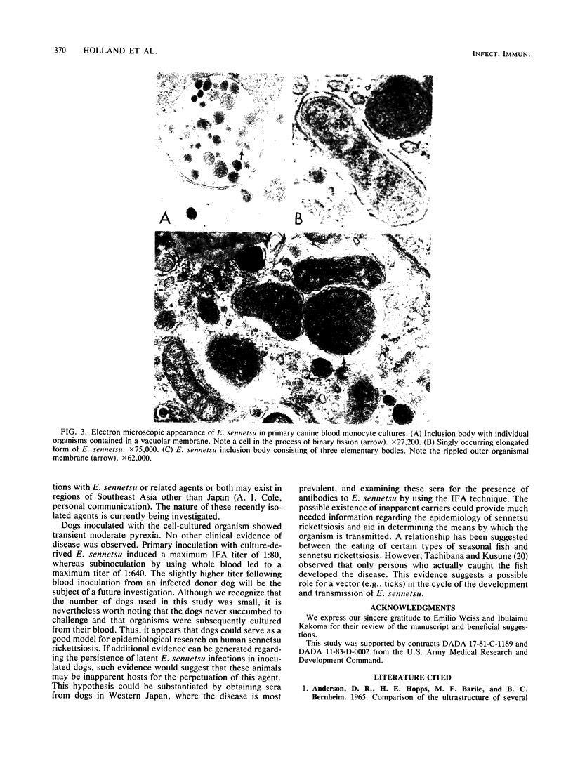
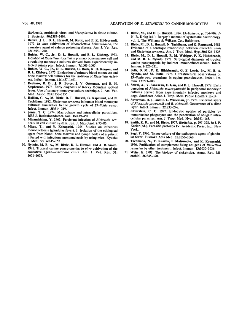
Ehrlichia sennetsu, the causative agent of human sennetsu rickettsiosis, was successfully propagated in primary canine blood monocyte cultures. The growth cycle of this organism appears to be similar to that of Ehrlichia canis. The antigen derived from our E. sennetsu cultures was used to develop an indirect fluorescent antibody test for detection and titration of serum antibodies to the organism. Using this test system, we found that five human serum samples obtained from patients clinically diagnosed as having sennetsu rickettsiosis were positive for anti-E. sennetsu antibodies. In addition, 29% of the serum samples obtained from 200 patients having a fever of unknown origin and residing in various regions of Malaysia were also serologically positive. All sera from apparently healthy individuals were negative in the test. Dogs inoculated with cell culture-adapted E. sennetsu developed a significant specific antibody titer to E. sennetsu, and the organism was subsequently isolated from their blood. These animals showed no clinical evidence of disease. The possibility of a higher prevalence of human sennetsu rickettsiosis in Southeast Asia and the potential usefulness of the canine model for studies of human sennetsu rickettsiosis are discussed.
Full text
PDF
Images in this article
Selected References
These references are in PubMed. This may not be the complete list of references from this article.
- Anderson D. R., Hopps H. E., Barile M. F., Bernheim B. C. Comparison of the ultrastructure of several rickettsiae, ornithosis virus, and Mycoplasma in tissue culture. J Bacteriol. 1965 Nov;90(5):1387–1404. doi: 10.1128/jb.90.5.1387-1404.1965. [DOI] [PMC free article] [PubMed] [Google Scholar]
- Brown J. L., Huxsoll D. L., Ristic M., Hildebrandt P. K. In vitro cultivation of Neorickettsia helminthoeca, the causative agent of salmon poisoning disease. Am J Vet Res. 1972 Aug;33(8):1695–1700. [PubMed] [Google Scholar]
- Buhles W. C., Huxsoll D. L., Ruch G., Kenyon R. H., Elisberg B. L. Evaluation ofprimary blood monocyte and bone marrow cell culture for the isolation of Rickettsia rickettsii. Infect Immun. 1975 Dec;12(6):1457–1463. doi: 10.1128/iai.12.6.1457-1463.1975. [DOI] [PMC free article] [PubMed] [Google Scholar]
- Buhles W. C., Jr, Huxsoll D. L., Elisberg B. L. Isolation of Rickettsia rickettsi in primary bone marrow cell and circulating monocyte cultures derived from experimentally infected guinea pigs. Infect Immun. 1973 Jun;7(6):1003–1005. doi: 10.1128/iai.7.6.1003-1005.1973. [DOI] [PMC free article] [PubMed] [Google Scholar]
- DeShazo R. D., Boyce J. R., Osterman J. V., Stephenson E. H. Early diagnosis of Rocky Mountain spotted fever. Use of primary monocyte culture technique. JAMA. 1976 Mar 29;235(13):1353–1355. [PubMed] [Google Scholar]
- Hoilien C. A., Ristic M., Huxsoll D. L., Rapmund G. Rickettsia sennetsu in human blood monocyte cultures: similarities to the growth cycle of Ehrlichia canis. Infect Immun. 1982 Jan;35(1):314–319. doi: 10.1128/iai.35.1.314-319.1982. [DOI] [PMC free article] [PubMed] [Google Scholar]
- Jones T. C. Macrophages and intracellular parasitism. J Reticuloendothel Soc. 1974 May;15(5):439–450. [PubMed] [Google Scholar]
- Minamishima Y. Persistent infection of Rickettsia sennetsu in cell culture system. Jpn J Microbiol. 1965 Jun;9(2):75–86. doi: 10.1111/j.1348-0421.1965.tb00277.x. [DOI] [PubMed] [Google Scholar]
- Nyindo M. B., Ristic M., Huxsoll D. L., Smith A. R. Tropical canine pancytopenia: in vitro cultivation of the causative agent--Ehrlichia canis. Am J Vet Res. 1971 Nov;32(11):1651–1658. [PubMed] [Google Scholar]
- Ristic M., Huxsoll D. L., Tachibana N., Rapmund G. Evidence of a serologic relationship between Ehrlichia canis and Rickettsia sennetsu. Am J Trop Med Hyg. 1981 Nov;30(6):1324–1328. doi: 10.4269/ajtmh.1981.30.1324. [DOI] [PubMed] [Google Scholar]
- Ristic M., Huxsoll D. L., Weisiger R. M., Hildebrandt P. K., Nyindo M. B. Serological diagnosis of tropical canine pancytopenia by indirect immunofluorescence. Infect Immun. 1972 Sep;6(3):226–231. doi: 10.1128/iai.6.3.226-231.1972. [DOI] [PMC free article] [PubMed] [Google Scholar]
- Sells D. M., Hildebrandt P. K., Lewis G. E., Jr, Nyindo M. B., Ristic M. Ultrastructural observations on Ehrlichia equi organisms in equine granulocytes. Infect Immun. 1976 Jan;13(1):273–280. doi: 10.1128/iai.13.1.273-280.1976. [DOI] [PMC free article] [PubMed] [Google Scholar]
- Shirai A., Sankaran V., Gan E., Huxsoll D. L. Early detection of Rickettsia tsutsugamushi in peripheral monocyte cultures derived from experimentally infected monkeys and dogs. Southeast Asian J Trop Med Public Health. 1978 Mar;9(1):11–14. [PubMed] [Google Scholar]
- Silverman D. J., Wisseman C. L., Jr, Waddell A. D., Jones M. External layers of Rickettsia prowazekii and Rickettsia rickettsii: occurrence of a slime layer. Infect Immun. 1978 Oct;22(1):233–246. doi: 10.1128/iai.22.1.233-246.1978. [DOI] [PMC free article] [PubMed] [Google Scholar]
- Silverstein S. C. Endocytic uptake of particles by mononuclear phagocytes and the penetration of obligate intracellular parasites. Am J Trop Med Hyg. 1977 Nov;26(6 Pt 2):161–169. doi: 10.4269/ajtmh.1977.26.161. [DOI] [PubMed] [Google Scholar]
- Tachibana N., Kusaba T., Matsumoto I., Kobayashi Y. Purification of complement-fixing antigens of Rickettsia sennetsu by ether treatment. Infect Immun. 1976 Apr;13(4):1030–1036. doi: 10.1128/iai.13.4.1030-1036.1976. [DOI] [PMC free article] [PubMed] [Google Scholar]
- Weiss E. The biology of rickettsiae. Annu Rev Microbiol. 1982;36:345–370. doi: 10.1146/annurev.mi.36.100182.002021. [DOI] [PubMed] [Google Scholar]