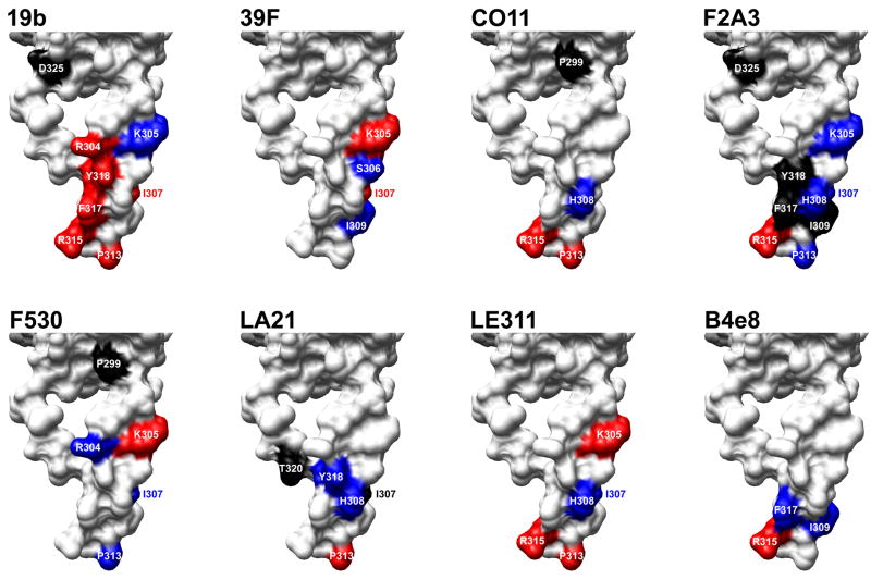
Fig. 2.
Effects of alanine substitutions on antibody binding mapped onto the structure of V3 in the context of gp120. Substitutions that significantly diminished or increased binding of the V3 mAbs tested here (Table 2) were mapped onto the structure of gp120JR-FL-core with V3 attached (Huang et al., 2005); only the V3 structure is depicted (surface rendering). The color scheme is the same as in Table 2. Molecular graphics images were produced using the UCSF Chimera package (http://www.cgl.ucsf.edu/chimera), then compiled and labeled in Adobe Photoshop.

