Abstract
The use of antiretroviral therapy in HIV type 1 (HIV-1)–infected patients does not lead to virus eradication. This is due, to a significant degree, to the fact that HIV-1 can establish a highly stable reservoir of latently infected cells. In this work, we describe an ex vivo experimental system that generates high levels of HIV-1 latently infected memory cells using primary CD4+ T cells. Using this model, we were able to dissect the T cell–signaling pathways and to characterize the long terminal repeat (LTR) cis-acting elements involved in reactivation of HIV-1 in memory CD4+ T cells. We conclude that Lck and nuclear factor of activated T cells (NFAT), but not NF-κB, are required for optimal latent virus reactivation in memory T cells. We also found that the cis-acting elements which are critical toward HIV-1 reactivation are the Sp1 and κB/NFAT transcription factor binding sites.
Introduction
HIV-1 persists in infected individuals even in the presence of highly active antiretroviral therapy (HAART). The principal reservoir of HIV-1 latency is thought to reside in resting, CD4+ memory T cells, which harbor integrated HIV-1.1 The low frequency of latently infected cells (1 in 106 resting CD4+ T cells2), for which known phenotypic markers are not available, poses a great challenge to the study of latency in vivo and ex vivo.
Previous studies on HIV-1 latency were based on the generation of chronically infected cell lines, such as the ACH2,3 JΔK,4 and J-Lat5 T-cell lines, and the U1 promonocytic cell line.6 In these systems, latency was defined as a state in which integrated proviruses failed to elicit detectable gene expression. However, these systems do not necessarily reflect the latency state in vivo because the lack of viral gene expression is due to mutations in tat (ACH2 and U13,6) or mutations in the long terminal repeat (LTR) (JΔK T-cell line4). While these latency models recapitulate a plethora of mechanisms that can lead to viral latency, we were interested in developing a more general model that would not rely on clonal proviral integration sites, and which used nontransformed, primary human T cells.
Recently, a model using human fetal liver and thymus tissues in severe combined immunodeficient (SCID-hu) mice has generated a great deal of interest in the field of HIV-1 latency.7 This model relies upon infection of thymocytes and the vast majority of latently infected cells in this system are mature, quiescent CD4+ single-positive naive T cells. This is in contrast with findings in HIV-1 patients, where the majority of latently infected cells are CD4+ memory T cells.1 Although naive and memory cells share the characteristic of being quiescent, a likely requirement for HIV-1 latency in T cells,1 there are important differences between these cell types that would impact the establishment of latency and reactivation.
In the present study, we describe the development of a novel HIV-1 latency model that uses human, primary cells and we use this model to dissect relevant signaling pathways involved in viral reactivation from latently infected memory CD4+ cells.
Methods
Reagents
The following reagents were obtained through the AIDS Research and Reference Reagent Program, Division of AIDS, National Institute of Allergy and Infectious Diseases (NIAID), National Institutes of Health (NIH): human rIL-2 from Dr Maurice Gately, Hoffmann-La Roche (Nutley, NJ)8; integrase inhibitor (118-D-24)9; and monoclonal antibody to HIV-1 p24 (AG3.0) from Dr Jonathan Allan (Southwest Foundation for Biomedical Research, San Antonio, TX).10
T cells
Peripheral blood mononuclear cells were obtained from unidentified, healthy donors. Naive CD4+ T cells were isolated by MACS microbead–negative sorting using the naive T-cell isolation kit (Miltenyi Biotec, Auburn, CA). The purity of the sort population was always higher than 95% with a phenotype CD4+CD45RA+CD45RO−CCR7+CD62L+CD27+.
Naive T cells were primed with beads coated with anti-CD3 and anti-CD28 (Dynal/Invitrogen, Carlsbad, CA) as previously described.11 Proliferating cells were expanded in medium containing 30 IU/mL IL-2, replacing media and IL-2 each 2 days.
Virus generation and viral infection
DHIV viruses were produced by transient transfection of HEK293T cells by calcium phosphate–mediated transfection.12 To normalize infections, p24 was analyzed in virus-containing supernatants by enzyme-linked immunosorbent assay (ELISA; ZeptoMetrix, Buffalo, NY). Cells were infected by spinoculation: 106 cells were infected with 500 ng/mL p24 during 2 hours at 2900 rpm and 37°C in 1 mL.
LTR mutants were generated by mutagenesis in DHIV using QuikChange II XL (Stratagene, Cedar Creek, TX). Mutations were confirmed by sequencing (the list of primers used is available on the Blood website; see the Supplemental Materials link at the top of the online article).
Flow cytometric analysis
To phenotype the cells, 2.5 × 10⁵ cells were stained with the following mAbs: phycoerythrin-conjugated (PE)–anti-CD4, PE–anti-CCR5, PE–anti-CD45RO, PE–anti-CD27, fluorescein isothiocyanate–conjugated (FITC)–anti-CCR7, TC–anti-CD45RA, or PE–anti-CXCR4 (Caltag, Burlingame, CA) followed by flow cytometric analysis in a FACSCalibur flow cytometer using the CellQuest Program (Becton Dickinson, Mountain View, CA).
To assess intracellular p24-gag expression, 5 × 10⁵ cells were fixed and permeabilized with Cytofix/Cytoperm during 30 minutes at 4°C (BD Biosciences, San Diego, CA). Cells were washed with Perm/Wash Buffer (BD Biosciences) and stained with a 1:40 dilution of anti-p24 antibody (AG3.0) in 100 μL Perm/Wash Buffer during 30 minutes at 4°C. Cells were washed with Perm/Wash Buffer and incubated with 1:100 Alexa Fluor 488 goat anti–mouse IgG (H + L) in 100 μL Perm/Wash Buffer during 30 minutes at 4°C. Cells were washed with Perm/Wash Buffer and samples were analyzed by flow cytometry. Forward versus side scatter plots were used to define the live population. In all the experiments, HIV p24-gag staining regions were set with uninfected cells treated in parallel.
Apoptosis was evaluated by simultaneous determination of phosphatidylserine (PS) exposure and mitochondrial membrane potential (ΔΨm) in the same cells as previously described.13
Reactivation assays
Cells (2.5 × 10⁵) were reactivated with beads coated with anti-CD3 and anti-CD28 during 72 hours in the presence of IL-2 (1 bead per cell).
For inhibition studies, cells were preincubated with the indicated inhibitor for 2 hours before stimulation. See Supplemental Materials for the concentration of each inhibitor or activator.
Integration analysis
Genomic DNA from 106 cells was isolated with the DNeasy Tissue Kit (QIAGEN, Valencia, CA). Genomic DNA (250 ng) was subjected to quantitative Alu-LTR polymerase chain reaction (PCR) for integrated provirus as previously described.14–16
Statistical methods
Statistical analyses were performed with SPSS12.0 for Windows (SPSS, Chicago, IL). The 2-tailed paired-samples t test analysis was used to calculate the P value (α = .05). Error bars in box-plots represent range.
Results
A novel ex vivo paradigm to study HIV-1 latency
To recapitulate the generation of memory cells ex vivo, we isolated human, primary naive CD4+ T cells using negative selection (Miltenyi Biotec; Figure 1). We then primed the naive cells toward differentiation into nonpolarized (NP), T-helper 1 (Th1), or T-helper 2 (Th2) as previously described.11
Figure 1.
Model of HIV-1 latency. Procedure used for the generation of human primary memory T cells and subsequent establishment of latent infections.
Phenotypic analysis confirmed the nature of the populations obtained in vitro (Figure S1). In vivo, memory CD4+ T cells fall into 2 main categories: central memory (TCM) and effector memory (TEM). The transcriptional profile of in vivo TCM cells closely resembles that of in vitro T cells stimulated in NP conditions.11,17 Specifically, both TCM and NP cells are characterized by simultaneous expression of CCR7 (a homing receptor for secondary lymphoid tissues) and CD27 (a coactivation molecule).11,17 Expression of CCR7 and CD27 is also found on naive cells, but is absent in TEM. We analyzed the expression of CCR7 and CD27 in the cells at 0 (naive), 7, 14, and 21 days after initial activation (Figure S1). As expected, naive CD4+ T cells expressed high levels of CCR7 and CD27, as did cells primed in NP conditions. In contrast to NP, priming under Th1- and Th2-polarizing conditions led to loss of CCR7 and CD27 expression, and generated populations with phenotypes that were characteristic of both TEM cells and TCM cells (Figure S1).
At day 7, cells from NP, Th1, and Th2 conditions were exposed to virus. A unique aspect of the model presented here is that the virus used in this model, DHIV,18 has a small out-of-frame deletion in the gp120-coding area that renders it defective in Env. To produce virus by transfection, HIV-1 Env is provided in trans in a separate plasmid.19 Due to the higher expression levels of CXCR4 compared with CCR5 after the cells were activated (Figure S1), we decided to produce a vector that consisted of the DHIV backbone pseudotyped with HIV-1LAI (an X4-tropic virus) Env. The engineered defect in Env in DHIV precludes the production of infectious progeny after a single round of infection and thus the virus is unable to spread and cause massive cell death, which would obscure the emergence of latency in vitro.
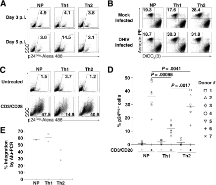
Once infected, cells were kept in culture in the presence of IL-2, and infection levels were estimated via intracellular expression of p24Gag at days 3 and 5 after infection. Intracellular p24Gag staining detects de novo–produced viral Gag protein, indicative of a productive viral infection. The maximal level of p24Gag expression was observed 5 days after infection, and this level was highest in Th1 cells (Figure 2A bottom panels). Mock-infected cultures displayed less than 0.5% background in intracellular p24Gag staining (data not shown).
Figure 2.
Generation of latently HIV-1–infected primary CD4+ T cells ex vivo. Cells were primed in NP, Th1-, or Th2-polarizing conditions and 7 days after activation cells were infected with DHIV. (A) At 3 and 5 days after infection, cells were assessed for intracellular p24 gag expression by flow cytometry. The percentage of p24-postive cells is indicated in each panel. The experiment shown is representative of 4 different experiments with 4 different donors. (B) At 5 days after infection, cells were assessed for annexin-PE and DiOC6(3) by flow cytometry. For each panel, the percentage of apoptotic cells (annexin-PE positive and DiOC6(3) low) is indicated. The experiment shown is representative of 3 different experiments with 3 different donors. (C) At 7 days after infection, cells were cultured without stimulation (untreated) or costimulated with antibodies to CD3 and CD28 for 3 days (CD3/CD28) and assessed for intracellular p24 gag expression by flow cytometry. The percentage of p24-postive cells is indicated in each panel for this representative experiment. Values corresponding to 7 different donors are shown in panel D, where each symbol represents a different donor and horizontal lines indicate media values. Significance by 2-tailed paired-samples t test analysis (P values provided). (E) Viral integration was analyzed by Alu-PCR 3 days after infection in donors 1 and 2. Horizontal lines indicate media values.
At day 5 after infection, apoptosis levels were measured by flow cytometry. Positive staining for annexin V and low staining for DiOC6(3) revealed the presence of apoptotic cells (Figure 2B). Apoptosis levels in DHIV-infected NP and Th2 cells were similar to those in mock-infected cells. In contrast, apoptosis in DHIV-infected Th1 cells was high. The higher level of apoptosis in Th1 (12.7% over mock) was in agreement with the higher level of productive infection measured in these cells (14.5%) relative to other subsets.
To induce reactivation of potential latent viruses, at day 7 after infection cells were restimulated for 3 days in the presence of beads coated with αCD3 and αCD28 antibodies (Figure 2C CD3/CD28). As a negative control, we incubated parallel cultures in the absence of beads (Figure 2C untreated). We detected low levels of p24Gag+ cells in the absence of restimulation (Figure 2C top panels). However, restimulation led to an increase in the percentage of p24Gag+ cells in all subsets (Figure 2C bottom panels). Remarkably, levels of p24Gag+ cells after reactivation were higher in NP cells than in Th1 or Th2 cells.
The results shown in Figure 2C correspond to a single blood donor (donor 1). To verify the generality of these findings in a broader population, we performed further experiments with 6 additional donors. The results, summarized in Figure 2D, confirm that NP cells and, to a lesser degree, Th2-polarized cells, can harbor high levels of HIV-1 latency, whereas Th1-polarized cells displayed low levels of latency.
To evaluate the levels of viral infection by a method that would be independent of viral gene expression we resorted to quantitative Alu-PCR.14–16 Quantitative Alu-PCR is specific for integrated viral DNA and should detect latent and productive infections with equal efficiency. We performed Alu-PCR at day 3 after infection for donors 1 and 2 (Figure 2E). Alu-PCR results for donor 1 (square symbols) correspond to Figure 2A,C,D, and show that the levels of integrated viruses in all 3 cell subsets greatly exceeded the frequency of p24Gag+ cells at day 3 or 5 after infection.
Th1 populations consistently contained lower levels of p24Gag+ cells than Th2 or NP upon reactivation. In addition, Th1 cells displayed levels of infection by Alu-PCR that were roughly equivalent or higher (Figure 2E) than those seen in Th2 or NP. Therefore, it appears that Th1 cells are able to sustain higher levels of initial productive infection (ie, p24Gag+; Figure 2A, day 5 after infection), followed by higher levels of apoptotic death, leading to less frequent latent infections.
Detection of viral gene expression can also be accomplished via reporter molecules, such as green fluorescence protein (GFP), with high sensitivity and specificity.5 To test whether our latency and reactivation system would produce similar results when using GFP as a reporter, we performed parallel infections with DHIV/X4 and DHIV-GFP/X4 (Figure S2). In DHIV-GFP, nef had been replaced by the GFP gene.5 Aside from the nef replacement with GFP, DHIV-GFP is identical to DHIV. Most GFP-positive cells were also positive for p24, and vice versa, both during the initial infection and also after reactivation (Figure S2). A small population of cells that are positive for GFP but negative for p24 can also be appreciated. These cells are, presumably, early in the infection or reactivation process and have not begun to produce viral late proteins. Therefore, intracellular p24 detection and GFP fluorescence can be used interchangeably in order to detect viral gene expression and our latency and reactivation model.
Signaling pathways involved in HIV-1 reactivation in TCM
Previous studies on cells from infected patients showed that central memory CD4+ cells contain the highest frequency of HIV-1 DNA, on average 10 times higher than that of effector memory cells.20 Based on transcriptional profiles, cytokine production, surface phenotype, and the ability to differentiate into effector memory cells upon secondary antigenic challenge, NP cells are considered the in vitro equivalents of TCM.11 Therefore, we focused further studies on latent infection and reactivation of NP cells.
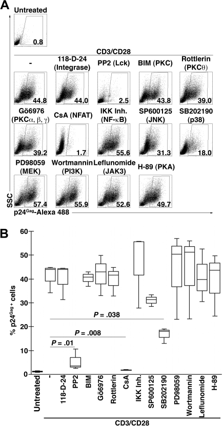
To begin to dissect potential signaling pathways leading to virus reactivation, we tested a panel of known signaling inhibitors. We reactivated DHIV-infected NP cells with αCD3/CD28, as shown in Figure 2C, in the presence or absence of pharmacologic inhibitors (Figure 3). As a control, and to confirm our expectation that the observed latent infections represent postintegration events, we tested the integrase inhibitor, 118-D-24 (Figure 3A). As expected, 118-D-24 did not have any effect on viral reactivation.
Figure 3.
Signaling antagonists and their effects on HIV-1 reactivation. NP cells were infected with DHIV and 7 days after infection cells were left untreated or costimulated with antibodies to CD3 and CD28 for 3 days (CD3/CD28) in the presence of the indicated inhibitor for the protein or pathway indicated between parentheses and assessed for intracellular p24 gag expression by flow cytometry. (A) Representative experiment. The percentage of p24-postive cells is indicated in each panel. (B) Box-plots corresponding to 3 different donors. Horizontal lines indicate median values and significance by 2-tailed paired-samples t test analysis (P values provided).
One of the proximal events after activation of T cells through CD3 and CD28 is activation of the tyrosine kinase, Lck (Figure S3; for a review, see Kane et al21). Blocking Lck activation with PP2 abrogated HIV-1 reactivation by about 96% (inhibition of reactivation = (1−[p24% with αCD3/CD28 and inhibitor − p24% untreated]/[p24% with αCD3/CD28 − p24% untreated] × 100; Figure 3A). Lck activation leads to PLCγ1 activation and production of the second messengers, diacylglycerol (DAG) and inositol 1,4,5-triphosphate (IP3). DAG activates various isoforms of protein kinase C (PKC).22 Specifically in T cells, PKCθ leads to phosphorylation and degradation of IκB, with the subsequent release and nuclear translocation of the transcription factor, NFκB.23 We probed the DAG-PKC–NFκB signaling axis by restimulating cells in the presence or absence of the general PKC inhibitor, BIM; we also tested an inhibitor of the classic isoforms of PKC (α, β, and γ), Gö6976; or Rottlerin, a specific inhibitor of PKCθ. None of these 3 compounds had a negative effect on viral reactivation (Figure 3A). To confirm the previous result, we used IκB kinase peptide inhibitor (IKK inh).24 When cells were reactivated in the presence of IKK inh, the levels of viral reactivation were not affected.
DAG also activates Ras guanyl-nulceotide-releasing protein (RasGRP).25 RasGRP and many isoforms of PKC activate Ras, leading to subsequent activation of the mitogen-activated protein (MAP) kinases, Erk1/2 (MEK), JNK, and p38. To probe the DAG-Ras-MAPK axis, we resorted to inhibition of JNK or Erk1/2 with SP600125 or PD98059, respectively. Neither SP600125 nor PD98059 inhibited viral reactivation. In contrast, an inhibitor of p38, SB202190, significantly diminished (66% inhibition; Figure 3A) HIV-1 reactivation.
The other second messenger generated by PLCγ1, IP3, activates calcineurin, which dephosphorylates and activates the transcription factor, NFAT.25 To probe the IP3-dependent signaling cascade, we restimulated latently infected cells in the presence or absence of the calcineurin inhibitor, cyclosporine A (CsA). CsA completely abolished (99.9%) viral reactivation (Figure 3A).
Stimulation through CD3/CD28 involves also recruitment and activation of PI3K, leading to the activation of the serin/threonin kinase Akt.26 PI3K is also involved in signal transduction downstream of γc cytokine receptor engagement (Figure S3). To ascertain the possible contribution of PI3K, we tested its inhibitor, wortmannin. Incubation with wortmanin had no effect on HIV-1 reactivation. To confirm the results with the γc-cytokine–dependent pathway, we tested the Janus-activated kinase-3 (JAK3) inhibitor, leflunomide. Leflunomide also failed to block viral reactivation.
Signaling cascades in T cells can also be initiated through engagement of G protein–coupled receptors (GPCRs; Figure S3). GPCR signals typically converge in the production of cyclic AMP (cAMP), which then activates protein kinase A (PKA), leading to the activation of the transcription factor CREB (for a review see Nordheim27). We incubated cells with the PKA inhibitor, H-89, which did not have any effect on viral reactivation (Figure 3A).
The above experiments with inhibitors were performed with 2 additional donors. The results with donors 1, 3, and 4, summarized in Figure 3B, denote striking similarities in the sensitivity of virus reactivation to drug inhibition across different individuals.
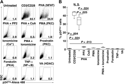
To complement the inhibitor studies, we undertook additional experimentation using agonists for the above signaling pathways. Latently infected cells were either left untreated or incubated with agonist. As a positive control, cells were treated with αCD3/CD28. The ability of the agonist to promote viral reactivation was then evaluated by detection of intracellular p24Gag as shown above (Figure 4). We first tested phytohemagglutinin (PHA), a lectin that binds nonspecifically to carbohydrate moieties on surface glycoproteins and acts as a potent polyclonal mitogen for T cells. PHA incubation efficiently reactivated viral gene expression (79%; reactivation efficiency = ([p24% with agonist − p24% untreated]/[p24% with aCD3/CD28 − p24% untreated] × 100; Figure 4A). The effects of PHA on T cells are mediated by NFAT activation (Figure S3). To confirm the role of NFAT in PHA-mediated reactivation, we coincubated cells with PHA and the Lck inhibitor, PP2, or the calcineurin inhibitor, CsA. PHA stimulation in the presence of PP2 or CsA resulted in extremely low viral reactivation (3% and 0%, respectively). These results are in complete agreement with those from inhibitor studies and confirm the central role of NFAT in HIV-1 reactivation in memory T cells.
Figure 4.
Signaling agonists and their effects on HIV-1 reactivation. NP cells were infected with DHIV and 7 days after infection cells were left untreated, costimulated (CD3/CD28) in the presence of the indicated agonist for the protein or pathway indicated between parentheses for 3 days, and assessed for intracellular p24 gag expression by flow cytometry. In the case of cells stimulated with PHA, cells were also costimulated in the presence of the inhibitors PP2 (Lck) or CsA (NFAT). (A) Representative experiment. The percentage of p24-postive cells is indicated in each panel. (B) Box-plots corresponding to 3 different donors. Horizontal lines indicate median values and significance by 2-tailed paired-samples t test analysis (P values provided).
To activate the DAG-PKC-NFκB signaling axis, we used PMA and, separately, prostratin (both direct activators of PKC). Neither compound was able to reactivate viral gene expression (Figure 4A).
Signaling downstream of IP3 involves an increase in intracellular levels of calcium. To directly stimulate calcium influx, we incubated cells with ionomycine, and this had no effect on viral reactivation. However, combination of PMA and ionomycine was able to induce minor, but significant, levels of reactivation (4%).
In agreement with the lack of effect of H-89 (an inhibitor of PKA; Figure 3A), incubation of cells with the PKA activator, forskolin, failed to reactivate virus gene expression.
In tumor cell lines harboring integrated, latent HIV-1, TNF-α can induce viral gene expression through the activation of NFκB.3,5,28,29 We tested the potential role of TNF-α in virus reactivation in latently infected memory T cells. We found that TNF-α failed to induce any degree of viral gene expression (Figure 4A).
Inhibition of histone deacetylases (HDACs) with valproic acid (VA) has previously been shown to induce viral reactivation.30–32 In our latent infection system, however, incubation with VA failed to reactivate viral gene expression (Figure 4A).
As with the inhibitor studies, results with signaling pathway agonists were very similar in 3 different donors (donors 1, 3, and 4), as shown in Figure 4B.
From the studies mentioned, we conclude that optimal HIV-1 reactivation in memory T cells requires a pathway that includes, upstream, the tyrosine kinase, Lck, and, downstream, the transcription factor, NFAT.
LTR sites required for HIV-1 reactivation
The HIV-1 latency model we present here uses a molecularly cloned virus and recapitulates a single virus replication cycle. Therefore, this system should allow us to ask which transcription factor binding sites in the viral promoter may be required for efficient reactivation. To that end, we engineered the DHIV viral construct to contain mutations in specific regions known to regulate LTR-driven transcription.33–39 These mutations were engineered in the U3 region of the 3′ LTR, such that the mutant promoter would be copied into the 5′ end of the virus after the first round of reverse transcription. We constructed mutants in the regions shown in Figure 5A (see also the specific mutations in Figure S4). NP cells were infected with mutant-promoter viruses, and cells were kept in vitro for an additional 7-day period. At this time point, immediately prior to restimulation, we isolated genomic DNA and quantitated viral integration by Alu-PCR (blue numbers in Figure 5B), to assess the levels of latent infection prior to reactivation. We then restimulated the cells with αCD3/CD28 and analyzed intracellular p24Gag expression 3 days later.
Figure 5.
Transcription factor binding sites involved in HIV-1 reactivation. (A) Scheme of HIV-1 LTR. (B) NP cells were infected with wt DHIV or with different LTR mutants. Mutations can be viewed in Figure S2. At 7 days after infection, cells were costimulated with antibodies to CD3 and CD28 for 3 days and assessed for intracellular p24 gag expression by flow cytometry. The percentage of p24-postive cells is indicated in each panel. Percentage of viral integration by Alu-PCR for each virus is indicated in blue (UD indicates undetectable). The experiment is representative of 3 different experiments with 3 different donors.
As shown in Figure 5B, mutation in the Sp1 sites abolished any ability of latent viruses to be reactivated (0.3%; efficiency of reactivation = [p24% of mutant with αCD3/CD28 − p24% of mutant without αCD3/CD28]/[p24% of wt virus with αCD3/CD28 − p24% of wt virus without αCD3/CD28] × 100). Likewise, mutation of both κB/NFAT sites almost completely abolished reactivation (9.5%). Mutation of the AP-2–binding site led to a reactivation efficiency of 64%. Mutation of both NF-IL6 I and NF-IL6 II–binding sites, USF, or TCF-1α had almost no effect (equal or higher than 85% reactivation efficiency).
The viral mutagenesis results together with the inhibitor and agonist studies indicate that the transcription factors NFAT and Sp1 are essential for reactivation in human NP memory CD4 T cells. Further dissection of signaling pathways and requirements for latency and reactivation can easily be pursued in the future, using this ex vivo system.
Discussion
HIV-1 latency reservoirs are small, but extremely long-lived. Latent infection is associated with low-to-null levels of viral gene expression and appears to be noncytopathic. However, upon reactivation, latent viruses enter an active mode of replication in which they are fully competent for spread and induction of disease. The current thinking in the field is that the combination of hypothetical drugs that will reactivate latent viruses, in combination with present-day antiretroviral drugs, is the desired approach toward viral eradication. However, we are limited by the scarcity of known drugs that can safely be used for viral reactivation. We are also limited by our poor understanding of the dynamics between establishment of latency and reactivation, and the cellular and viral factors that govern these processes. Our work describes the development of a novel method that recapitulates latent and productive viral infections in the laboratory. This method is easy to perform, powerful and, most importantly, lends itself to molecular analysis.
One key question about HIV-1 latency is what specific cell type(s) can harbor long-lived, latent proviruses. Previous work by several groups (reviewed in Douek et al40 and Persaud et al41) suggests that in vivo, quiescent memory T cells constitute the most long-lived viral reservoir, whose decay constant ranges from months to years. Memory cells, in vivo, are subdivided into various subsets whose biology can faithfully be recapitulated in vitro.11 We tested the relative abilities of NP, Th1, and Th2 cells to harbor latent viruses and found that, while latent infections could be induced in all subsets, NP cells were consistently more permissive for latent infection and accordingly less able to sustain productive infection. Conversely, Th1 cells were exquisitely sensitive to productive infection, and latent infection of these cells was significantly lower. It is tempting to speculate that the higher permissiveness to productive infection of Th1 cells was the cause of enhanced levels of apoptosis.
A unique aspect of the method presented here is that the virus is defective in Env. When the virus is produced, Env is provided in trans. Thus, upon infection, viral particles contain the full protein complement of HIV-1, and are fully infectious and competent for entry, reverse transcription, integration, and viral gene expression. However, the engineered defect in Env precludes the production of infectious progeny. Cells undergoing productive infection die within 3 to 5 days due to virus-mediated apoptosis and only uninfected and latently infected cells survive after the first week in culture.
A second unique feature of this system is the intrinsic ability of the virus to drive wild-type levels of gene expression. This is a crucial aspect of our model, as latent viruses in vivo, when reactivated, are fully capable of replicating and causing disease. This is an important distinction with previous models of latency in which lack of viral gene expression was shown to be associated with mutations in the virus or in the host cell29,42–45 or with specific sites of virus integration in heterochromatin.5
HIV-1 latency appears to be related to intrinsic activation and/or development characteristics of CD4+ T cells, rather than to the presence of latency-promoting genes in the virus. Thus, it is important to dissect, at a molecular level, the T cell–signaling pathway(s) that underlie the establishment, maintenance, and reactivation of latent infections. In the present work, we used a 3-prong approach to dissect signaling events leading to reactivation in primary memory cells. We used agonists and antagonists of cellular processes, and mutagenesis of viral cis-acting elements. The Sp1 and κB/NFAT promoter elements were critical toward reactivation.
Although Sp1 has been considered a ubiquitous and constitutive transcription factor, an emerging body of evidence indicates that the activity of Sp1 is regulated through the cell cycle.46,47 Sp1 is phosphorylated and inactive in quiescent cells. Upon entry into the cell division cycle, PP2A dephosphorylates Sp1, which becomes active and tightly associated with the chromatin.46,47 Our finding that Sp1 is absolutely required for reactivation of latent HIV-1 is in agreement with the idea that a latently infected cell in vivo may be quiescent, and reactivation of the virus is concomitant with entry into the cell division cycle.
The κB/NFAT-binding sites, also a stringent requirement for viral reactivation, are not separable by mutagenesis because NFκB and NFAT bind identical elements on the LTR.48 The potential roles played by NFAT and NFκB in reactivation are of paramount importance in our studies, as it has recently been shown that naive cells contain very low levels of NFATc1 and NFATc2, whereas memory cells contain high levels of such transcription factors.49 This explains why both naive and memory T cells rapidly induce IL-2 (whose promoter contains a κB/NFAT binding site) transcription upon T-cell receptor ligation, but the responsible transcription factors differ, being NFκB for naive cells, and NFAT for memory cells.49 Therefore, one would predict that in memory cells NFAT, but not NFκB, would be essential for viral reactivation. The results from our inhibitor studies clearly confirm this prediction, as CsA incubation completely blocked reactivation whereas IKK Inh or PKC inhibitors had no effect. In further support for the lack of a role of NFκB in viral reactivation in memory cells, agonists or stimuli that function through NFκB, such as PMA, prostratin, and TNF-α, failed to induce any degree of reactivation.
We tested 2 agonists of NFAT activation, PHA and ionomycin. It is intriguing that PHA induced reactivation almost as efficiently as αCD3/CD28 treatment, while ionomycin produced no detectable reactivation. PHA is a promiscuous mitogen that activates multiple pathways. However the observation that addition of CsA or PP2 completely blocked reactivation by PHA further supports that the required signaling axis dowstream of PHA is Lck-calcineurin-NFAT. In this context, the failure of ionomycin to induce reactivation was surprising, and could underlie important mechanistic details. Two possible explanations could be formulated. First, ionomycin alone is unable to induce cell proliferation. In the absence of cell proliferation, Sp1 would fail to be dephosphorylated and activated,46,47 as we discussed above. In partial support of this idea is, perhaps, the observation that addition of PMA to ionomycin treatment did stimulate reactivation, albeit to a small degree, concomitant with cell proliferation (data not shown). A second possible explanation is that both αCD3/CD28 and PHA induce activation of calcineurin (Figure S3) through Ca2+ release concomitantly with p38 activation. In contrast, ionomycin only induces Ca2+ release.
Inhibition of p38 with SB202190 had a significant effect (66% inhibition) on viral reactivation. p38 participates in 2 signaling events that may be relevant to viral reactivation (Figure S3). The classical p38 activation pathway requires signaling through DAG-RasGRP/PKC-Ras, whose inhibition did not affect reactivation. In recent years, an alternative p38 activation pathway has been described, which uses a scaffold protein known as Dlgh1.50 Dlgh1 is devoid of any known enzymatic activity, but can modify the signaling emerging from TCR engagement, transmitted through ZAP70 and Lck, to facilitate p38 activation and subsequent activation of NFAT in a calcineurin-dependent manner (Figure S3). Likely, this alternative pathway50 of p38 activation is responsible for viral reactivation in our system. Further experimentation will be required to confirm this model.
Our results are similar, although with important differences, to those reported earlier using a SCID-hu mouse model of HIV-1 latency.51 The model by Brooks et al and our results agree on the requirements for Lck and NFAT toward viral reactivation but disagree on the requirement of NFκB.51 A key difference in the model proposed by Brooks et al is the use of CD4 single-positive thymocytes, which may bare characteristics of naive T cells rather than memory T cells.
In a Jurkat model of postintegration latency, it was found that latently infected cells frequently contained HIV-1 integrated in the proximity of alphoid repeat elements in heterochromatin.5 Reactivation of these latent viruses could be accomplished with PMA or TNF-α. PMA and TNF-α failed to induce any detectable reactivation in our latency system. The differences between our studies and the studies of Jordan et al5 may be attributed to the use of a Jurkat cell line. It is well known that Jurkat and primary T cells shared some but not all T-cell signaling pathways.52 Since integration in our system is likely polyclonal, analysis of the characteristics of integration sites will require careful analysis.
Other means of inducing reactivation of latent proviruses have been proposed, based on pharmacologic modification of the “histone code” with histone deacetylase inhibitors, such as valproic acid.32 Valproic acid was incapable of inducing reactivation in our model. However, in future studies we will test other HDAC inhibitors for their potential use in reactivation.
To our knowledge this is the first report to document efficient generation of HIV-1 latently infected memory T cells ex vivo. There are many outstanding questions that this model may help answer. For example, future experiments should address the mechanisms that lead to a productive infection versus a latent infection. It would also be important to test whether cells polarized in other conditions, such as Th17 or Treg, have different abilities to sustain or reactivate latent infections.
Supplementary Material
Acknowledgments
We thank Dr Jonathan Allan for providing the monoclonal antibody to HIV-1 p24 (AG3.0). We would like to acknowledge the excellent technical assistance of Tara Mleynek.
This work was supported by NIH grant AI49057 to V.P.; and by a Postdoctoral Fellowship from Ministerio de Educacion y Cultura, Spain to A.B.
Footnotes
The online version of this article contains a data supplement.
The publication costs of this article were defrayed in part by page charge payment. Therefore, and solely to indicate this fact, this article is hereby marked “advertisement” in accordance with 18 USC section 1734.
Authorship
Contribution: A.B. designed and performed the research, analyzed the data, and wrote the manuscript; and V.P. designed the research, analyzed the data, and wrote the manuscript.
Conflict-of-interest disclosure: The authors declare no competing financial interests.
Correspondence: Vicente Planelles, University of Utah School of Medicine, 15 North Medical Dr East #2100, Rm 2520, Salt Lake City, Utah 84112; e-mail: vicente.planelles@path.utah.edu.
References
- 1.Finzi D, Hermankova M, Pierson T, et al. Identification of a reservoir for HIV-1 in patients on highly active antiretroviral therapy. Science. 1997;278:1295–1300. doi: 10.1126/science.278.5341.1295. [DOI] [PubMed] [Google Scholar]
- 2.Chun TW, Carruth L, Finzi D, et al. Quantification of latent tissue reservoirs and total body viral load in HIV-1 infection. Nature. 1997;387:183–188. doi: 10.1038/387183a0. [DOI] [PubMed] [Google Scholar]
- 3.Folks TM, Clouse KA, Justement J, et al. Tumor necrosis factor alpha induces expression of human immunodeficiency virus in a chronically infected T-cell clone. Proc Natl Acad Sci U S A. 1989;86:2365–2368. doi: 10.1073/pnas.86.7.2365. [DOI] [PMC free article] [PubMed] [Google Scholar]
- 4.Antoni BA, Rabson AB, Kinter A, Bodkin M, Poli G. NF-kappa B-dependent and -independent pathways of HIV activation in a chronically infected T cell line. Virology. 1994;202:684–694. doi: 10.1006/viro.1994.1390. [DOI] [PubMed] [Google Scholar]
- 5.Jordan A, Bisgrove D, Verdin E. HIV reproducibly establishes a latent infection after acute infection of T cells in vitro. Embo J. 2003;22:1868–1877. doi: 10.1093/emboj/cdg188. [DOI] [PMC free article] [PubMed] [Google Scholar]
- 6.Folks TM, Justement J, Kinter A, Dinarello CA, Fauci AS. Cytokine-induced expression of HIV-1 in a chronically infected promonocyte cell line. Science. 1987;238:800–802. doi: 10.1126/science.3313729. [DOI] [PubMed] [Google Scholar]
- 7.Brooks DG, Kitchen SG, Kitchen CM, Scripture-Adams DD, Zack JA. Generation of HIV latency during thymopoiesis. Nat Med. 2001;7:459–464. doi: 10.1038/86531. [DOI] [PubMed] [Google Scholar]
- 8.Lahm HW, Stein S. Characterization of recombinant human interleukin-2 with micromethods. J Chromatogr. 1985;326:357–361. doi: 10.1016/s0021-9673(01)87461-6. [DOI] [PubMed] [Google Scholar]
- 9.Svarovskaia ES, Barr R, Zhang X, et al. Azido-containing diketo acid derivatives inhibit human immunodeficiency virus type 1 integrase in vivo and influence the frequency of deletions at two-long-terminal-repeat-circle junctions. J Virol. 2004;78:3210–3222. doi: 10.1128/JVI.78.7.3210-3222.2004. [DOI] [PMC free article] [PubMed] [Google Scholar]
- 10.Simm M, Shahabuddin M, Chao W, Allan JS, Volsky DJ. Aberrant Gag protein composition of a human immunodeficiency virus type 1 vif mutant produced in primary lymphocytes. J Virol. 1995;69:4582–4586. doi: 10.1128/jvi.69.7.4582-4586.1995. [DOI] [PMC free article] [PubMed] [Google Scholar]
- 11.Messi M, Giacchetto I, Nagata K, Lanzavecchia A, Natoli G, Sallusto F. Memory and flexibility of cytokine gene expression as separable properties of human T(H)1 and T(H)2 lymphocytes. Nat Immunol. 2003;4:78–86. doi: 10.1038/ni872. [DOI] [PubMed] [Google Scholar]
- 12.Zhu Y, Gelbard HA, Roshal M, Pursell S, Jamieson BD, Planelles V. Comparison of cell cycle arrest, transactivation, and apoptosis induced by the simian immunodeficiency virus SIVagm and human immunodeficiency virus type 1 vpr genes. J Virol. 2001;75:3791–3801. doi: 10.1128/JVI.75.8.3791-3801.2001. [DOI] [PMC free article] [PubMed] [Google Scholar]
- 13.Gomez-Benito M, Balsas P, Bosque A, Anel A, Marzo I, Naval J. Apo2L/TRAIL is an indirect mediator of apoptosis induced by interferon-alpha in human myeloma cells. FEBS Lett. 2005;579:6217–6222. doi: 10.1016/j.febslet.2005.10.007. [DOI] [PubMed] [Google Scholar]
- 14.Vandegraaff N, Kumar R, Hocking H, et al. Specific inhibition of human immunodeficiency virus type 1 (HIV-1) integration in cell culture: putative inhibitors of HIV-1 integrase. Antimicrob Agents Chemother. 2001;45:2510–2516. doi: 10.1128/AAC.45.9.2510-2516.2001. [DOI] [PMC free article] [PubMed] [Google Scholar]
- 15.Butler SL, Hansen MS, Bushman FD. A quantitative assay for HIV DNA integration in vivo. Nat Med. 2001;7:631–634. doi: 10.1038/87979. [DOI] [PubMed] [Google Scholar]
- 16.Dehart JL, Andersen JL, Zimmerman ES, et al. The ataxia telangiectasia-mutated and Rad3-related protein is dispensable for retroviral integration. J Virol. 2005;79:1389–1396. doi: 10.1128/JVI.79.3.1389-1396.2005. [DOI] [PMC free article] [PubMed] [Google Scholar]
- 17.Rivino L, Messi M, Jarrossay D, Lanzavecchia A, Sallusto F, Geginat J. Chemokine receptor expression identifies Pre-T helper (Th)1, Pre-Th2, and nonpolarized cells among human CD4+ central memory T cells. J Exp Med. 2004;200:725–735. doi: 10.1084/jem.20040774. [DOI] [PMC free article] [PubMed] [Google Scholar]
- 18.Andersen JL, DeHart JL, Zimmerman ES, et al. HIV-1 Vpr-induced apoptosis is cell cycle dependent and requires Bax but not ANT. PLoS Pathog. 2006;2:e127. doi: 10.1371/journal.ppat.0020127. [DOI] [PMC free article] [PubMed] [Google Scholar]
- 19.Challita-Eid PM, Klimatcheva E, Day BT, et al. Inhibition of HIV type 1 infection with a RANTES-IgG3 fusion protein. AIDS Res Hum Retroviruses. 1998;14:1617–1624. doi: 10.1089/aid.1998.14.1617. [DOI] [PubMed] [Google Scholar]
- 20.Brenchley JM, Hill BJ, Ambrozak DR, et al. T-cell subsets that harbor human immunodeficiency virus (HIV) in vivo: implications for HIV pathogenesis. J Virol. 2004;78:1160–1168. doi: 10.1128/JVI.78.3.1160-1168.2004. [DOI] [PMC free article] [PubMed] [Google Scholar]
- 21.Kane LP, Lin J, Weiss A. Signal transduction by the TCR for antigen. Curr Opin Immunol. 2000;12:242–249. doi: 10.1016/s0952-7915(00)00083-2. [DOI] [PubMed] [Google Scholar]
- 22.Isakov N, Mally MI, Scholz W, Altman A. T-lymphocyte activation: the role of protein kinase C and the bifurcating inositol phospholipid signal transduction pathway. Immunol Rev. 1987;95:89–111. doi: 10.1111/j.1600-065x.1987.tb00501.x. [DOI] [PubMed] [Google Scholar]
- 23.Bauer B, Krumbock N, Ghaffari-Tabrizi N, et al. T cell expressed PKCtheta demonstrates cell-type selective function. Eur J Immunol. 2000;30:3645–3654. doi: 10.1002/1521-4141(200012)30:12<3645::AID-IMMU3645>3.0.CO;2-#. [DOI] [PubMed] [Google Scholar]
- 24.Swaroop N, Chen F, Wang L, Dokka S, Toledo D, Rojanasakul Y. Inhibition of nuclear transcription factor-kappaB by specific IkappaB kinase peptide inhibitor. Pharm Res. 2001;18:1631–1633. doi: 10.1023/a:1013051019098. [DOI] [PubMed] [Google Scholar]
- 25.Lin J, Weiss A. T cell receptor signalling. J Cell Sci. 2001;114:243–244. doi: 10.1242/jcs.114.2.243. [DOI] [PubMed] [Google Scholar]
- 26.Franke TF, Yang SI, Chan TO, et al. The protein kinase encoded by the Akt proto-oncogene is a target of the PDGF-activated phosphatidylinositol 3-kinase. Cell. 1995;81:727–736. doi: 10.1016/0092-8674(95)90534-0. [DOI] [PubMed] [Google Scholar]
- 27.Nordheim A. Transcription factors. CREB takes CBP to tango. Nature. 1994;370:177–178. doi: 10.1038/370177a0. [DOI] [PubMed] [Google Scholar]
- 28.Osborn L, Kunkel S, Nabel GJ. Tumor necrosis factor alpha and interleukin 1 stimulate the human immunodeficiency virus enhancer by activation of the nuclear factor kappa B. Proc Natl Acad Sci U S A. 1989;86:2336–2340. doi: 10.1073/pnas.86.7.2336. [DOI] [PMC free article] [PubMed] [Google Scholar]
- 29.Duh EJ, Maury WJ, Folks TM, Fauci AS, Rabson AB. Tumor necrosis factor alpha activates human immunodeficiency virus type 1 through induction of nuclear factor binding to the NF-kappa B sites in the long terminal repeat. Proc Natl Acad Sci U S A. 1989;86:5974–5978. doi: 10.1073/pnas.86.15.5974. [DOI] [PMC free article] [PubMed] [Google Scholar]
- 30.Ylisastigui L, Archin NM, Lehrman G, Bosch RJ, Margolis DM. Coaxing HIV-1 from resting CD4 T cells: histone deacetylase inhibition allows latent viral expression. Aids. 2004;18:1101–1108. doi: 10.1097/00002030-200405210-00003. [DOI] [PubMed] [Google Scholar]
- 31.Simon G, Moog C, Obert G. Valproic acid reduces the intracellular level of glutathione and stimulates human immunodeficiency virus. Chem Biol Interact. 1994;91:111–121. doi: 10.1016/0009-2797(94)90031-0. [DOI] [PubMed] [Google Scholar]
- 32.Lehrman G, Hogue IB, Palmer S, et al. Depletion of latent HIV-1 infection in vivo: a proof-of-concept study. Lancet. 2005;366:549–555. doi: 10.1016/S0140-6736(05)67098-5. [DOI] [PMC free article] [PubMed] [Google Scholar]
- 33.Tesmer VM, Rajadhyaksha A, Babin J, Bina M. NF-IL6-mediated transcriptional activation of the long terminal repeat of the human immunodeficiency virus type 1. Proc Natl Acad Sci U S A. 1993;90:7298–7302. doi: 10.1073/pnas.90.15.7298. [DOI] [PMC free article] [PubMed] [Google Scholar]
- 34.Sheridan PL, Sheline CT, Cannon K, et al. Activation of the HIV-1 enhancer by the LEF-1 HMG protein on nucleosome-assembled DNA in vitro. Genes Dev. 1995;9:2090–2104. doi: 10.1101/gad.9.17.2090. [DOI] [PubMed] [Google Scholar]
- 35.Ruocco MR, Chen X, Ambrosino C, et al. Regulation of HIV-1 long terminal repeats by interaction of C/EBP(NF-IL6) and NF-kappaB/Rel transcription factors. J Biol Chem. 1996;271:22479–22486. doi: 10.1074/jbc.271.37.22479. [DOI] [PubMed] [Google Scholar]
- 36.Perkins ND, Agranoff AB, Duckett CS, Nabel GJ. Transcription factor AP-2 regulates human immunodeficiency virus type 1 gene expression. J Virol. 1994;68:6820–6823. doi: 10.1128/jvi.68.10.6820-6823.1994. [DOI] [PMC free article] [PubMed] [Google Scholar]
- 37.Jones KA, Kadonaga JT, Luciw PA, Tjian R. Activation of the AIDS retrovirus promoter by the cellular transcription factor, Sp1. Science. 1986;232:755–759. doi: 10.1126/science.3008338. [DOI] [PubMed] [Google Scholar]
- 38.d'Adda di Fagagna F, Marzio G, Gutierrez MI, Kang LY, Falaschi A, Giacca M. Molecular and functional interactions of transcription factor USF with the long terminal repeat of human immunodeficiency virus type 1. J Virol. 1995;69:2765–2775. doi: 10.1128/jvi.69.5.2765-2775.1995. [DOI] [PMC free article] [PubMed] [Google Scholar]
- 39.Bohnlein E, Lowenthal JW, Siekevitz M, Ballard DW, Franza BR, Greene WC. The same inducible nuclear proteins regulates mitogen activation of both the interleukin-2 receptor-alpha gene and type 1 HIV. Cell. 1988;53:827–836. doi: 10.1016/0092-8674(88)90099-2. [DOI] [PubMed] [Google Scholar]
- 40.Douek DC, Picker LJ, Koup RA. T cell dynamics in HIV-1 infection. Annu Rev Immunol. 2003;21:265–304. doi: 10.1146/annurev.immunol.21.120601.141053. [DOI] [PubMed] [Google Scholar]
- 41.Persaud D, Zhou Y, Siliciano JM, Siliciano RF. Latency in human immunodeficiency virus type 1 infection: no easy answers. J Virol. 2003;77:1659–1665. doi: 10.1128/JVI.77.3.1659-1665.2003. [DOI] [PMC free article] [PubMed] [Google Scholar]
- 42.Kim YK, Bourgeois CF, Pearson R, et al. Recruitment of TFIIH to the HIV LTR is a rate-limiting step in the emergence of HIV from latency. Embo J. 2006;25:3596–3604. doi: 10.1038/sj.emboj.7601248. [DOI] [PMC free article] [PubMed] [Google Scholar]
- 43.Chen BK, Saksela K, Andino R, Baltimore D. Distinct modes of human immunodeficiency virus type 1 proviral latency revealed by superinfection of nonproductively infected cell lines with recombinant luciferase-encoding viruses. J Virol. 1994;68:654–660. doi: 10.1128/jvi.68.2.654-660.1994. [DOI] [PMC free article] [PubMed] [Google Scholar]
- 44.Cannon P, Kim SH, Ulich C, Kim S. Analysis of Tat function in human immunodeficiency virus type 1-infected low-level-expression cell lines U1 and ACH-2. J Virol. 1994;68:1993–1997. doi: 10.1128/jvi.68.3.1993-1997.1994. [DOI] [PMC free article] [PubMed] [Google Scholar]
- 45.Butera ST, Roberts BD, Lam L, Hodge T, Folks TM. Human immunodeficiency virus type 1 RNA expression by four chronically infected cell lines indicates multiple mechanisms of latency. J Virol. 1994;68:2726–2730. doi: 10.1128/jvi.68.4.2726-2730.1994. [DOI] [PMC free article] [PubMed] [Google Scholar]
- 46.Vicart A, Lefebvre T, Imbert J, Fernandez A, Kahn-Perles B. Increased chromatin association of Sp1 in interphase cells by PP2A-mediated dephosphorylations. J Mol Biol. 2006;364:897–908. doi: 10.1016/j.jmb.2006.09.036. [DOI] [PubMed] [Google Scholar]
- 47.Lacroix I, Lipcey C, Imbert J, Kahn-Perles B. Sp1 transcriptional activity is up-regulated by phosphatase 2A in dividing T lymphocytes. J Biol Chem. 2002;277:9598–9605. doi: 10.1074/jbc.M111444200. [DOI] [PubMed] [Google Scholar]
- 48.Giffin MJ, Stroud JC, Bates DL, von Koenig KD, Hardin J, Chen L. Structure of NFAT1 bound as a dimer to the HIV-1 LTR kappa B element. Nat Struct Biol. 2003;10:800–806. doi: 10.1038/nsb981. [DOI] [PubMed] [Google Scholar]
- 49.Dienz O, Eaton SM, Krahl TJ, et al. Accumulation of NFAT mediates IL-2 expression in memory, but not naive, CD4+ T cells. Proc Natl Acad Sci U S A. 2007;104:7175–7180. doi: 10.1073/pnas.0610442104. [DOI] [PMC free article] [PubMed] [Google Scholar]
- 50.Round JL, Humphries LA, Tomassian T, Mittelstadt P, Zhang M, Miceli MC. Scaffold protein Dlgh1 coordinates alternative p38 kinase activation, directing T cell receptor signals toward NFAT but not NF-kappaB transcription factors. Nat Immunol. 2007;8:154–161. doi: 10.1038/ni1422. [DOI] [PubMed] [Google Scholar]
- 51.Brooks DG, Hamer DH, Arlen PA, et al. Molecular characterization, reactivation, and depletion of latent HIV. Immunity. 2003;19:413–423. doi: 10.1016/s1074-7613(03)00236-x. [DOI] [PubMed] [Google Scholar]
- 52.Abraham RT, Weiss A. Jurkat T cells and development of the T-cell receptor signalling paradigm. Nat Rev Immunol. 2004;4:301–308. doi: 10.1038/nri1330. [DOI] [PubMed] [Google Scholar]
Associated Data
This section collects any data citations, data availability statements, or supplementary materials included in this article.


