Abstract
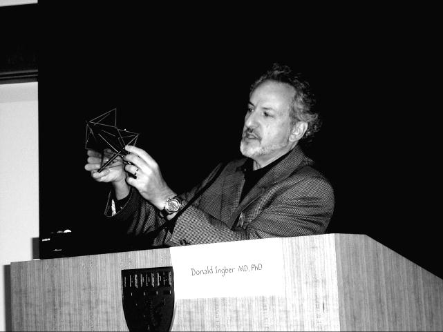
Anyone who is skilled in the art of physical therapy knows that the mechanical properties, behavior and movement of our bodies are as important for human health as chemicals and genes. However, only recently have scientists and physicians begun to appreciate the key role that mechanical forces play in biological control at the molecular and cellular levels. This article provides a brief overview of a lecture presented at the 1st International Fascia Research Congress that convened at Harvard Medical School in Boston, MA on October 4, 2007. (see figure 1) In this lecture, I described what we have learned over the past thirty years as a result of our research focused on the molecular mechanisms by which cells sense mechanical forces and convert them into changes in intracellular biochemistry and gene expression – a process called “mechanotransduction”. This work has revealed that molecules, cells, tissues, organs, and our entire bodies use “tensegrity” architecture to mechanically stabilize their shape, and to seamlessly integrate structure and function at all size scales. Through use of this tension-dependent building system, mechanical forces applied at the macroscale produce changes in biochemistry and gene expression within individual living cells. This structure-based system provides a mechanistic basis to explain how application of physical therapies might influence cell and tissue physiology.
Keywords: tensegrity, mechanotransduction, cytoskeleton, integrins, cell tension, physical therapy
To understand how physical manipulation or movement of our bodies influence cellular biochemistry and tissue physiology, it is necessary to understand how our tissues and organs are structured across multiple size scales. The compression-resistant bones of our skeleton are smaller elements of a larger supporting framework, the musculoskeletal system, which is comprised of an interconnected network of bones, muscles, cartilage, ligaments, tendons, and fascia. This is a physically integrated framework that supports the weight of our bodies, allows us to rapidly adjust to resist external forces, and permits us to move freely in our environment. But without the aid of surrounding tension-generating muscles and tension-resisting tendons, ligaments and fascia, bones and cartilage would do little to support our upright forms.
Engineering teaches us that the stability of a structural network is determined by the material properties of the building elements, their arrangement, and the “play” (free movement) in the joints that interlink these elements. Our bodies stabilize these critical joint regions, by imposing an internal tension or “prestress” to reduce the play in the system; this ensures immediate mechanical responsiveness (i.e., that movement of one element is felt by all others) and reduces impact fatigue at the joint. Thus, opposing muscles and bones establish a mechanical force balance and place our entire musculoskeletal system in a state of isometric tension, so that they experience this type of stabilizing prestress. Hence, the shape stability of our arm or leg (whether it is stiff or floppy) depends on the level of tension or “tone” in our muscles. Architects call this type of prestressed structural network, composed of opposing tension and compression elements that self-stabilizes its shape through establishment of a mechanical force balance, a tensegrity (tensional-integrity) structure.
When we move our muscles and bones, we add mechanical energy to this mechanical equilibrium that already exists in our bodies. This results in stress channeling through the load-bearing elements, which induces physical distortion of the living tissues that comprise these organs. To understand how this physical process might impact human health, we must take into account that living organisms, such as man, are constructed from tiers of systems within a system within a system. Each organ, such as a whole muscle, is constructed from tissues (e.g., muscle fibers, endothelium, tendon, nerve), which are composed of living cells that are linked together by extracellular matrix (ECM) scaffolds. Cells adhere to these ECMs (composed of collagens, glycoproteins, and proteoglycans) via binding of specific cell surface receptors, known as “integrins”.
Integrins span the cell’s surface membrane and form a multi-molecular bridge to the internal “cytoskeleton” of the cell – an internal molecular framework composed of actin microfilaments, microtubules and intermediate filaments that gives shape to the cell. Many of the actin filaments closely associate with myosin filaments, which slide along each other, shorten and thereby, generate mechanical tension that is distributed to all elements of the cell, as well as to the external ECM, via their integrin contact points. Importantly, this is observed in the cytoskeleton of all cells (e.g., epithelial cells, nerve cells, immune cells, bone cells and fibroblasts) and not just in muscle cells.
The tensional forces that are generated in contractile acto-myosin filaments in the cytoskeleton are resisted by the cells external tethers to the ECM, and by other cytoskeletal filaments that resist being compressed (e.g., microtubules, cross-linked actin filament bundles) by these inward-directed forces. For this reason, all cells in our tissues also exist in a state of isometric tension, and it is because of this internal prestress that surgeons need to suture together wounds when they incise living organs. Thus, tensegrity is used to stabilize the shape of living cells, tissues and organs, as well as our whole bodies.
We and others have developed theoretical models based on tensegrity that effectively predict mechanical behaviors of various types of cells, as well as individual molecules, viruses, tissues and whole organs. Moreover, we have carried out many experimental studies that confirm the prediction of these models. These studies have revealed that because our bodies use tensegrity, mechanical forces applied at the macroscale are channeled through discrete load-bearing ECMs of our organs and tissues, and focused on cell adhesion sites where they physically couple their contractile cytoskeleton to the ECM.
For this reason, integrins have been shown in a wide variety of tissues to act as mechanoreceptors – they are among the first molecules on the cell surface to sense a mechanical signal, and they transmit it across the cell surface via a specific molecular pathway. Moreover, forces transmitted over these receptors, but not other transmembrane receptors in the same cells, are converted into changes in intracellular biochemistry and gene expression. Some of this is accomplished directly at the site where the integrins are mechanically coupled to the ends of the contractile acto-myosin filaments on the inner surface of the membrane within macromolecular anchorage complexes called “focal adhesions”. When mechanical stresses are focused on these sites, a subset of the molecules that make up these anchoring structures change their shape and biochemical activities. For example, focal adhesions undergo self-assembly and grow in size when stresses are increased, whereas they disassemble when stress is dissipated. At the same time, signal transduction molecules in the focal adhesion, that also bear these mechanical loads, change their activities as well; this results in activation of calcium influx through stress-sensitive ion channels, protein phosphorylation, and increased signaling through the cAMP, large G protein, and small GTPase pathways.
These intracellular chemical signals and second messengers can alter gene expression, protein synthesis and other aspects of metabolism, much in the way they do when hormones, cytokines and growth factors bind their cell surface receptors and elicit production of similar chemical signals. However, because cells use tensegrity to stabilize themselves, mechanical stresses that are conveyed across integrins also can be channeled through load-bearing cytoskeletal elements in the cytoplasm and nucleus. Importantly, mechanical signals are transmitted much more quickly through these discrete load-bearing elements, than are chemical signals (e.g., wave propagation is much faster than chemical diffusion). In fact, experiments have confirmed that mechanical stress application to surface integrin receptors results in almost immediate changes in molecular structure deep in the cytoplasm and nucleus, as well as activation of signaling events at these distant sites (e.g., Src kinase activation, nuclear calcium signaling) almost as rapidly as those that occur at the cell surface.
Our work on tensegrity in cells and other living materials has revealed that use of this architectural system for structural organization provides a mechanism to physically integrate part and whole. Every time we move our arms, the muscles contract, the bones compress, and the skin stretches without any irreversible injury. This is made possible because most of the load-bearing elements of the discrete cellular and ECM networks that comprise living tissues rearrange in response to stress, and then return to their original position when it is released, as is observed in all tensegrities. If stresses are excessive or sustained, then our bodies remodel themselves through “mechanochemistry”, i.e., force-dependent changes in molecular polymerization-depolymerization dynamics or alterations molecular biochemistry. In this way, tensegrity governs how mechanical forces influence the form and function of the living cells that inhabit all of our tissues.
In conclusion, physical therapies, dance, exercise and various forms of movement can indeed influence cellular activities, including cell growth, differentiation, and potentially even immune cell responses that are critical to human health. This is an obvious fact to anyone who has ever exercised regularly or has experienced the wonderful healing powers of physical therapy. Chemicals and genes are absolutely of critical importance; however, they are governed by mechanical forces, as well as chemical cues.
Relevant Publications
Ingber, DE. 1998 (Jan) The Architecture of Life. Scientific American 278:48–57.
Chen CS, Ingber DE. 1999 Tensegrity and mechanoregulation: from skeleton to cytoskeleton. Osteoarthritis Cartilage, 7(1):81–94.
Ingber DE. 2003 Cellular tensegrity revisited I. Cell structure and hierarchical systems biology. J. Cell Sci 116:1157–1173.
Ingber DE. 2003 Mechanobiology and diseases of mechanotransduction. Ann. Med 35:564–577.
Ingber DE. 2006 Cellular mechanotransduction: putting all the pieces together again. FASEB J. 20:811–827.
Ingber DE 2008 Tensegrity-based mechanosensing from macro to micro. Progress Biophys. Mol. Biol. 2008 Feb 13; [Epub ahead of print]
Figure 1.
Caption: Donald Ingber with a tensegrity model, Fascia Congress, Boston, October 2007
Footnotes
Publisher's Disclaimer: This is a PDF file of an unedited manuscript that has been accepted for publication. As a service to our customers we are providing this early version of the manuscript. The manuscript will undergo copyediting, typesetting, and review of the resulting proof before it is published in its final citable form. Please note that during the production process errors may be discovered which could affect the content, and all legal disclaimers that apply to the journal pertain.


