Abstract
The species constituting the genus Malassezia are considered to be emergent opportunistic yeasts of great importance. Characterized as lipophilic yeasts, they are found in normal human skin flora and sometimes are associated with different dermatological pathologies. We have isolated seven Malassezia species strains that have a different Tween assimilation pattern from the one typically used to differentiate M. furfur, M. sympodialis, and M. slooffiae from other Malassezia species. In order to characterize these isolates of Malassezia spp., we studied their physiological features and conducted morphological and molecular characterization by PCR-restriction fragment length polymorphism and sequencing of the 26S and 5.8S ribosomal DNA-internal transcribed spacer 2 regions in three strains from healthy individuals, four clinical strains, and eight reference strains. The sequence analysis of the ribosomal region was based on the Blastn algorithm and revealed that the sequences of our isolates were homologous to M. furfur sequences. To support these findings, we carried out phylogenetic analyses to establish the relationship of the isolates to M. furfur and other reported species. All of our results confirm that all seven strains are M. furfur; the atypical assimilation of Tween 80 was found to be a new physiological pattern characteristic of some strains isolated in Colombia.
The genus Malassezia comprises lipophilic yeasts found in the normal flora of human skin and other mammals. These yeasts were described by Eichstedt in 1848 as being associated with pityriasis versicolor (PV) lesions (13). The taxonomy and nomenclature of the genus Malassezia was controversial for many decades. Indeed, until 1990 only three species were recognized: M. furfur, M. sympodialis, and M. pachydermatis, a non-lipid-dependent species (17, 21, 38). The species M. globosa, M. restricta, M. obtusa, and M. slooffiae were described in 1995 by morphology, ultrastructure, physiology, and molecular techniques (18, 20). In the last few years, M. dermatis, M. japonica, M. nana, and M. yamatoensis (26, 40-42) have been reported as Malassezia species. Recently, Cabañes et al. described two new species, M. equina and M. caprae, which were isolated from domestic animals (4).
Nine of the 13 species within the genus, M. furfur, M. sympodialis, M. globosa, M. restricta, M. slooffiae, M. obtusa, M. dermatis, M. japonica, and M. yamatoensis, are associated with normal human flora and pathologies. Four species, M. pachydermatis, M. nana, M. equina, and M. caprae, are associated with animals (4, 6, 11, 17, 18, 20, 21, 26, 34, 38, 40-42).
Malassezia species have been associated with diverse dermatological pathologies, including PV, seborrheic dermatitis dandruff, atopic dermatitis, folliculitis, psoriasis, onicomycosis, and blepharitis. M. furfur and M. pachydermatis have been associated with systemic infections in patients with underlying diseases and those receiving intravenous lipid emulsions (6, 7, 9-11, 16, 29, 33, 34).
Although the role of Malassezia species in the development of these diseases is not clear, some authors suggest that M. globosa is the causal agent of PV, while others have found a greater percentage of isolates of M. sympodialis associated with the disease. Differences in diagnosis might be due to sampling methods and differences between the culture media used, leading to controversies in clinical studies of these dermatological pathologies (1, 7, 9, 10).
New physiological patterns for identification have been described, and recently the availability of molecular biology and sequencing techniques has allowed the species to be distinguished more clearly (17-21). Despite the difficulty in isolating, maintaining, and identifying these yeasts, different characteristics of the genus, such as macroscopic and microscopic morphology and some physiological aspects (e.g., the presence/absence of catalase, selective growth on Cremophor EL, β-glucosidase activity, and growth and pigment production on a pigment-producing medium [p-agar]), allow them to be differentiated (18, 22, 30, 31). The assimilation of Tween 20, 40, 60, and 80 by M. furfur, M. sympodialis, and M. slooffiae yields a specific pattern that easily differentiates them from other species (18, 22).
In a previous study, we isolated a total of 154 strains of Malassezia spp., 7 of which were omitted and remained uncharacterized because of an atypical Tween assimilation pattern (35). The aim of the present study was to characterize these seven isolates by investigating their physiological features and conducting morphological and molecular characterization by PCR-restriction fragment length polymorphism (RFLP) and the sequencing of the 26S and 5.8S ribosomal DNA-internal transcribed spacer 2 (rDNA-ITS2) regions for all of the strains. Establishing a relationship with reported species through phylogenetic analyses will support our findings.
MATERIALS AND METHODS
Strains and growth conditions.
The strains of Malassezia spp. with a Tween assimilation pattern different from that of the other strains were obtained as uncharacterized isolates from a previous study (35). All strains were preserved in 10% skim milk at −80°C (8) and recovered in modified Dixon agar by incubation for 4 to 5 days at 32°C (18, 22).
Phenotypic characterization.
The morphological characteristics of the colonies were recorded, and Gram staining was performed. The morphology of the stained cells was assessed by light microscopy. Physiological characteristics were assessed as previously described. The following tests were performed five times: assimilation (Tween 20, 40, 60, and 80 and Cremophor EL), enzyme production (β-glucosidase and catalase), pigment production on tryptophan-based medium, and growth (on Sabouraud, Sabouraud plus 10% Tween 20, and Dixon agar at 37 and 42°C) (18, 22, 30, 31).
Molecular characterization. (i) DNA extraction.
Colonies grown on Dixon agar at 32°C for 4 to 5 days were transferred to 600 μl sterile distilled water and centrifuged at 10,000 rpm for 5 min. DNA was extracted as previously described (36).
(ii) PCR-RFLP of 26S and 5.8S rDNA-ITS2 regions.
The 26S rDNA region was PCR amplified using forward and reverse primers as previously described (32). The 5.8S rDNA-ITS2 regions were PCR amplified using primers ITS3 and ITS4 (15). All PCRs were performed in a final volume of 25 μl (46).
The amplified 26S rDNA product was digested with 10 U of the restriction enzymes CfoI (Sigma-Aldrich) and BstF5I (SibEnzyme, Novosibirsk, Russia) by incubation for 4 h at 37 and 65°C, respectively. The amplified 5.8S rDNA-ITS2 product was digested with 10 U of AluI (Promega) by incubation at 37°C for 4 h. The products were visualized by 2% agarose gel electrophoresis using Quantity One software (Bio-Rad, Hercules, CA).
(iii) Sequencing and phylogenetic analysis.
Single electrophoretic bands from the PCR products were sequenced using a 3730xl DNA analyzer (PE Applied Biosystems). Sequence assembly and editing were performed manually on the software CLC DNA Workbench.
For each region (26S and 5.8S rDNA-ITS2), a data set was formed containing sequences from reported Malassezia species in GenBank. Each data set was aligned using the ClustalW algorithm (45) under default settings in the BIODEDIT package (25). To assess phylogenetic relationships, maximum-parsimony analysis was conducted on the aligned data sets using PAUP* 4.0b8 software under default settings (43), and Filobasidiella neoformans was used as the outgroup for both analyses. A bootstrap resampling was conducted for 1,000 replicates to assess relative branch support (14).
Nucleotide sequence accession number.
The nucleotide sequences (26S and 5.8S rDNA-ITS2 regions) obtained in this study have been deposited in GenBank with accession numbers EU815303 to EU815322 (National Center for Biotechnology Information, Bethesda, MD).
RESULTS
To characterize the isolates of Malassezia spp., we studied their physiological features and conducted morphological and molecular characterization by PCR-RFLP and the sequencing of the 26S and 5.8S rDNA-ITS2 regions in three M. furfur strains from healthy individuals, four M. furfur clinical strains, and eight Malassezia spp. reference strains (Table 1).
TABLE 1.
Sources and origins of strains from Malassezia species used in this study
| Malassezia species | Strain | Origin | Accession no.
|
|
|---|---|---|---|---|
| D1/D2 26S rDNA | 5.8S rDNA-ITS2 | |||
| M. furfur (n = 3) | 13 NL | Healthy individuals | EU815312 | EU815321 |
| 28 NL | Healthy individuals | EU815306 | EU815315 | |
| 39 NL | Healthy individuals | EU815304 | EU815320 | |
| M. furfur (n = 2) | 1828 PV | PV patient | EU815309 | EU815319 |
| 45 PV | PV patient | EU815311 | EU815322 | |
| M. furfur | 12 DSHIV+ | Seborrheic dermatitis- and human immunodeficiency virus-positive patient | EU815303 | EU815313 |
| M. furfur | 4 DS | Seborrheic dermatitis patient | EU815310 | EU815318 |
| M. furfur | CBS 1878b | Human pityriasis capitis | EU815307 | EU815316 |
| M. furfur | CBS 7019b | Human PV | EU815308 | EU815317 |
| M. furfur | CBS 6094 | Normal skin (of the rump) | EU815305 | EU815314 |
| M. globosa | CBS 7966a | Human PV | AY743604 | AY387133-AY38713335 |
| M. restricta | CBS 7877a | Human normal skin | AY743067 | AY387143-AY38714345 |
| M. pachydermatis | CBS 1879b | Dog otitis externa | DQ915500-DQ915502, AY743605 | AY387139-AY38713942 |
| M. slooffiae | CBS 7956a | Healthy ear of pig | AY743606, AJ249956 | AY387146-AY38714649 |
| M. sympodialis | CBS 7222a | Human normal skin | AY743615, AY743627, AY743628 | AY743638-AY743640 |
Type strain.
Neotype strain.
Phenotypic characterization.
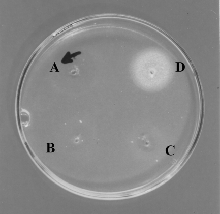
All M. furfur colonies were smooth, opaque, and umbonate. Microscopically, all M. furfur isolates appeared as small ovoid cells (1 to 1.5 by 2 to 2.5 μm), and buds were formed on a broad base. The physiological tests other than Tween assimilation showed the results expected for M. furfur (Table 2). The Tween assimilation test result was different from the standard patterns so far reported for different Malassezia species but coincided with that of the M. furfur CBS 6094 strain. Tween 80 was the only Tween assimilated, as shown in Fig. 1. However, M. furfur CBS 6094 does not produce pigment at 25, 30, 37, or 42°C.
TABLE 2.
Physiological characteristics of Malassezia isolates included in this study
| Species | Utilization of :
|
β-Glucosidase activity | Growth with 10% Tween 20 | Catalase reaction | Growth in Sabouraud | Growth and pigment production in p-agar at:
|
Growth in Dixon agar at:
|
||||||||
|---|---|---|---|---|---|---|---|---|---|---|---|---|---|---|---|
| Tween 20 | Tween 40 | Tween 60 | Tween 80 | Cremophor EL | 25°C | 30°C | 37°C | 42°C | 37°C | 40°C | |||||
| M. furfur 13 NLa | − | − | − | + | + | − | − | + | − | + | + | + | − | + | + |
| M. furfur 28 NLa | − | − | − | + | + | − | − | + | − | + | + | + | − | + | − |
| M. furfur 39 NLa | − | − | − | + | + | − | − | + | − | + | + | + | − | + | − |
| M. furfur 1828 PVa | − | − | − | + | + | − | − | + | − | + | + | + | − | + | − |
| M. furfur 45 PVa | − | − | − | + | + | − | − | + | − | + | + | + | − | + | − |
| M. furfur 12 DSHIV+a | − | − | − | + | + | − | − | + | − | + | + | + | − | + | − |
| M. furfur 4 DSa | − | − | − | + | + | − | − | + | − | + | + | + | − | + | − |
| M. furfur CBS 1878 | + | + | + | + | + | − | + | + | − | + | + | + | − | + | + |
| M. furfur CBS 7019 | + | + | + | + | + | − | + | + | − | + | + | + | − | + | + |
| M. furfur CBS 6094a | − | − | − | + | + | − | − | + | − | − | − | − | − | + | + |
| M. globosa CBS 7966 | − | − | − | − | − | − | − | + | − | − | − | − | − | − | − |
| M. restricta CBS 7877 | − | − | − | − | − | − | − | − | − | − | − | − | − | − | − |
| M. pachydermatis CBS 1879 | + | + | + | + | + | + | + | + | + | − | − | − | − | + | + |
| M. slooffiae CBS 7956 | + | + | + | + | − | − | + | + | − | − | − | − | − | + | − |
| M. sympodialis CBS 7222 | − | + | + | + | − | + | − | + | − | − | − | − | − | + | + |
Isolates with an atypical pattern of Tween assimilation.
FIG. 1.
Atypical Tween 80 assimilation pattern of a Malassezia sp. isolate. (A) Tween 20; (B) Tween 40; (C) Tween 60; (D) Tween 80.
Molecular characterization.
The 26S rDNA PCR amplified an ∼580-bp product; two bands of ∼180 and ∼400 bp were obtained after BstF51 digestion, and three bands of ∼260, ∼130, and ∼70 bp were obtained after CfoI digestion. The 5.8S-ITS2 PCR amplified an ∼500-bp product. AluI digestion yielded two bands of ∼290 and 250 bp (Table 3).
TABLE 3.
RFLP 26S rDNA and 5.8S rDNA-ITS2 PCR products generated by BstF51, CfoI, and AluI, respectively, for Malassezia isolates included in this study
| Malassezia isolate(s) | 26SrDNA amplification product (bp) | 5.8S rDNA-ITS2 amplification product (bp) | BstF51 digest (bp) | CfoI digest (bp) | AluI digest (bp) |
|---|---|---|---|---|---|
| Strains with atypical Tween patterna | 580 | 500 | 400, 180 | 260, 130, 70 | 290, 250 |
| M. furfur CBS 1878 | 580 | 500 | 400, 180 | 260, 130, 70 | 290, 250 |
| M. furfur CBS 7019 | 580 | 500 | 400, 180 | 260, 130, 70 | 290, 250 |
| M. furfur CBS 6094 | 580 | 500 | 400, 180 | 260, 130, 70 | 290, 250 |
| M. globosa CBS 7966 | 580 | 410 | 480, 100 | 480,100 | 240, 170 |
| M. restricta CBS 7877 | 580 | 410 | 500, 70 | NRSb | NRS |
| M. pachydermatis CBS 1879 | 580 | 470 | 500, 70 | 250, 230, 100 | 360, 100 |
| M. slooffiae CBS 7956 | 580 | 430 | NRS | 250, 110, 100, 70 | 320, 110 |
| M. sympodialis CBS 7222 | 580 | 400 | 400, 180 | 390, 190 | NRS |
Isolates with the atypical pattern of Tween assimilation were 13 NL, 28 NL, 39 NL, 1828 PV, 45PV, 12 DSHIV, and 4DS.
NRS, no restriction site.
Phylogenetic analysis.
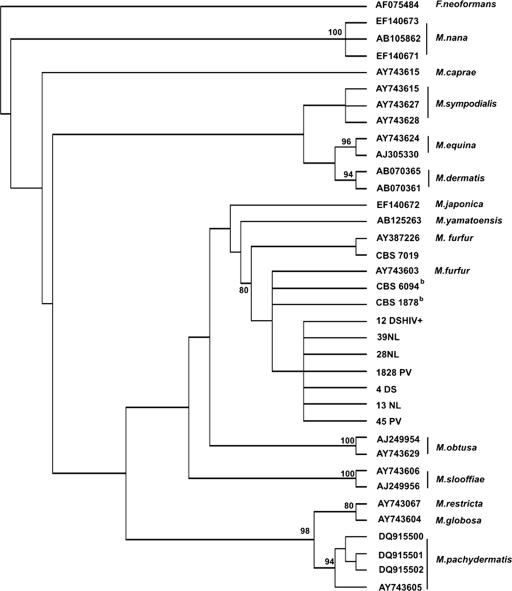
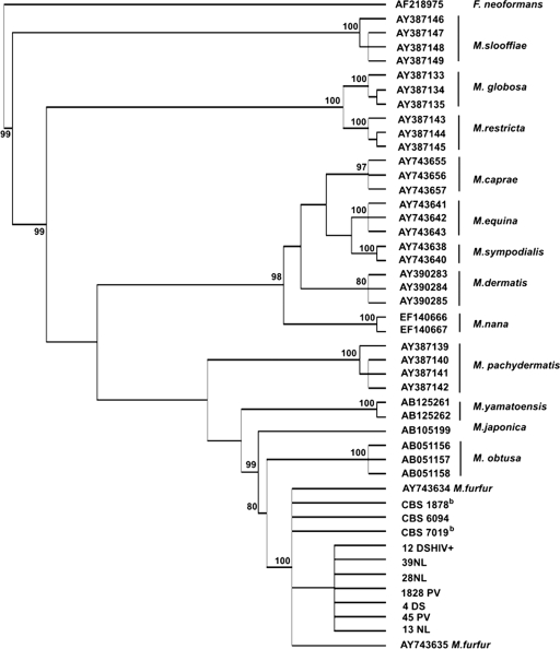
The isolates with the atypical Tween assimilation pattern formed a single cluster that contained the reference M. furfur sequences, with high bootstrap support values of 80 and 100% for the 26S and 5.8S rDNA-ITS2 phylogeny, respectively (Fig. 2 and 3). Although the topologies differed somewhat in support values, the 5.8 rDNA-ITS2 topology showed higher bootstrap support values overall than the 26S topology, suggesting that this region provides a better resolution of the Malassezia spp. Taking both topologies into account, it remains unclear which is the sister group of M. furfur: the 26S topology places this group next to M. yamatoensis, while the 5.8S rDNA-ITS2 topology places it next to M. obtusa.
FIG. 2.
Maximum-parsimony phylogeny of the genus Malassezia based on the D1 and D2 sequences from the 26S rDNA gene. The majority-rule consensus tree is shown. Values above branches correspond to parsimony bootstrap proportions (>80) of 1,000 replicates.
FIG. 3.
Phylogenetic relationships of the genus Malassezia based on the sequences of the 5.8S ITS-rDNA region. The majority-rule consensus tree is shown. Values above branches correspond to parsimony bootstrap proportions (>80) of 1,000 replicates.
DISCUSSION
The Malassezia species are difficult microorganisms to identify and to maintain in culture (9). To overcome these difficulties, several physiological and molecular techniques have been implemented recently that allow the species to be differentiated and new ones to be described (2, 15, 17, 18, 20-24, 27, 38, 44). In this work, we thoroughly characterized seven isolates with an atypical Tween 80 assimilation pattern, and our results support the view that these isolates correspond to M. furfur.
Because these species are lipophilic, the Tween assimilation pattern allows us to identify different Malassezia species (18, 22). This test is based on the ability of Malassezia spp. to use different fatty acids. Some authors attribute this characteristic to lipase activity and propose that it is related to the adaptation of Malassezia spp. to host body regions rich in fatty acids under given conditions (3, 12, 37, 47). Brunke et al. described the lipophilic activity of MfLip in M. furfur; this extracellular lipase apparently is involved in cellular growth processes and related to pathogenicity mechanisms (3).
On the other hand, M. furfur has been described as showing high phenotypic and genotypic variability. Indeed, colonies of this species vary in size and form and show a high degree of cellular pleomorphism, including oval, cylindrical, and spherical cells (18, 22). Techniques such as randomly amplified polymorphic DNA and amplified fragment length polymorphism have demonstrated high intraspecific variability in this species (2, 5, 44). The atypical assimilation pattern of our isolates, involving an inability to utilize Tween 20, 40, or 60, reveals a metabolic variation probably resulting from alternate gene expression.
Such variability has been observed in related organisms such as Candida spp. Lan et al. found a relationship between phenotypic variation and metabolic flexibility in the genus Candida, which increases its selection according to the availability of nutrients in certain body regions (28). Phenotypic and genotypic variability such as the atypical Tween assimilation pattern in the case of M. furfur may, like the variability in Candida spp., be a determining factor in the infection strategy used by the microorganism, which in turn could be related to the expression of genes involved in pathogenic processes and colonization (39).
Shifts in metabolic processes could be related to a change of condition from commensal to pathogenic. The resulting high variability also may be influenced by host responses such as the generation of adverse microenvironments, which perturb the host-pathogen balance (7, 9, 16, 29, 33). Nonetheless, the variation in the Tween assimilation pattern observed in this research (based on the seven atypical strains) was not related to a particular dermatological condition, since it was present both in patients with a dermatological condition and those without apparent injury.
The expected results for 26S (32) and 5.8S rDNA-ITS2 (15) were obtained with all 15 isolates, including the reference ones. The PCR products and the products of digestion revealed no differences between the isolates with the atypical Tween assimilation pattern and the reference strains M. furfur CBS 1878 and CBS 7019. Our sequence analyses based on homology searches of the ribosomal regions identified our isolates as M. furfur. Furthermore, the phylogenetic analyses for 26S and 5.8S rDNA-ITS2 grouped our isolates and reference strains together in a single cluster with high bootstrap values (Fig. 2 and 3). The phylogenies obtained in this study support our findings and the identity of the isolates as M. furfur; the atypical assimilation of Tween 80 was found to be a new physiological pattern characteristic of certain strains isolated in Colombia.
In conclusion, we suggest further studies that will explain the genetic background of the atypical Tween assimilation pattern and its relationship to the establishment of the yeast in certain host body regions. The evaluation of the metabolic pathways and the gene products involved in the fatty acid degradation, and even the relationship between gene expression and diverse host conditions, would produce a better understanding of this unique Tween assimilation pattern, which constitutes a new parameter for the identification of M. furfur.
Acknowledgments
We express our acknowledgment to the Science Faculty of the Universidad de los Andes for the financial support of this project.
Footnotes
Published ahead of print on 29 October 2008.
REFERENCES
- 1.Ashbee, H. R. 2007. Update on the genus Malassezia. Med. Mycol. 45287-303. [DOI] [PubMed] [Google Scholar]
- 2.Boekhout, T., M. Kamp, and E. Gueho. 1998. Molecular typing of Malassezia species with PFGE and RAPD. Med. Mycol. 36365-372. [DOI] [PubMed] [Google Scholar]
- 3.Brunke, S., and B. Hube. 2006. MfLIP1, a gene encoding an extracellular lipase of the lipid-dependent fungus Malassezia furfur. Microbiology 152547-554. [DOI] [PubMed] [Google Scholar]
- 4.Cabañes, F. J., B. Theelen, G. Castella, and T. Boekhout. 2007. Two new lipid-dependent Malassezia species from domestic animals. FEMS Yeast Res. 71064-1076. [DOI] [PubMed] [Google Scholar]
- 5.Celis, A. M., and M. C. Cepero de Garcia. 2005. Genetic polymorphism of Malassezia spp. yeast isolates from individuals with and without dermatological lesions. Biomedica 25481-487. [PubMed] [Google Scholar]
- 6.Chryssanthou, E., U. Broberger, and B. Petrini. 2001. Malassezia pachydermatis fungaemia in a neonatal intensive care unit. Acta Paediatr. 90323-327. [PubMed] [Google Scholar]
- 7.Crespo Erchiga, V., A. Ojeda Martos, A. Vera Casano, A. Crespo Erchiga, and F. Sanchez Fajardo. 2000. Malassezia globosa as the causative agent of pityriasis versicolor. British J. Dermatology 143799-803. [DOI] [PubMed] [Google Scholar]
- 8.Crespo, M. J., M. L. Abarca, and F. J. Cabanes. 2000. Evaluation of different preservation and storage methods for Malassezia spp. J. Clin. Microbiol. 383872-3875. [DOI] [PMC free article] [PubMed] [Google Scholar]
- 9.Crespo, V., and V. Delgado. 2002. Malassezia species in skin diseases. Current Opinion in Infectious Diseases. 210. [DOI] [PubMed] [Google Scholar]
- 10.Crespo-Erchiga, V., and V. D. Florencio. 2006. Malassezia yeasts and pityriasis versicolor. Curr. Opin. Infect. Dis. 19139-147. [DOI] [PubMed] [Google Scholar]
- 11.Dankner, W. M., S. A. Spector, J. Fierer, and C. E. Davis. 1987. Malassezia fungemia in neonates and adults: complication of hyperalimentation. Rev. Infect. Dis. 9743-753. [DOI] [PubMed] [Google Scholar]
- 12.DeAngelis, Y. M., C. W. Saunders, K. R. Johnstone, N. L. Reeder, C. G. Coleman, J. R. Kaczvinsky, Jr., C. Gale, R. Walter, M. Mekel, M. P. Lacey, T. W. Keough, A. Fieno, R. A. Grant, B. Begley, Y. Sun, G. Fuentes, R. S. Youngquist, J. Xu, and T. L. Dawson, Jr. 2007. Isolation and expression of a Malassezia globosa lipase gene, LIP1. J. Investig. Dermatol 1272138-2146. [DOI] [PubMed] [Google Scholar]
- 13.Eichstedt, C. 1846. Pilzbildung in der Pityriasis versicolor. Frorip Neue Notizen aus dem Gebeite der Naturkunde Heilkinde 39270-271. [Google Scholar]
- 14.Felsenstein, J. 1985. Phylogenies and the comparative method. Am. Naturalist 1251-15. [Google Scholar]
- 15.Gaitanis, G., A. Velegraki, E. Frangoulis, A. Mitroussia, A. Tsigonia, A. Tzimogianni, A. Katsambas, and N. J. Legakis. 2002. Identification of Malassezia species from patient skin scales by PCR-RFLP. Clin. Microbiol. Infect. 8162-173. [DOI] [PubMed] [Google Scholar]
- 16.Guého, E., T. Boekhout, H. R. Ashbee, J. Guillot, A. Van Belkum, and J. Faergemann. 1998. The role of Malassezia species in the ecology of human skin and as pathogens. Med. Mycol. 36(Suppl. 1)220-229. [PubMed] [Google Scholar]
- 17.Guého, E., and S. A. Meyer. 1989. A reevaluation of the genus Malassezia by means of genome comparison. Antonie van Leeuwenhoek 55245-251. [DOI] [PubMed] [Google Scholar]
- 18.Guého, E., G. Midgley, and J. Guillot. 1996. The genus Malassezia with description of four new species. Antonie van Leeuwenhoek 69337-355. [DOI] [PubMed] [Google Scholar]
- 19.Guillot, J., M. Deville, M. Berthelemy, F. Provost, and E. Gueho. 2000. A single PCR-restriction endonuclease analysis for rapid identification of Malassezia species. Lett. Appl. Microbiol. 31400-403. [DOI] [PubMed] [Google Scholar]
- 20.Guillot, J., and E. Gueho. 1995. The diversity of Malassezia yeasts confirmed by rRNA sequence and nuclear DNA comparisons. Antonie van Leeuwenhoek 67297-314. [DOI] [PubMed] [Google Scholar]
- 21.Guillot, J., E. Gueho, and R. Chermette. 1995. Confirmation of the nomenclatural status of Malassezia pachydermatis. Antonie van Leeuwenhoek 67173-176. [DOI] [PubMed] [Google Scholar]
- 22.Guillot, J., E. Guého, M. Lesourd, G. Midgley, G. Chevrier, and B. Dupont. 1996. Identification of Malassezia species. J. Mycol. Med. 6103-110. [Google Scholar]
- 23.Gupta, A. K., T. Boekhout, B. Theelen, R. Summerbell, and R. Batra. 2004. Identification and typing of Malassezia species by amplified fragment length polymorphism and sequence analyses of the internal transcribed spacer and large-subunit regions of ribosomal DNA. J. Clin. Microbiol. 424253-4260. [DOI] [PMC free article] [PubMed] [Google Scholar]
- 24.Gupta, A. K., Y. Kohli, and R. C. Summerbell. 2000. Molecular differentiation of seven Malassezia species. J. Clin. Microbiol. 381869-1875. [DOI] [PMC free article] [PubMed] [Google Scholar]
- 25.Hall, T. A. 1999. BioEdit: a user-friendly biological sequence alignment editor and analysis program for Windows 95/98/NT. Nucleic Acids Symp. Ser. 4195-98.
- 26.Hirai, A., R. Kano, K. Makimura, E. R. Duarte, J. S. Hamdan, M. A. Lachance, H. Yamaguchi, and A. Hasegawa. 2004. Malassezia nana sp. nov., a novel lipid-dependent yeast species isolated from animals. Int. J. Syst. Evol. Microbiol. 54623-627. [DOI] [PubMed] [Google Scholar]
- 27.Kaneko, T., K. Makimura, M. Abe, R. Shiota, Y. Nakamura, R. Kano, A. Hasegawa, T. Sugita, S. Shibuya, S. Watanabe, H. Yamaguchi, S. Abe, and N. Okamura. 2007. Revised culture-based system for identification of Malassezia species. J. Clin. Microbiol. 453737-3742. [DOI] [PMC free article] [PubMed] [Google Scholar]
- 28.Lan, C. Y., G. Newport, L. A. Murillo, T. Jones, S. Scherer, R. W. Davis, and N. Agabian. 2002. Metabolic specialization associated with phenotypic switching in Candida albicans. Proc. Natl. Acad. Sci. USA 9914907-14912. [DOI] [PMC free article] [PubMed] [Google Scholar]
- 29.Marcon, M. J., and D. A. Powell. 1992. Human infections due to Malassezia spp. Clin. Microbiol. Rev. 5101-119. [DOI] [PMC free article] [PubMed] [Google Scholar]
- 30.Mayser, P., P. Haze, C. Papavassilis, M. Pickel, K. Gruender, and E. Gueho. 1997. Differentiation of Malassezia species: selectivity of Cremophor EL, castor oil and ricinoleic acid for M. furfur. Br. J. Dermatol. 137208-213. [DOI] [PubMed] [Google Scholar]
- 31.Mayser, P., A. Tows, H. J. Kramer, and R. Weiss. 2004. Further characterization of pigment-producing Malassezia strains. Mycoses 4734-39. [DOI] [PubMed] [Google Scholar]
- 32.Mirhendi, H., K. Makimura, K. Zomorodian, T. Yamada, T. Sugita, and H. Yamaguchi. 2005. A simple PCR-RFLP method for identification and differentiation of 11 Malassezia species. J. Microbiol. Methods 61281-284. [DOI] [PubMed] [Google Scholar]
- 33.Nakabayashi, A., Y. Sei, and J. Guillot. 2000. Identification of Malassezia species isolated from patients with seborrhoeic dermatitis, atopic dermatitis, pityriasis versicolor and normal subjects. Med. Mycol. 38337-341. [DOI] [PubMed] [Google Scholar]
- 34.Nicholls, J. M., K. Y. Yuen, and H. Saing. 1993. Malassezia furfur infection in a neonate. Br. J. Hosp. Med. 49425-427. [PubMed] [Google Scholar]
- 35.Rincón, S., A. Celis, L. Sopo, A. Motta, and M. C. Cepero de Garcia. 2005. Malassezia yeast species isolated from patients with dermatologic lesions. Biomedica 25189-195. [PubMed] [Google Scholar]
- 36.Sambrook, J., E. F. Fritsch, and T. Maniatis. 1989. Molecular cloning: a laboratory manual, 2nd ed. Cold Spring Harbor Laboratory Press, Cold Spring Harbor, NY.
- 37.Shibata, N., N. Okanuma, K. Hirai, K. Arikawa, M. Kimura, and Y. Okawa. 2006. Isolation, characterization and molecular cloning of a lipolytic enzyme secreted from Malassezia pachydermatis. FEMS Microbiol. Lett. 256137-144. [DOI] [PubMed] [Google Scholar]
- 38.Simmons, R. B., and E. Guého. 1990. A new species of Malassezia. Mycol. Res. 944. [Google Scholar]
- 39.Staib, P., S. Wirsching, A. Strauss, and J. Morschhauser. 2001. Gene regulation and host adaptation mechanisms in Candida albicans. Int. J. Med. Microbiol. 291183-188. [DOI] [PubMed] [Google Scholar]
- 40.Sugita, T., M. Tajima, M. Takashima, M. Amaya, M. Saito, R. Tsuboi, and A. Nishikawa. 2004. A new yeast, Malassezia yamatoensis, isolated from a patient with seborrheic dermatitis, and its distribution in patients and healthy subjects. Microbiol. Immunol. 48579-583. [DOI] [PubMed] [Google Scholar]
- 41.Sugita, T., M. Takashima, M. Kodama, R. Tsuboi, and A. Nishikawa. 2003. Description of a new yeast species, Malassezia japonica, and its detection in patients with atopic dermatitis and healthy subjects. J. Clin. Microbiol. 414695-4699. [DOI] [PMC free article] [PubMed] [Google Scholar]
- 42.Sugita, T., M. Takashima, T. Shinoda, H. Suto, T. Unno, R. Tsuboi, H. Ogawa, and A. Nishikawa. 2002. New yeast species, Malassezia dermatis, isolated from patients with atopic dermatitis. J. Clin. Microbiol. 401363-1367. [DOI] [PMC free article] [PubMed] [Google Scholar]
- 43.Swofford, D. L. 2002. PAUP*: Phylogenetic analysis using parsimony (and other methods), 4.0 ed. Sinauer Associates, Sunderland, MA.
- 44.Theelen, B., M. Silvestri, E. Gueho, A. van Belkum, and T. Boekhout. 2001. Identification and typing of Malassezia yeasts using amplified fragment length polymorphism (AFLP), random amplified polymorphic DNA (RAPD) and denaturing gradient gel electrophoresis (DGGE). FEMS Yeast Res. 179-86. [DOI] [PubMed] [Google Scholar]
- 45.Thompson, J. D., D. G. Higgins, and T. J. Gibson. 1994. CLUSTAL W: improving the sensitivity of progressive multiple sequence alignment through sequence weighting, position-specific gap penalties and weight matrix choice. Nucleic Acids Res. 224673-4680. [DOI] [PMC free article] [PubMed] [Google Scholar]
- 46.Williams, J. G., A. R. Kubelik, K. J. Livak, J. A. Rafalski, and S. V. Tingey. 1990. DNA polymorphisms amplified by arbitrary primers are useful as genetic markers. Nucleic Acids Res. 186531-6535. [DOI] [PMC free article] [PubMed] [Google Scholar]
- 47.Xu, J., C. W. Saunders, P. Hu, R. A. Grant, T. Boekhout, E. E. Kuramae, J. W. Kronstad, Y. M. Deangelis, N. L. Reeder, K. R. Johnstone, M. Leland, A. M. Fieno, W. M. Begley, Y. Sun, M. P. Lacey, T. Chaudhary, T. Keough, L. Chu, R. Sears, B. Yuan, and T. L. Dawson, Jr. 2007. Dandruff-associated Malassezia genomes reveal convergent and divergent virulence traits shared with plant and human fungal pathogens. Proc. Natl. Acad. Sci. USA 10418730-18735. [DOI] [PMC free article] [PubMed] [Google Scholar]


