Abstract
Objective
To describe the magnetic resonance (MR) appearance of intact posterior cruciate ligament (PCL) grafts.
Materials and Methods
Thirty-one postoperative MR examinations were performed in 21 grafts of 20 patients after PCL reconstruction. All 21 grafts were proven to be intact on second-look arthroscopic examination. Two musculoskeletal radiologists retrospectively analyzed the MR findings and reached decisions by consensus. The signal intensity (SI) of the graft on proton density-weighted and T2-weighted images, as well as the shapes, locations, and segments of increased SI were recorded. The graft thickness was also recorded and correlated to elapsed time since reconstructive surgery.
Results
The SI of the graft was high (15/31, 48%), intermediate (10/31, 32%), or low (6/31, 19%) on proton density-weighted images, and high (9/31, 29%), intermediate (6/31, 19%), or low (16/31, 52%) on T2-weighted images. The graft SI decreased significantly as postoperative time elapsed. The shape of the increased SI within the grafts was band-like (14/25, 56%) or focal (11/25, 44%). The increased SI was located in the proximal (18/25, 72%), middle (21/25, 82%), and distal (12/25, 48%) segments. In the axial plane, the location of increased SI was intrasubstance (19/25, 76%) or peripheral (10/25, 40%). A 'focal' shape of increased SI was found significantly more in Achilles tendon allografts, while a band-like shape was more frequent in autogenous double-loop hamstring tendon grafts. Graft thickness ranged from 5-15 mm. The difference in graft thickness relative to postoperative time was not statistically significant (p = 0.79).
Conclusion
Stable PCL grafts commonly showed an increased SI at any segment or location, even though they were stable. The shape of increased SI differed according to allograft donor sites. However, SI tended to decrease as time elapsed.
Keywords: Reconstructive surgical procedure, Posterior cruciate ligament, Magnetic resonance (MR)
Although posterior cruciate ligament (PCL) injuries are much less common than anterior cruciate ligament (ACL) injuries, loss of the PCL has a significant impact on knee joint mechanics, including posterior subluxation of the tibia on the femur (1, 2). However, surgical indications for PCL reconstruction remain controversial. Most patients with grade I and II PCL laxity do well with conservative treatment (3). In contrast, patients with grade III PCL laxity, persistent symptoms, chronic instability and osteoarthritis have led physicians to increasingly consider surgical reconstruction (3-6). Surgical treatment is advocated for patients with bone avulsion fractures, acutely symptomatic knees with substantial PCL laxity (grade III), combined ligament injuries, or chronic symptomatic PCL laxity (2, 7-10).
Until recently, the results of PCL reconstruction have been less satisfactory than those for ACL (2). The reasons for PCL graft failure are multifactorial, including surgical technique, graft selection, graft fixation, and postoperative management (11). MR imaging is a widely used noninvasive means of evaluating the status of ACL grafts. However, as fewer PCL reconstructions have been performed in the past, few studies have described the MR appearance of the postoperative PCL (12-15). Thus, the purpose of this study was to describe the MR appearance of intact PCL grafts.
MATERIALS AND METHODS
Patients
From March 1997 to December 2003, a total of 58 PCL arthroscopic reconstructions were performed in 57 patients by a single experienced orthopedic surgeon. Among these 58 PCL grafts, postoperative MR examinations were performed for 50 patients with 51 grafts as part of the postoperative follow-up protocol, and 20 patients with 21 grafts underwent both postoperative MR examination and second-look arthroscopic examination; these 20 patients comprised the study population. Second-look arthroscopy was performed when the patient either had knee pain related to the hardware or wanted the hardware removed. Thus, 16 men and four women (age range, 14-61; mean age, 31 years) were enrolled in our MR analysis. The chief complaints prior to surgery were chronic posterior instability (n = 16), pain and limping (n = 2), and posterolateral instability (n = 3). The investigational review board at our hospital approved this study protocol. Informed consent was waived because of the retrospective nature of this study, which included evaluation of MR images and medical records. All arthroscopic PCL reconstruction surgeries were performed through a posterior trans-septal portal using a transtibial technique. The graft materials included twelve Achilles tendon allografts and nine autogenous double-loop hamstring tendons. The graft material was selected according to the patient's choice after appropriate counseling. Neither the remnant bundle of the PCL nor the meniscofemoral ligaments were debrided during reconstruction. The same surgeon performed the second-look arthroscopic examinations in all 20 patients in order to evaluate graft status and to remove the fixation screw. The interval between PCL reconstruction surgery and second-look arthroscopic examination in these patients ranged from 119 days to 1,598 days (mean, 520 days). All grafts had intact continuity, good tension, and abundant synovialization and vascularization on second-look arthroscopic examinations.
MR Examination and Image Analysis
Thirty-one sets of postoperative MR examinations were obtained from 21 grafts (2 sets of postoperative MR examinations for 8 grafts, 3 sets of postoperative MR examinations for 1 graft). The interval between PCL reconstruction and postoperative MR examination in these patients ranged from 114 days to 1,129 days (mean, 475 days). All patients were examined with a 1.5-T MR imaging system (Signa; GE, Milwaukee, WI) and knee coil (Quadrature coil; GE, Milwaukee, WI). The MR imaging protocols included proton density-weighted fast spin-echo images (TR/TE 2000/20 msec) and fast spin-echo T2-weighted images (TR/TE 2000/80 msec) in the coronal and sagittal planes as double-echo sequences with the following parameters: field of view, 14 cm; excitation number, 4; echo train length, 4; matrix number, 256 × 192; slice thickness, 3 mm; and intersection gap, 1 mm.
Two musculoskeletal radiologists, with three and 10 years of experience, respectively, in the interpretation of knee MR images, independently and retrospectively analyzed the MR images; final decisions on the findings were reached by consensus. The signal intensity (SI) of the intra-articular portion of the grafts and graft thickness were evaluated. The graft SI was evaluated in both coronal and sagittal images. The graft SI was recorded separately on proton density-weighted images and T2-weighted images and classified into grades of low, intermediate, or high compared to the SI of muscles. If there was a region of intermediate or high SI in the intra-articular portion of the graft, its segment (proximal, mid, or distal), location (intrasubstance or periphery), and shape (band-like or focal) were recorded. The thickness of each graft was measured at the mid-point of the intra-articular graft in the sagittal plane and defined as the mean of three measurements.
Statistical Analysis
Statistical analyses were performed using commercially available software (SAS 8.2; SAS Institute, Cary, NC). The following differences were tested: graft thickness relative to time since reconstruction; the frequency of intermediate or high SI between proton density-weighted and T2-weighted images; the shape of intermediate or high SI according to the type of graft and the time since reconstruction; the frequency of intermediate or high SI according to segment and location; and the frequency of intermediate or high SI by segment or location according to time since reconstruction using a mixed model. Differences of the graft SI relative to time since reconstruction were tested using the GEE (Generalized Estimating Equation). Statistical significance was assigned at a p-value < 0.05.
RESULTS
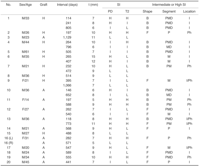
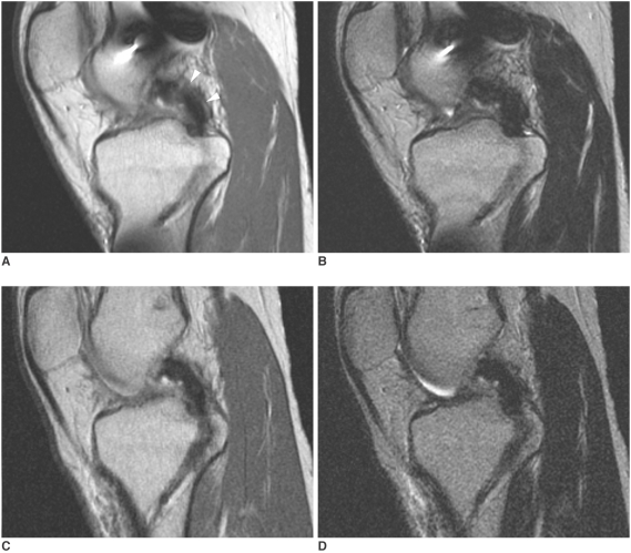
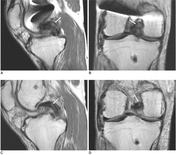
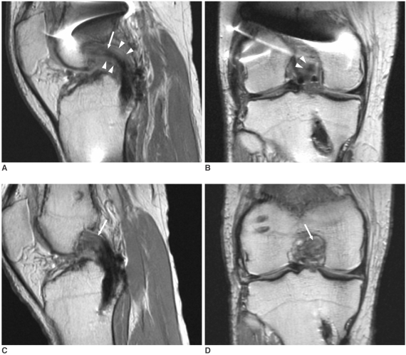
Patient data and results are summarized in Table 1. Fifteen (48%), ten (32%), and six (19%) grafts exhibited high, intermediate, and low SI on proton density-weighted images, respectively, compared with nine (29%), six (19%), and sixteen (52%) on T2-weighted images, respectively. The graft SI decreased significantly as time following reconstruction increased (p < 0.0001 for proton density-weighted images, p = 0.0004 for T2-weighted images) (Fig. 1). Intermediate or high SI within grafts was either band-like (14/25, 56%) (Fig. 2) or focal (11/25, 44%) (Fig. 3). The band-like shape was seen in five MR examinations of three Achilles tendon allografts and nine MR examinations of five autogenous double-loop hamstring tendon grafts. Focal increased graft SI was seen in nine MR examinations of eight Achilles tendon allografts and two MR examinations of two autogenous double-loop hamstring tendons. The shape of intermediate or high SI within grafts differed significantly according to the type of graft (p = 0.04), but was not different according to the time since reconstruction (p = 0.28). The involved segment with intermediate or high SI was proximal in 18 of 25 (72%, 11 MR examinations of 10 Achilles tendon allografts and 7 MR examinations of 5 autogenous double-loop hamstring tendon grafts), middle in 21 of 25 (72%, 11 MR examinations of 7 Achilles tendon allografts and 10 MR examinations of 6 autogenous double-loop hamstring tendon grafts), and distal in 12 of 25 (46%, 6 MR examinations of 5 Achilles tendon allografts and 6 MR examinations of 3 autogenous double-loop hamstring tendon grafts). The incidence of intermediate or high SI was significantly different (p = 0.04) according to the involved segment. The differences in incidence of the involved intermediate or high SI segment according to both graft type and time after reconstruction were not significant (p = 0.54, p = 0.18). On the axial plane, the intermediate or high SI location was intrasubstance (19/25, 76%, 10 MR examinations of 7 Achilles tendon allografts and 9 MR examinations of 5 autogenous double-loop hamstring tendon grafts) and peripheral (10/25, 40%, 7 MR examinations of 5 Achilles tendon allografts and 3 MR examinations of 3 autogenous double-loop hamstring tendon grafts). The incidence of the involved location of intermediate or high SI was significantly different (p = 0.0007) The differences in incidence of the involved location of intermediate or high SI according to both graft type and time after reconstruction were not significant (p = 0.50, p = 0.72). The graft thickness ranged from 5-mm to 15-mm (mean, 8.3-mm). The differences in graft thickness relative to time following reconstruction were not statistically significant (p = 0.79) (Figs. 2, 3).
Table 1.
Summary of MR Findings in Stable PCL Grafts
Note.-Graft (H = autogenous double-loop hamstring tendon, A = Achilles tendon allograft), Interval (time interval between surgery and MR examination), t (thickness), Signal intensity (SI, H = high SI, I = intermediate SI, L = low SI), Shape (B = band-like, F = focal), Segment (P = proximal, M = middle, D = distal), Location (I = intrasubstance, Ph = periphery)
Fig. 1.
Proton density-weighted sagittal (A, C) (TR/TE, 2000/20 msec; echo train length, 4) and T2-weighted sagittal (B, D) (TR/TE, 2000/80 msec; echo train length, 4) images of a 21-year-old man who received posterior cruciate ligament reconstruction using autogenous double-loop hamstring tendon (patient #7). Proton density-weighted image obtained 232 days after posterior cruciate ligament reconstruction shows high band-like peripheral signal intensity (arrowheads) in the proximal and middle segments (A). The high signal intensity has disappeared, and the graft shows homogeneous low signal intensity on proton density-weighted image obtained 472 days after posterior cruciat ligament reconstruction (C). The graft shows homogenous low signal intensity on T2-weighted images (B, D).
Fig. 2.
Proton density-weighted sagittal (A, C) and coronal (B, D) images (TR/TE, 2000/20 msec; echo train length, 4) of a 35-year-old man who received posterior cruciate ligament reconstruction using autogenous double-loop hamstring tendon graft (patient #6). MR images obtained 265 days after posterior cruciate ligament reconstruction (A, B) show high band-like intrasubstance signal intensity (arrows) in the middle segment. The high signal intensity (arrows) persists on MR images obtained 407 days after posterior cruciate ligament reconstruction (C, D). The graft thickness decreases from 15 mm (A) to 12 mm (C).
Fig. 3.
Proton density-weighted sagittal (A, C) and coronal (B, D) images (TR/TE, 2000/20 msec; echo train length, 4) of a 36-year-old man who received posterior cruciate ligament reconstruction using Achilles tendon allograft (patient #13). MR images obtained 118 days after posterior cruciate ligament reconstruction (A, B) show high band-like intrasubstance (arrow) and peripheral signal intensity (arrowheads) in the proximal, middle, and distal segments. MR images obtained 773 days after posterior cruciate ligament reconstruction (C, D) show high focal intrasubstance and peripheral signal intensity (arrows) in the proximal and middle segments. The graft thickness increases from 8 mm (A) to 11 mm (C).
DISCUSSION
An intact PCL graft is presumed to have MR findings analogous to intact ACL grafts (12). Many researchers have stated that increased SI in such grafts might be related to impingement, but it could also be seen in unimpinged, clinically stable grafts (16-21). Autografts or allografts go through four stages following transplantation: necrosis, revascularization, cellular proliferation, and remodeling (22). These stages may explain the changes in graft SI on MR imaging. In previous studies, both clinically stable ACL and PCL grafts primarily exhibited low SI bands with increased SI in the intrasubstance and along the periphery of the graft (10, 14-16, 21, 23); these tended to diminish with time following reconstruction (19, 21, 23, 24). In our study, SI was increased in 26 of 32 grafts (81%) and tended to decrease with time following reconstruction, as expected.
Furthermore, in our study, increased SI was more common intrasubstance and in the middle segment. The intrasubstance increase in SI was more easily detected than that in the periphery. Greater tissue contrast is one possible explanation for this. In addition, the 'magic angle artifact' may play a role in increasing the signal intensity of middle segments. We recorded the overlap of the involved segments, such as proximal and middle segments or middle and distal segments. Therefore, a large number of cases had increased SI in more than one segment, which may influence the overall rate of increased SI in the middle segment. Mariani et al. (15) observed slower graft healing in the area exiting the tibial tunnel, with localized increased SI disappearing at long-term evaluation. They postulated that the sharp angle at the tibial tunnel entrance, the 'killer turn,' may produce abnormal graft stress that results in slower healing, even though increased SI within the graft does not always indicate graft impingement or damage. This hypothesis corresponds well with our results, which showed increased SI in the distal segment of 46% of intact PCL grafts. In terms of the shape, the increased SI was more commonly focal in Achilles tendon allografts and band-like in autogenous double-loop hamstring tendon grafts. Murakami et al. (23) described the transitional findings of stable ACL grafts using double-loop hamstring autografts. These findings initially showed high SI soft tissue surrounding the graft; the surrounding tissue then gradually invaded between the bundles, resulting in inter-bundle high SI. These findings may also correspond to our observation of band-like increased SI in this type of graft. The relatively wide surface area of the 'double loop' graft may lead to abundant synovialization and revascularization, which may cause the band-like increase in SI.
Sherman et al. (14) reported that PCL grafts appeared thicker on MR imaging in the early postoperative period, and that over time the thickness of the grafts gradually decreased. In our study, however, the graft thickness did not correlate to elapsed time after surgery. Remnants from the original PCL bundle and meniscofemoral ligaments in our patients may lead to this discrepancy. Generally, to allow for easier passage of the graft, both the remnant bundle of the PCL and the meniscofemoral ligaments are debrided during reconstruction (8, 9). However, we believe that preserving these structures significantly contributes to posterior stability of the joint and promotes healing of the graft (8, 25).
There are some limitations to this study, including the retrospective design and the small number of patients. Also, because the PCL grafts included in this study were all intact, we could not determine any differences between intact and failed grafts. An insufficient follow-up period is another limitation. A long-term follow-up study of the fate of increased SI in PCL reconstruction and an experimental investigation that includes MR imaging and histological comparison is needed to clarify the clinical significance of this SI.
In conclusion, postoperative MR of stable PCL grafts commonly showed increased SI within PCL grafts at any segment or location, even though the PCL grafts were stable. The shape of the increased SI differed according to the graft type. However, this SI tended to decrease as time elapsed.
References
- 1.Höher J, Harner CD, Vogrin TM, Baek GH, Carlin GJ, Woo SL. In situ forces in the posterolateral structures of the knee under posterior tibial loading in the intact and posterior cruciate ligament-deficient knee. J Orthop Res. 1998;16:675–681. doi: 10.1002/jor.1100160608. [DOI] [PubMed] [Google Scholar]
- 2.Dowd GS. Reconstruction of the posterior cruciate ligament. Indications and results. J Bone Joint Surg. (Br) 2004;86:480–491. [PubMed] [Google Scholar]
- 3.Parolie JM, Bergfeld JA. Long-term results of nonoperative treatment of isolated posterior cruciate ligament injuries in the athlete. Am J Sports Med. 1986;14:35–38. doi: 10.1177/036354658601400107. [DOI] [PubMed] [Google Scholar]
- 4.Covey CD, Sapega AA. Injuries of the posterior cruciate ligament. J Bone Joint Surg Am. 1993;75:1376–1386. doi: 10.2106/00004623-199309000-00014. [DOI] [PubMed] [Google Scholar]
- 5.Richter M, Kiefer H, Hehl G, Kinzl L. Primary repair for posterior cruciate ligament injuries. An eight-year followup of fifty-three patients. Am J Sports Med. 1996;24:298–305. doi: 10.1177/036354659602400309. [DOI] [PubMed] [Google Scholar]
- 6.Berg EE. Posterior cruciate ligament tibial inlay reconstruction. Arthroscopy. 1995;11:69–76. doi: 10.1016/0749-8063(95)90091-8. [DOI] [PubMed] [Google Scholar]
- 7.Miller MD, Bergfeld JA, Fowler PJ, Harner CD, Noyes FR. The posterior cruciate ligament injured knee: principles of evaluation and treatment. Instr Course Lect. 1999;48:199–207. [PubMed] [Google Scholar]
- 8.Ahn JH, Chung YS, Oh I. Arthroscopic posterior cruciate ligament reconstruction using the posterior trans-septal portal. Arthroscopy. 2003;19:101–107. doi: 10.1053/jars.2003.50017. [DOI] [PubMed] [Google Scholar]
- 9.Fanelli GC, Giannotti BF, Edson CJ. The posterior cruciate ligament arthroscopic evaluation and treatment. Arthroscopy. 1994;10:673–688. doi: 10.1016/s0749-8063(05)80067-2. [DOI] [PubMed] [Google Scholar]
- 10.Sanders TG. MR imaging of postoperative ligaments of the knee. Semin Musculoskelet Radiol. 2002;6:19–33. doi: 10.1055/s-2002-23161. [DOI] [PubMed] [Google Scholar]
- 11.Burns WC, 2nd, Draganich LF, Pyevich M, Reider B. The effect of femoral tunnel position and graft tensioning technique on posterior laxity of the posterior cruciate ligament-reconstructed knee. Am J Sports Med. 1995;23:424–430. doi: 10.1177/036354659502300409. [DOI] [PubMed] [Google Scholar]
- 12.Munk PL, Vellet AD, Fowler PJ, Miniaci T, Crues JV., 3rd Magnetic resonance imaging of reconstructed knee ligaments. Can Assoc Radiol J. 1992;43:411–419. [PubMed] [Google Scholar]
- 13.Irizarry JM, Recht MP. MR imaging of the knee ligaments and the postoperative knee. Radiol Clin North Am. 1997;35:45–76. [PubMed] [Google Scholar]
- 14.Sherman PM, Sanders TG, Morrison WB, Schweitzer ME, Leis HT, Nusser CA. MR imaging of the posterior cruciate ligament graft: initial experience in 15 patients with clinical correlation. Radiology. 2001;221:191–198. doi: 10.1148/radiol.2211010105. [DOI] [PubMed] [Google Scholar]
- 15.Mariani PP, Margheritini F, Camillieri G, Bellelli A. Serial magnetic resonance imaging evaluation of the patellar tendon after posterior cruciate ligament reconstruction. Arthroscopy. 2002;18:38–45. doi: 10.1053/jars.2002.29937. [DOI] [PubMed] [Google Scholar]
- 16.Hong SJ, Ahn JM, Ahn JH, Park SW. Postoperative MR findings of the healthy ACL grafts: correlation with second look arthroscopy. Clin Imaging. 2005;29:55–59. [PubMed] [Google Scholar]
- 17.Howell SM, Berns GS, Farley TE. Unimpinged and impinged anterior cruciate ligament grafts: MR signal intensity measurements. Radiology. 1991;179:639–643. doi: 10.1148/radiology.179.3.2027966. [DOI] [PubMed] [Google Scholar]
- 18.Cheung Y, Magee TH, Rosenberg ZS, Rose DJ. MRI of anterior cruciate ligament reconstruction. J Comput Assist Tomogr. 1992;16:134–137. [PubMed] [Google Scholar]
- 19.Yamato M, Yamagishi T. MRI of patellar tendon anterior cruciate ligament autografts. J Comput Assist Tomogr. 1992;16:604–607. doi: 10.1097/00004728-199207000-00021. [DOI] [PubMed] [Google Scholar]
- 20.Horton LK, Jacobson JA, Lin J, Hayes CW. MR imaging of anterior cruciate ligament reconstruction graft. AJR Am J Roentgenol. 2000;175:1091–1097. doi: 10.2214/ajr.175.4.1751091. [DOI] [PubMed] [Google Scholar]
- 21.Jansson KA, Karjalainen PT, Harilainen A, Sandelin J, Soila K, Tallroth K, et al. MRI of anterior cruciate ligament repair with patellar and hamstring tendon autografts. Skeletal Radiol. 2001;30:8–14. doi: 10.1007/s002560000288. [DOI] [PubMed] [Google Scholar]
- 22.Canale ST. Campbell's Operative Orthopaedics. 10th ed. St.Louis: Mosby, Inc; 2003. p. 2166. [Google Scholar]
- 23.Murakami Y, Sumen Y, Ochi M, Fujimoto E, Adachi N, Ikuta Y. MR evaluation of human anterior cruciate ligament autograft on oblique axial imaging. J Comput Assist Tomogr. 1998;22:270–275. doi: 10.1097/00004728-199803000-00021. [DOI] [PubMed] [Google Scholar]
- 24.Trattnig S, Rand T, Czerny C, Stocker R, Breitenseher M, Kainberger F, et al. Magnetic resonance imaging of the postoperative knee. Top Magn Reson Imaging. 1999;10:221–236. doi: 10.1097/00002142-199908000-00004. [DOI] [PubMed] [Google Scholar]
- 25.Buess E, Imhoff AB, Hodler J. Knee evaluation in two systems and magnetic resonance imaging after operative treatment of posterior cruciate ligament injuries. Arch Orthop Trauma Surg. 1996;115:307–312. doi: 10.1007/BF00420321. [DOI] [PubMed] [Google Scholar]


