Abstract
Sclerosing angiomatoid nodular transformation of the spleen is a recently described benign pathologic entity that is characterized by round shaped vascular spaces that are lined by endothelial cells, and the spaces are circumscribed by granulomatoid structures. Microscopically, all the reported cases had multiple angiomatoid nodules in a fibrosclerotic stroma. Each angiomatoid nodule was made up of slit-like, round or irregular shaped vascular spaces that were lined by endothelial cells and interspersed ovoid cells. We present here the CT and dynamic gadolinium-enhanced MR findings of a patient with sclerosing angiomatoid nodular transformation. The spoke-wheel pattern that was observed on MRI in this case may be an important imaging clue for making the correct diagnosis of this benign lesion.
Keywords: Computed tomography (CT), Magnetic resonance (MR), Spleen
The secondary involvement of the spleen in different diseases is more widespread when compared to the spleen's primary lesions (1). Sclerosing angiomatoid nodular transformation (SANT) of the spleen is one of the primary benign diseases of the spleen that has recently been described (2). Although it is defined by its morphological details, any data regarding its radiological appearance is extremely scarce. Microscopically, all the previously reported cases had multiple angiomatoid nodules in a fibrosclerotic stroma. Each angiomatoid nodule was made up of slit-like, round or irregular shaped vascular spaces that were lined by endothelial cells and interspersed ovoid cells. To date, only the CT findings were briefly reported from one case (3). In this article, we present the CT and dynamic MR findings of a case with SANT of the spleen. A spoke-wheel pattern was observed on MRI, which may be important to diagnose this benign condition.
CASE REPORT
A 44-year-old male patient was admitted to our hospital with vague pelvic pain and questionable prostate symptoms. His vital signs and physical examination, as well as the basic biochemical and complete blood count examinations, were within normal limits. His past medical and family history were also unremarkable.
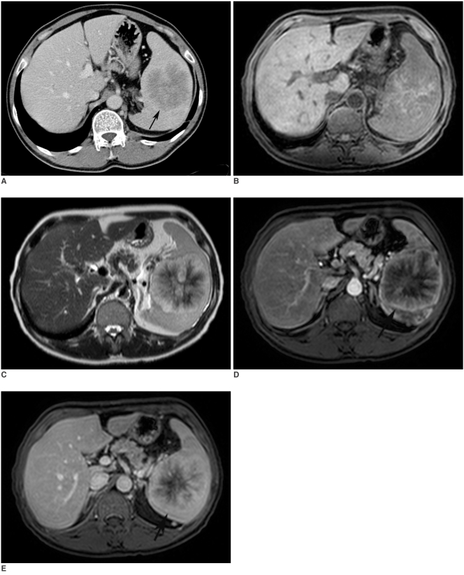
The patient was referred to our department by the attending physician for a CT examination to check for abdominal pathology. CT examination revealed a centrally hypodense lesion that measured 8cm in diameter with well-defined borders and an enhancing peripheral portion that radiated centrally to almost completely fill the splenic parenchyma (Fig. 1A). MRI study confirmed the CT findings, revealing a lesion with heterogeneous intensity with a centrally scattered hyperintense signal on the precontrast fat saturated T1-weighted images; this was suggestive of hemorrhagic components, which were confirmed by pathologic analysis (Fig. 1B). On the T2-weighted images (Fig. 1C), the lesion appeared peripherally hyperintense and centrally hypointense with radiating enhanced areas towards the center of the lesion, which appeared hyperintense. The early arterial phase T1-weighted gradient echo images (Figs. 1D, E) showed peripheral enhancement with progressive central progression in a radiating fashion on the late phase images, and this resembled a spoke-wheel pattern. The central portion was not completely filled on the late phase images, and this was consistent with fibrosis, which was confirmed by the pathology report. There was no lympadenaopathy or any other focal solid organ lesion that was suggestive of metastasis.
Fig. 1.
Sclerosing angiomatoid nodular transformation in 44-year-old man.
A. Axial CT image shows predominantly hypodense mass with lobulated contours. Also note rim-style contrast enhancement of external borders of lesion (arrow).
B. Fat-saturated precontrast T1-weighted image shows central hyperintensity that is consistent with hemorrhage.
C. T2-weighted MR image shows spoke-wheel pattern of lesion that is predominantly hyperintense with central hypointense areas. Note hyperintense radiations towards center of lesion.
D, E Postcontrast arterial (D) and delayed venous phase (E) T1 weighted MR images clearly show progressive enhancement from periphery to center of lesion, which is similar to spoke wheel pattern (arrows).
The patient underwent splenectomy. The gross examination revealed an enlarged spleen weighing 650 gr. A whitish firm nodule with hemorrhagic spiculations extending towards the normal splenic parenchyma was seen on the gross cut section. Microscopic examination depicted that the lesion was primarily composed of multiple angiomatoid nodules that were separated by sclerotic fibrous stroma. Round-shaped vascular spaces lined by endothelial cells were noted to be centrally circumscribed by granulomatoid structures; these were the most prominent morphological features of these nodules. The cells within the nodules had vesicular nuclei and they rarely contain eosinophilic nucleoli with rare mitosis. Hyalinization and fibrosis were noted in the central area of the lesion. Immunohistochemical studies of the nodules revealed CD34-positive, CD31-positive and CD8-negative cells in the peripheral sector, while the central portion was mainly comprised of CD31-positive, CD34- and CD8- negative cells. The final diagnosis was SANT.
The patient did well during the post-surgical period and he was discharged four days after the surgery without any complications.
DISCUSSION
The spleen may be affected during several disease processes, and particularly lymphomas. Non-lymphomatoid tumors of the spleen are unusual and these are primarily of a vascular origin (4). Most of these lesions are comprised of cavernous hemangiomas, while other disorders of this class, namely, hemangioendothelioma, Littoral cell angiomas and hamartomas are rare. SANT of the spleen is a newly defined distinct pathological vascular disorder of the spleen by Martel et al. (2) in 2004; the pathological analysis of this new entity was described in detail in their series of 25 patients. They also concluded in their study that SANT of the spleen is a benign disorder with no tendency for recurrence, and splenectomy offered complete cure to patients. In another manuscript by Li et al. (3), the CT findings were described together with an in-depth pathological analysis of a patient. The reported lesion was hypodense, as in our patient, with an internal calcific focus that became indistinguishable from the surrounding splenic parenchyma on the late portal phase images.
In our patient, the lesion demonstrated diffuse peripheral enhancement on the early arterial phase with progressive centripedal filling in a radiating pattern, while its center remained hypodense and hypointense on the late phase images of CT and MRI, respectively. To the best of our knowledge, the MRI findings of this rare entity have not been published before.
Although rare, SANT should be considered in the differential diagnosis for hypervascular lesions of the spleen. SANT was reported to cause no symptoms in the affected patients and it is generally incidentally detected. Its clinical prognosis was reported to be excellent with complete cure after splenectomy (2).
Making the differential diagnosis among the other benign and malignant vascular lesions of the spleen may be difficult. Angiosarcoma, the most prevalent primary malignant vascular entity, may sometimes be difficult to differentiate from other benign lesions. However, considering their highly aggressive biological nature, it would be highly unusual to see an angiosarcoma of this size to be restricted to only the spleen without any distant metastasis. Littorall cell angioma may be differentiated due to its typical multiple hypodense nodules and considering the solitary appearance of SANT of the spleen (2). Although lymphoma must also be kept in mind for splenic lesions, the absence of splenomegaly, the solitary character of the lesion and the absence of any associated intraabdominal lympadenopathy may be helpful clues for making the differential diagnosis (2). Also, the general well-being of the patient and the absence of any other clinical symptoms regarding lymphoma may be other supportive findings against the diagnosis of lymphoma. It may be difficult or impossible to confidently differentiate SANT of the spleen from other benign disorders of the spleen like hamartoma and inflammatory pseudotumor of the spleen with using only the images. The scarcity of data on the imaging characteristics of the SANT of the spleen make this even more problematic. So, we think that pathological examination and invasive treatment may be almost always indicated for making the correct diagnosis with the currently available data.
In conclusion, SANT is a rare vascular lesion of the spleen and its incidence may increase, after its recent description as a separate clinico-pathologic entity. MRI may be helpful for the diagnosis of splenic nodular angiomatoid transformation by demonstrating a spoke-wheel pattern on MRI with centripedal filling in a radiating pattern, and the splenic mass may contain fibrotic and hemorrhagic components. The description of the radiological and clinical features of this entity in a large series may also increase the recognition of SANT by practicing radiologists.
References
- 1.Elsayes KM, Narra VR, Mukundan G, Lewis JS, Jr, Menias CO, Heiken JP. MR imaging of the spleen: spectrum of abnormalities. Radiographics. 2005;25:967–982. doi: 10.1148/rg.254045154. [DOI] [PubMed] [Google Scholar]
- 2.Martel M, Cheuk W, Lombardi L, Lifschitz-Mercer B, Chan JK, Rosai J. Sclerosing angiomatoid nodular transformation (SANT): report of 25 cases of a distinctive benign splenic lesion. Am J Surg Pathol. 2004;28:1268–1279. doi: 10.1097/01.pas.0000138004.54274.d3. [DOI] [PubMed] [Google Scholar]
- 3.Li L, Fisher DA, Stanek AE. Sclerosing angiomatoid nodular transformation (SANT) of the spleen: addition of a case with focal CD68 staining and distinctive CT features. Am J Surg Pathol. 2005;29:839–841. doi: 10.1097/01.pas.0000160441.23523.d1. [DOI] [PubMed] [Google Scholar]
- 4.Rosai J. Rosai and Ackerman's surgical pathology. 9th ed. Edinburgh: Mosby; 2004. p. 2035. [Google Scholar]


