Abstract
We describe here a case of intraductal tubular carcinoma of the main pancreatic duct. Gadolinium-enhanced pancreas magnetic resonance (MR) imaging showed an enhancing mass that was confined in the dilated main pancreatic duct of the pancreatic body, along with dilatation of the upstream main pancreatic duct and chronic pancreatitis that was due to obstruction. MR cholangiopancreatography and an endoscopic retrograde pancreatogram showed a filling defect that was due to an intraductal mass of the pancreatic body, along with dilatation of the upstream main pancreatic duct and no dilatation of the downstream main pancreatic duct. The pathological findings demonstrated an intraductal nodular appearance without papillary projection or mucin hypersecretion.
Keywords: Intraductal tubular tumor, Intraductal tubular carcinoma, Computed tomography (CT), Magnetic resonance (MR), Pancreas
As defined by the World Health Organization (WHO) definition and grading system, an intraductal tubular carcinoma (ITC) of the pancreas is classified as a subtype of intraductal papillary mucinous neoplasms (IPMNs) (1). However, in the General Rules for the study of Pancreatic Cancer published by the Japan Pancreas Society (2), intraductal tumors of the pancreas are classified into IPMNs and intraductal tubular tumors (ITTs). ITTs are further divided into intraductal tubular adenomas (ITAs) and ITCs. To date, only 12 cases of ITCs have been reported in the clinical literature (3-9). We describe here the imaging findings of a case of intraductal tubular carcinoma of the pancreas that presented as an intraductal mass in the dilated main pancreatic duct.
CASE REPORT
A 63-year-old woman presented with abdominal pain and diarrhea for two weeks. The patient had undergone transabdominal ultrasonography at a local hospital, and the procedure incidentally demonstrated a dilatated main pancreatic duct in the pancreatic tail; she was then referred to our hospital for further evaluation. There was no personal or family history of pancreatic disease. A physical examination revealed no unusual findings. No elevation of the levels of serum amylase and lipase and such tumor markers as carbohydrate antigen 19-9 (CA 19-9) were noted on the laboratory tests.
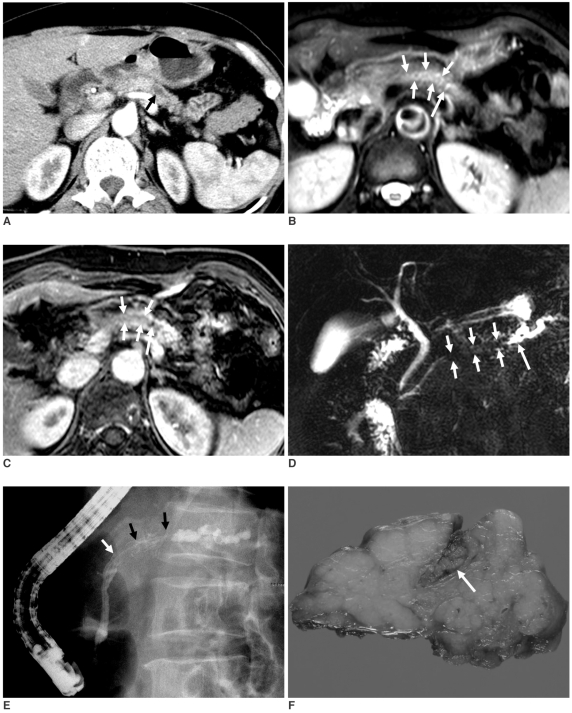
Contrast-enhanced pancreas computed tomography (CT) showed a moderate dilatation of the main pancreatic duct in the pancreatic tail without an obstructive mass (Fig. 1A). No peripancreatic fatty infiltration or lymph node enlargement was noted. Magnetic resonance (MR) imaging with MR cholangiopancreatography (MRCP) was performed to exclude any lesion that could cause obstruction proximal to the dilated main pancreatic duct. The unenhanced and gadolinium-enhanced pancreas MR images (Figs. 1B, C) showed an enhancing mass in the dilated main pancreatic duct of the pancreatic body, which resulted in obstruction and dilatation of the upstream main pancreatic duct, and this caused chronic pancreatitis. MRCP showed a filling defect due to an intraductal mass with dilatation of the upstream main pancreatic duct with no dilatation of the downstream main pancreatic duct (Fig. 1D). An endoscopic retrograde pancreatogram (Fig. 1E) showed a filling defect of the contrast media due to a mass in the dilated, yet patent main pancreatic duct. No hypersecretion of mucin was noted.
Fig. 1.
63-year-old woman with intraductal tubular carcinoma of pancreas that presented as intraductal mass.
A. Contrast-enhanced pancreas CT scan obtained at arterial phase shows only moderate dilatation of main pancreatic duct (arrow) in tail of pancreas, and there is no evidence of obstructive mass. Atrophy with decreased enhancement of pancreatic tail is due to chronic pancreatitis.
B, C. T2-weighted (B) and gadolinium-enhanced (C) pancreas MR images show enhancing intraductal mass (short arrows) of main pancreatic duct in pancreatic body with dilatation of upstream main pancreatic duct (long arrow).
D. MR cholangiopancretography shows filling defect (short arrows) that is due to intraductal mass with dilatation of upstream main pancreatic duct (long arrow) and there is no dilatation of downstream main pancreatic duct.
E. Endoscopic retrograde pancreatogram shows filling defect (arrows) of contrast media that is due to mass in main pancreatic duct in pancreatic body.
F. Photograph of resected specimen shows mass (arrow) in dilated main pancreatic duct.
Under the impression of a mass confined to the main pancreatic duct of the pancreatic body, the patient underwent a subtotal pancreatectomy. Macroscopically, the tumor was 2.5 cm in the maximum diameter and it filled the lumen of the main pancreatic duct (Fig. 1F). Microscopically, the tumor was confined to the main pancreatic duct with focal stromal invasion and the tumor had an intraductal nodular appearance with a monotonous tubular growth pattern, and there was no papillary projection or mucin hypersecretion. On the immunohistochemical staining, the tumor cells were diffusely positive for MUC-1 and p53, and they were negative for MUC-2 and MUC-5AC. The final diagnosis was a T1N0M0 stage ITC.
DISCUSSION
An ITC is an extremely rare pancreatic tumor (2) in the pancreatic duct that shows a nodular or polypoid appearance with a monotonous tubular growth pattern and it is without papillary projection or mucin hypersecretion. On immunohistochemical staining, ITC cells are positive for MUC-1 and they are negative for MUC-2 and MUC-5AC. However, intraductal papillary mucinous carcinoma cells are negative for MUC-1 and most cells are positive for MUC-2 and MUC-5AC (3). There may be two different pathways for the development of an ITC: the "de novo" and "adenoma-carcinoma sequence" pathways (4).
Although the prognosis of an ITT is unclear, ITTs and IPMNs are considered to have similar malignant potential (9). ITTs may have an invasive component and/or they have involvement in lymph node metastasis, and so the prognosis of ITT is ghastly (9). ITCs are considered to be more favorable when compared with a pancreatic ductal adenocarcinoma (9). Therefore, early detection and appropriate surgical resection are crucial to achieve long-term survival in patients with an ITC.
As in the present case, MR imaging with MRCP together with CT and an endoscopic retrograde pancreatogram are very useful for diagnosing an intraductal tumor of the pancreas. However, on the imaging findings, making the diagnosis of ITC from the imaging findings can be confused with that of an IPMN, and especially for the main duct type IPMN because of the similar imaging findings such as dilatation of the pancreatic duct and the presence of an intraductal mass, in addition to the pancreatitis-like symptoms (10). Based on the imaging findings of the present case, the imaging finding of an IPMN shows the dilatation of mainly the downstream main pancreatic duct, which is caused by the hypersecretion of mucin. In contrast ITC results in obstructive dilatation of the upstream main pancreatic duct with no radiological or endoscopic evidence of mucin hypersecretion. For the radiological differentiation of ITCs from ITAs, intraductal growth that completely occupies the dilated portion of the pancreatic duct with large size may favor an ITC. Yet it was difficult to differentiate between ITAs and ITCs in the present case and this was also difficult to do in the previous reports (4, 6, 10). A further study with a large series is needed to resolve this issue.
In conclusion, an ITC should be recognized as another entity of an intraductal tumor of the pancreas that is morphologically and immunohistochemically different from an IPMN. An ITC should be included in the differential diagnosis when an intraductal mass that causes obstructive dilatation of the upstream main pancreatic duct has no radiological or endoscopic evidence of mucin hypersecretion.
References
- 1.Longnecker DS, Adler G, Hruban RH, Kloppel G. Intraductal papillary mucinous neoplasms of the pancreas. In: Hamilton SR, Aaltonen LA, editors. Pathology and genetics of tumours of the digestive system. WHO classification of tumours. Lyon: IARC Press; 2000. pp. 237–240. [Google Scholar]
- 2.Japan Pancreas Society. General Rules for the Study of Pancreatic Cancer. 2nd ed. Tokyo: Kanehara; 2002. [Google Scholar]
- 3.Tajiri T, Tate G, Inagaki T, Kunimura T, Inoue K, Mitsuya T, et al. Intraductal tubular neoplasms of the pancreas: histogenesis and differentiation. Pancreas. 2005;30:115–121. doi: 10.1097/01.mpa.0000148513.69873.4b. [DOI] [PubMed] [Google Scholar]
- 4.Itatsu K, Sano T, Hiraoka N, Ojima H, Takahashi Y, Sakamoto Y, et al. Intraductal tubular carcinoma in an adenoma of the main pancreatic duct of the pancreas head. J Gastroenterol. 2006;41:702–705. doi: 10.1007/s00535-006-1821-2. [DOI] [PubMed] [Google Scholar]
- 5.Tajiri T, Tate G, Kunimura T, Inoue K, Mitsuya T, Yoshiba M, et al. Histologic and immunohistochemical comparison of intraductal tubular carcinoma, intraductal papillary-mucinous carcinoma, and ductal adenocarcinoma of the pancreas. Pancreas. 2004;29:116–122. doi: 10.1097/00006676-200408000-00006. [DOI] [PubMed] [Google Scholar]
- 6.Ito K, Fujita N, Noda Y, Kobayashi G, Kimura K, Horaguchi J, et al. Intraductal tubular adenocarcinoma of the pancreas diagnosed before surgery by transpapillary biopsy: case report and review. Gastrointest Endosc. 2005;61:325–329. doi: 10.1016/s0016-5107(04)02634-3. [DOI] [PubMed] [Google Scholar]
- 7.Thirot-Bidault A, Lazure T, Ples R, Dimet S, Dhalluin-Venier V, Fabre M, et al. Pancreatic intraductal tubular carcinoma: a subgroup of intraductal papillary-mucinous tumors or a distinct entity? A case report and review of the literature. Gastroenterol Clin Biol. 2006;30:1301–1304. doi: 10.1016/s0399-8320(06)73538-2. [DOI] [PubMed] [Google Scholar]
- 8.Hisa T, Nobukawa B, Suda K, Ohkubo H, Shiozawa S, Ishigame H, et al. Intraductal carcinoma with complex fusion of tubular glands without macroscopic mucus in main pancreatic duct: dilemma in classification. Pathol Int. 2007;57:741–745. doi: 10.1111/j.1440-1827.2007.02163.x. [DOI] [PubMed] [Google Scholar]
- 9.Suda K, Hirai S, Matsumoto Y, Mogaki M, Oyama T, Mitsui T, et al. Variant of intraductal carcinoma (with scant mucin production) is of main pancreatic duct origin: a clinicopathological study of four patients. Am J Gastroenterol. 1996;91:798–800. [PubMed] [Google Scholar]
- 10.Albores-Saavedra J, Sheahan K, O'Riain C, Shukla D. Intraductal tubular adenoma, pyloric type, of the pancreas: additional observations on a new type of pancreatic neoplasm. Am J Surg Pathol. 2004;28:233–238. doi: 10.1097/00000478-200402000-00011. [DOI] [PubMed] [Google Scholar]


