Abstract
We report here on a 64-year-old woman with extramedullary plasmacytoma involving the bilateral adrenal glands. Primary adrenal extramedullary plasmacytoma is extremely rare and only three cases of extramedullary plasmacytoma in the unilateral adrenal gland have currently been reported on. This case is of interest in that the bilateral adrenals were involved. In this article, we present the MRI findings and we briefly review the relevant literature.
Keywords: Adrenal neoplasm, Extramedullary plasmacytoma, Magnetic resonance (MR)
Extramedullary plasmacytoma (EMP) is an uncommon tumor arising from clonal proliferation of atypical plasma cells, and it commonly develops outside the bone. Although EMP frequently affects the head and neck areas, any extraosseous organ may also be involved (1, 2). Adrenal involvement is a rare finding. To the best of our knowledge, only three cases of EMP in the unilateral adrenal gland have been currently reported in the literature (3-5). We report here on a rare case of EMP that involved the bilateral adrenal glands on the MR images.
CASE REPORT
A 64-year-old woman complained of back pain that had persisted for five months. The history confirmed the patient had been in good health, and there was no family history of cancer. The physical examination results were unremarkable. The complete blood count, electrolytes plasma electrophoresis and creatinine in the serum were all within the normal range. The tests for vanillylmandelic acid and Bence-Jones protein were negative. On the endocrinological examination, the results were negative for the aldosterone; the basal levels of cortisol and ACTH (adrenal corticotropic hormone) in the plasma were normal. Bone marrow biopsy of the iliac crest showed no evidence of multiple myeloma and we found less than 3% plasma cells of the total bone marrow cells.
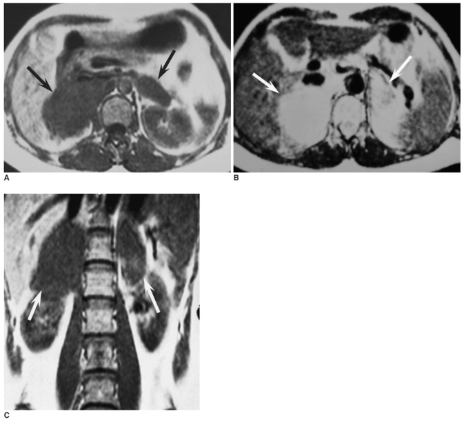
Chest radiography showed no abnormalities in both lungs. Plain film of the skeletal system failed to demonstrate evidence of multiple myeloma. The abdominal MR imaging (Elscint Gyrex V) revealed two large soft-tissue masses in the bilateral adrenal regions. The diameters of the masses in the right and left adrenal regions were 5 × 6 × 7 cm and 3 × 4 × 4 cm, respectively. The masses were homogeneous hypointense to the liver on the T1-weighted images (TR/TE = 510/12), and they were hyperintense on the T2-weighted images (TR/TE = 2200/65). The right mass invaded the inferior vein cava and upper polar of the right kidney. The right crus of the diaphragm was thickened and could not be differentiated from the mass. The well-defined left mass could be differentiated from the left kidney by the perirenal adipose tissue (Figs. 1A-C).
Fig. 1.
Abdominal MRI demonstrates that the masses in the bilateral adrenal glands are homogeneous hypointense to the liver (black arrow) on the axial T1-weighted spin echo image (A) and hyperintense (white arrow) on the axial T2-weight spin echo image (B). The mass in the right adrenal region has invaded the inferior vein cava (A, B) and upper polar of the right kidney (white arrow) on the coronal T1-weighted spin echo image (C).
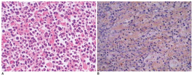
Surgical excision of the masses was recommended because malignant lesion was suspected due to the right adrenal mass invading the inferior vena cava and the upper polar of the right kidney on the MR images. After laparotomy, it was revealed that the right mass infiltrated the inferior vena cava and the upper polar of the right kidney, so right adrenalectomy was performed. The mass on the left adrenal region was removed simultaneously. The surgically excised masses were solid without any presence of hemorrhage and necrosis. Light microscopy revealed that the masses were composed of poorly-differentiated plasma cells and cells with multiple nuclei (Fig. 2A). The immunohistochemical staining was positive for monoclonality of IgG-lambda (Fig. 2B), but it was negative for kappa light chain. Based on these findings, the masses were diagnosed as extramedullary plasmacytoma arising from the bilateral adrenal glands.
Fig. 2.
A. Photomicrography of the histology specimen shows the tumor consists of poorly-differentiated plasma cells with large and hyperchromatic nuclei (Haematoxylin & Eosin, × 300).
B. Immunohistochemical staining of the resected masses is positive for the monoclonality of IgG-lambda (Immunohistochemical staining, × 200).
DISCUSSION
Plasma cell tumors can be divided into four categories: multiple myeloma, plasma cell leukemia, solitary plasmacytoma of the bone and EMP. The clinical manifestations are mostly nonspecific. Local recurrence is observed in 30% of the cases, and distant metastasis and transformation to multiple myeloma occasionally occur. Before EMP is diagnosed, multiple myeloma must be excluded by performing such imagings and laboratory tests as serum protein electrophoresis, bone marrow biopsy and skeletal imaging examinations. Bone marrow biopsy should show no evidence of multiple myeloma and less than 3% of plasma cells. Monoclonal bands of serum protein and Bence-Jones protein in the urine can sometimes be detected (1, 2). EMP can involve any extraosseous organs, but it predominantly affects the head and neck areas (1, 2). Primary EMP involving the adrenal gland is extremely uncommon. In 2001, Kahara et al. (3) reported the first case of EMP arising from a right adrenal gland.
To the best of our knowledge, only three cases of adrenal plasmacytomas to date have been reported in the literature. All of these cases involved tumor that arose from the right adrenal gland. Two of them were nonfunctioning tumors and they were found incidentally by imaging examinations (3, 4). However, Fujikata et al. (5) reported a case of adrenal plasmacytoma with functional abnormalities. Kahara et al. (3) reported a nonfunctioning plasmacytoma with a round shape, heterogeneous density and a smooth margin on abdominal CT, and it showed a heterogeneous signal on the T1- and T2-weighted images. Rogers et al. (4) reported on a tumor with heterogeneous enhancement on the contrast-enhanced MRI and heterogeneous hyperintensity was seen on the T2-weighted images. In contrast, our case is rare in that primary plasmacytoma involved the bilateral adrenals. Another remarkable characteristic is that the lesions appeared as homogeneous signals on the T1- and T2-weighted images. However, most of the lesions in the previous reports showed heterogeneous intensity on the MR images. These findings indicate adrenal plasmacytomas can appear as masses of variable intensity on MR images. As far as primary tumors involving bilateral adrenal glands are concerned, pheochromocytoma should be considered first because pheochromocytoma is frequently located bilaterally (about 10%) among the primary tumors (6). Second, bilateral adrenal metastases from other primary tumors can not be excluded when bilateral adrenal masses are revealed, but most of these cases are easily diagnosed because the primary malignancies are known. In this case, unfortunately, the differential diagnosis among pheochromocytoma, metastases of an unknown primary location and other uncommon disorders was very difficult before surgery because of the non-specific features seen on the MR images. A combination of the clinical and laboratory findings and the imaging features may be helpful to make a definite diagnosis.
In conclusion, we report here on the first documented case of extramedullary plasmacytoma involving the bilateral adrenal glands. Although the morphological appearance of EMP on MRI is nonspecific and the preoperative diagnosis may be difficult, MRI or CT scanning is often needed to obtain more information before making an appropriate therapeutic decision.
References
- 1.Fernandez LA, Couban S, Sy R, Miller R. An unusual presentation of extramedullary plasmacytoma occurring sequentially in the testis, subcutaneous tissue, and heart. Am J Hematol. 2001;67:194–196. doi: 10.1002/ajh.1106. [DOI] [PubMed] [Google Scholar]
- 2.Kondo M, Hashimoto S, Inuyama Y, Okamoto R, Yamada F. Extramedullary plasmacytoma of the sinonasal cavities: CT evaluation. J Comput Assist Tomogr. 1986;10:841–844. doi: 10.1097/00004728-198609000-00026. [DOI] [PubMed] [Google Scholar]
- 3.Kahara T, Nagai Y, Yamashita H, Nohara E, Kobayashi K, Takamura T. Extramedullary plasmacytoma in the adrenal incidentaloma. Clin Endocrinol (Oxf) 2001;55:267–270. doi: 10.1046/j.1365-2265.2001.01191.x. [DOI] [PubMed] [Google Scholar]
- 4.Rogers CG, Pinto PA, Weir EG. Extraosseous (extramedullary) plasmacytoma of the adrenal gland. Arch Pathol Lab Med. 2004;128:e86–e88. doi: 10.5858/2004-128-e86-EEPOTA. [DOI] [PubMed] [Google Scholar]
- 5.Fujikata S, Tanji N, Aoki K, Ohoka H, Hojo N, Yokoyama M. Extramedullary plasmacytoma arising from an adrenal gland. Urology. 2002;60:514. doi: 10.1016/s0090-4295(02)01833-2. [DOI] [PubMed] [Google Scholar]
- 6.Kawashima A, Sandler CM, Fishman EK, Charnsangavej C, Yasumori K, Honda H, et al. Spectrum of CT findings in nonmalignant disease of the adrenal gland. Radiographics. 1998;18:393–412. doi: 10.1148/radiographics.18.2.9536486. [DOI] [PubMed] [Google Scholar]


