Abstract
Mycoplasma mycoides subspecies mycoides small colony (SC) is the aetiologic agent of contagious bovine pleuropneumonia (CBPP), a respiratory disease causing important losses in cattle production. The publication of the genome sequence of M. mycoides subsp. mycoides SC should facilitate the identification of putative virulence factors. However, real progress in the study of molecular mechanisms of pathogenicity also requires efficient molecular tools for gene inactivation. In the present study, we have developed a transposon-based approach for the random mutagenesis of M. mycoides subsp. mycoides SC. A PCR-based screening assay enabled the characterization of several mutants with knockouts of genes potentially involved in pathogenicity. The initial transposon was further improved by combining it with the transposon γδ TnpR/res recombination system to allow the production of unmarked mutations. Using this approach, we isolated a mutant free of antibiotic-resistance genes, in which the gene encoding the main lipoprotein LppQ was disrupted. The mutant was found to express only residual amounts of the truncated N-terminal end of LppQ. This approach opens the way to study virulence factors and pathogen-host interactions of M. mycoides subsp. mycoides SC and to develop new, genetically defined vaccine strains.
INTRODUCTION
Mycoplasma mycoides subspecies mycoides biotype small colony (SC) is the causal agent of contagious bovine pleuropneumonia (CBPP), a respiratory disease of cattle that is widespread in most of sub-Saharan Africa and causes important economic losses in the cattle industry. Although the disease was eradicated from the USA, Canada and most of Europe in the 19th century (for review, see Provost et al., 1987), several outbreaks occurred in southern European countries during the 1980s and 1990s, and molecular typing studies have demonstrated that the isolated strains were from a lineage distinct from the African ones (Bischof et al., 2006; Cheng et al., 1995; Lorenzon et al., 2003; Vilei et al., 2000).
Although CBPP was controlled in Europe by the systematic slaughter of infected herds, such a strategy is difficult to propose in many African countries because of the negative effects on short-term economics and population health (Boonstra et al., 2001). Consequently, the main method recommended to control the spread of CBPP is vaccination with live-attenuated vaccine strains. Several vaccine strains have been produced since the 1990s by serial culture of the organism in liquid medium or in embryonated eggs (Thiaucourt et al., 2003, 2004). The first vaccine strains, V5 and KH3J, conferred only a low level of protection upon the vaccinated animals and were replaced by derivatives of the T1 strain. The T1/44 strain was obtained after 44 passages in embryonated eggs, and subsequent passages resulted in the streptomycin-resistant variant T1sr. Although both vaccine strains confer a certain level of protection, frequent inoculations are required to obtain satisfactory results. The mutations leading to the attenuation are unknown for all of these strains. The precise characterization of the mutation would improve the biological safety of live vaccine strains significantly, and is required for licensing of recombinant live bacterial vaccine strains in most countries (Frey, 2007). Marked post-vaccine reactions have been observed with the T1/44 strain, suggesting that it may have retained significant virulence, which has been confirmed by the report of CBPP cases following T1/44 vaccine inoculation via the endobronchial route (Mbulu et al., 2004).
New genetically defined vaccine strains should be developed to confer long-term protection, but they should be exempt from any residual pathogenicity or risk of reversion towards virulence. Only a few molecular determinants of M. mycoides subsp. mycoides SC have been proposed as virulence factors. Arguably the best-characterized factor is the glycerol catabolism system, which includes an ATP-binding cassette (ABC) transporter (GtsABC) and a membrane-bound glycerol-phosphate oxidase (GlpO) that results in the production of hydrogen peroxide (Vilei & Frey, 2001). High amounts of H2O2 are released by virulent African strains, but not by less-virulent European strains, which has been directly associated with cytotoxic effects (Pilo et al., 2005). However, this is not the only virulence attribute of mycoplasma, because vaccine strains, including T1/44, also show high cytotoxicity and high H2O2 release (Bischof et al., 2008). As in most mycoplasma species, the M. mycoides subsp. mycoides SC genome encodes various lipoproteins that are suspected to play an important role in the interaction with the host (Westberg et al., 2004). Some of these lipoproteins have been characterized, including LppB (Vilei & Frey, 2001) and LppQ, which has been described as a major antigen recognized during the immune response (Abdo et al., 2000; Bonvin-Klotz et al., 2008). A specific ELISA based on the detection of LppQ-antibodies has been developed to identify infected or vaccinated animals (Bruderer et al., 2002). However, the specific role of the LppB and LppQ lipoproteins in pathogenicity is unknown, due to the lack of genetic tools available to generate mutants. Ideally, such mutants would be devoid of antibiotic-resistance markers, allowing experimental field trials for the evaluation of virulence.
The first step in the development of efficient genetic tools was the construction of oriC plasmids for M. mycoides subsp. mycoides SC, M. mycoides subsp. mycoides large colony (LC) and Mycoplasma capricolum subsp. capricolum (Lartigue et al., 2003). These plasmids harbour the replicative origin of the chromosome and have been shown to replicate in mycoplasma cells. The use of these oriC plasmids for efficient gene targeting in M. capricolum subsp. capricolum and M. mycoides subsp. mycoides LC has been demonstrated (Janis et al., 2005). Taking into account the phylogenetic proximity of the SC and LC biotypes, our first attempts to inactivate genes in M. mycoides subsp. mycoides SC were based on the same strategy. However, no recombination events were detected, despite several attempts and the choice of various target genes, including lppQ, glpO and the gtsABC operon (unpublished results). Similar difficulties have hampered the isolation of Mycoplasma pulmonis mutants using the same strategy (Cordova et al., 2002).
Consequently, another strategy based on the use of a modified transposon was chosen. We took advantage of the development of a Tn4001 derivative (pMT85) used in Mycoplasma pneumoniae (Zimmerman & Herrmann, 2005). In the present work, pMT85 was first evaluated as a molecular tool for efficient gene mutagenesis in M. mycoides subsp. mycoides SC. In a second step, we further modified pMT85 to be able to eliminate the antibiotic resistance gene after selection of transformants. Using this new strategy, a lppQ mutant of M. mycoides subsp. mycoides SC strain T1/44 was isolated and characterized.
METHODS
Bacterial strains and culture conditions
The vaccine strain T1/44 (CIRAD-EMVT/PANVAC; Batch 002) of M. mycoides subsp. mycoides SC was grown at 37 °C in modified Hayflick medium (Freund, 1983) without thallium acetate and supplemented with BBL IsoVitalex Enrichment (Becton Dickinson). For cloning procedures and propagation of plasmids, the Escherichia coli DH10B strain (Stratagene) was used; gentamicin (100 μg ml−1) or tetracycline (5 μg ml−1) was added when required. For selection of mycoplasma transformants, gentamicin and tetracycline were used at 300 and 20 μg ml−1, respectively.
Plasmid constructions
Plasmid pMT85 is a plasposon harbouring a transposon derived from Tn4001 (Zimmerman & Herrmann, 2005). This plasposon contains a colE1 replicative origin for propagation in E. coli and the aacA-aphD gene that confers resistance to gentamicin, tobramycin and kanamycin (Fig. 1). This selection marker is located between the two inverted repeats (IRs) that define the extremities of the transposed fragment. In contrast, the transposase-encoding gene (tnpA) is located outside the transposed fragment.
Fig. 1.
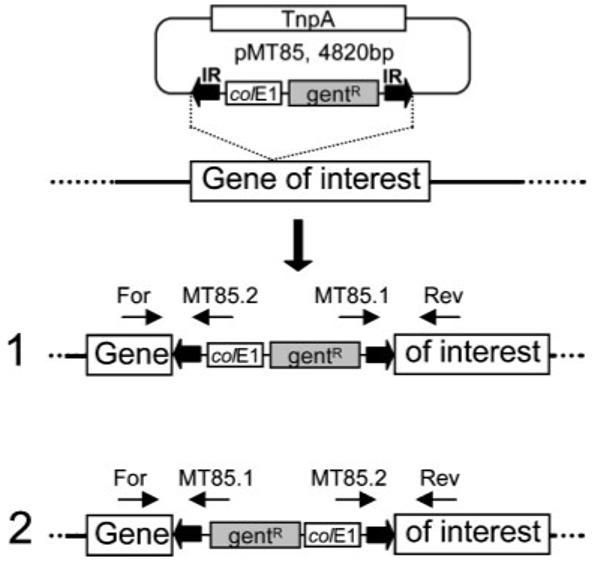
Schematic outline of the PCR-based screening procedure for transposon insertions. Thick black arrows indicate the IRs of the transposon. colE1, replication origin in E. coli; gentR, gentamicin-resistance marker encoded by the aacA-aphD gene. The transposase-encoding gene (TnpA) is located outside the transposed region of the pMT85 plasposon. The locations of primers used for the PCR-based screening are shown by thin arrows. The forward and reverse primers are specific to the gene of interest; the MT85.1 and MT85.2 primers are complementary to transposon sequences. Two orientations of the transposed elements are possible, as indicated by (1) and (2).
The res sequences of the γδ transposon were retrieved from the pSD6 plasmid (Duret et al., 2005). The first res sequence was PCR-amplified with the res7/res8 primers (Table 1) and cloned in the pMT85 plasmid after digestion with NheI and Acc65I. The second res sequence was PCR-amplified with the res5/res6 primers (Table 1) and cloned in the intermediate plasmid after digestion with SpeI and BamHI. The resulting plasmid was named pMT85/2res.
Table 1.
Primers used in this study
| Primer | Sequence 5′ -3′ | Tm (°C) |
|---|---|---|
| Primers used for library screening and junction sequencing | ||
| MT85.1 | ACAGTAATTGCGGGTGGATC | 60 |
| MT85.2 | TTACCGCCTTTGAGTGAGCT | 60 |
| GlpO6′ | GCTCTTGCACTTGTTGTTCC | 60 |
| GlpO7 | ACAGATTGCCACTGCTCATTT | 60 |
| GtsA3 | ATGGATCCGCATGCTAGGACCTTCTGGGAGTGGA | 62 |
| GtsB3 | GCTCAGCTGCCGACGCTCTAATAAGGCTGCTGCAAATAAAAT | 58 |
| GtsB1 | GATCGCATGCAAGATTTGCACCAAATGAAGC | 58 |
| GtsC4 | ATCTGCAGGCATGCCATAGCACCAGCTGCTTGAA | 58 |
| LppQ1 | GATCGCATGCAAAAATGAGTGACAAAAACAAGAT | 60 |
| LppQ2 | ATATGCATGCTGTGTCAACCAAAGGCATC | 58 |
| Primers used for the construction of pCJ13, pCJ15 and pMT85/2res | ||
| LppQ7 | ATATGCATGCTGGTGTTGCTACTGAAATTTGT | 60 |
| LppQ8 | ATATGCATGCCTGTTCCTCTTGCCCATCAT | 60 |
| PSresR | ATATGCATGCATTAAGCAATTTATTTGGAAAATC | 56 |
| PSresL | ATATGCATGCTTAGTTGCTTTCATTTATTACTT | 56 |
| res5 | ATATACTAGTGGATCTCATAAAAATGTATCCTA | 60 |
| res6 | ATATGGATCCGCAACCGTCCGAAATATTATAA | 60 |
| res7 | ATATGCTAGCGGATCTCATAAAAATGTATCCTA | 60 |
| res8 | ATATGGTACCGCAACCGTCCGAAATATTATAA | 60 |
The oriC plasmid pMYSO1 is a plasmid that replicates in M. mycoides subsp. mycoides SC cells and confers tetracycline resistance due to the tet(M) gene (Lartigue et al., 2003). For the complementation of the lppQ mutant, the wild-type lppQ gene and surrounding sequences were PCR-amplified using the primers LppQ7 and LppQ8 (Table 1) and cloned at the SphI site of the pMYSO1 plasmid to generate pCJ13. The sequence of the cloned lppQ gene was verified by DNA sequencing.
The γδ resolvase gene (tnpR) was PCR-amplified from the pSD25 plasmid (Duret et al., 2005) using the PSresR and PSresL primers (Table 1) and cloned into the SphI site of the pMYSO1 plasmid; the resulting plasmid was named pCJ15. The sequence of the resolvase gene was verified by DNA sequencing.
Construction of transposon insertion libraries
PEG-mediated transformation of M. mycoides subsp. mycoides SC was performed as described elsewhere (King & Dybvig, 1994), using 30 μg pMT85 (or pM85/2res). Transformed cells were resuspended in 500 μl prewarmed Hayflick medium and incubated for 3 h at 37 °C. A 2 ml volume of medium supplemented with gentamicin (300 μg ml−1) was then added and serial dilutions were performed in selective medium to determine the transformation efficiency. For the pMT85-based library, the volume of the culture was increased to 10 ml with selective medium after 1-3 days, and further incubated for 1-3 days. One millilitre was kept as a stock at −80 °C and the remaining 9 ml was used for DNA extraction. For the pMT85/2res-based library, the 2.5 ml culture was incubated for 17-24 h at 37 °C and then plated onto solid medium supplemented with gentamicin (300 μg ml−1). Isolated colonies were picked after 7-10 days and cells were grown in 1.5 ml gentamicin-containing liquid medium. From each culture, 750 μl was stored at −80 °C and the remaining 750 μl was used for DNA extraction.
DNA isolation, PCR screening of the library and Southern blot hybridization
Genomic DNA was prepared from each pool of transformants using the NucleoSpin Tissue DNA purification kit (Macherey-Nagel). Genomic DNAs from the pools of transformants generated by each of the 122 transformations were gathered into 14 superpools for a first round of PCR. A second round was performed for each pool contained in a positive superpool. PCRs (25 μl) were performed according to the recommendations of the Taq DNA polymerase supplier (Promega).
For Southern blot hybridization, 2 μg genomic DNA or 30 ng plasmid DNA was digested with EcoRI, and hybridization was performed in the presence of DIG-labelled DNA probes (20 ng ml−1). Detection of hybridized probes was achieved using anti-DIG antibodies coupled to alkaline phosphatase, and the fluorescent substrate CDP-star (Roche Molecular Biochemicals).
Protein analysis, and Western and colony blotting
Mycoplasma cells were collected from exponential-phase cultures by centrifugation (8000 g, 10 min, 4 °C). Whole-cell antigens of strains T1/44, T1lppQ−RSP1 and Afadé were prepared by mixing the concentrated cells in PBS with one volume SDS sample buffer [0.06 M Tris/HCl, pH 6.8, 2 % SDS, 10 % glycerol (v/v), 2 % β-mercaptoethanol, 0.025 % Bromophenol Blue], and boiling for 5 min. The antigens were separated by SDS-PAGE using 12 or 10 % polyacrylamide gels and then blotted onto a nitrocellulose membrane with a pore size of 0.2 μm (Bio-Rad). The membrane was then blocked with blocking buffer (1 % milk powder, 0.1 M Tris/HCl, pH 8.0, 1 M NaCl, 0.5 % Tween 20, 0.1 % merthiolate) for 1 h at room temperature, and incubated with the sera in blocking buffer (dilutions of 1: 500 and 1: 1000 for mouse sera and rabbit sera, respectively) for 1 h at room temperature and washed with deionized H2O. The membranes were then incubated for 1 h at room temperature with alkaline phosphatase-labelled conjugates, which were diluted in blocking buffer. The conjugates used were polyclonal antibody anti-mouse IgG (H + L) (Kirkegaard & Perry Laboratories) diluted 1: 2000 and polyclonal antibody anti-rabbit IgG (H + L) (Kirkegaard & Perry Laboratories) diluted 1: 2000. The membranes were then washed with deionized H2O. The colour reaction was initiated by adding 0.3 mg ml−1 nitro blue tetrazolium (NBT) (Roche Diagnostics), followed by 0.15 mg ml−1 5-bromo-4-chloro-3-indolyl phosphate (BCIP) (Roche Diagnostics) in alkaline buffer (100 mM Tris/HCl, pH 9.5, 5 mM MgCl2), and was stopped with deionized H2O.
For immunodetection of LppQ at the surface of mycoplasma cells, colonies were lifted from solid medium onto nitrocellulose membranes. After 5 min air drying, membranes were incubated for 1 h at room temperature in blocking buffer (25 mM Tris/HCl pH 8.0, 125 mM NaCl, 0.1 % Tween 20, 5 % defatted dry milk). Anti-LppQ-N’ serum (Abdo et al., 2000) (diluted 1: 500) was then added for 1 h incubation at room temperature; the subsequent steps of the immunoblotting procedure were as described above.
Immunization of mice
Seven BALB/c mice, two males and five females, were immunized subcutaneously with 5×10⁸ live cells of M. mycoides subsp. mycoides SC. The two male mice and two of the female mice were immunized with strain T1/44 and three female mice were immunized with strain T1lppQ−RSP1. After 18 days, the immunizations were repeated, and after another 18 days, blood was collected from the animals. Blood samples were centrifuged at 1000 g for 5 min at 4 °C to recover the serum.
RESULTS
Construction of a library of M. mycoides subsp. mycoides SC mutants using pMT85
The plasposon pMT85 used in this study harbours a transposon derived from Tn4001 which confers gentamicin resistance (Zimmerman & Herrmann, 2005). We first determined the MIC for gentamicin of the T1/44 strain of M. mycoides subsp. mycoides SC. This MIC ranged from 15.6 to 32.2 μg ml−1, and a concentration of 300 μg ml−1 was chosen for the selection of transformants. The yield of each transformation of M. mycoides subsp. mycoides SC with pMT85 was found to vary for unknown reasons, generating between 10 and 1000 mutants. A set of 122 transformation experiments was conducted to generate the transposon library. From each experiment, the transformants were amplified in liquid medium as a pool, before DNA extraction. The total number of mutants in the library was estimated to be in the range of 40 000-400 000. Considering that 1016 genes were identified in the genome of the type strain PG1 of M. mycoides subsp. mycoides SC (Westberg et al., 2004), each non-essential gene should be targeted several times by the transposon in this library of mutants, assuming random insertions of the transposon.
PCR-based detection of mutants of interest within the transposon insertion library
To evaluate the diversity of mutants within the library, transposon insertion sites were screened using a PCR-based method. Three loci with genes potentially involved in the pathogenicity of M. mycoides subsp. mycoides SC were searched: the gtsABC operon, the glpO gene and the lppQ gene. For the first two targets, there is experimental evidence showing that the genes are not essential. Indeed, the gtsABC operon is partly deleted and therefore not functional in European strains of M. mycoides subsp. mycoides SC (Vilei & Frey, 2001), and a glycerol-phosphate oxidase-deficient strain has been described (Wadher et al., 1990).
The PCR-based strategy for screening mutants of interest is described in Fig. 1. Briefly, two primers flanking the locus of interest were used in combination with two other primers directed against specific sequences of the transposon, MT85.1 and MT85.2 (Table 1). Accordingly, a PCR product should be generated with two of the four possible pairs of primers whenever a transposon is inserted between the positions of the target-specific oligonucleotides.
To limit the number of PCR reactions, genomic DNAs from the pools of transformants generated by each of the 122 transformations were gathered into 14 superpools. For each of the three targeted loci, a first round of PCR was performed on each superpool. Amplification products compatible with the locus size were observed for each target in at least one superpool. For example, in the search for lppQ mutants, a fragment of about 1300 bp was amplified from the DNA superpool SP1 in the reaction mix containing the LppQ2/MT85.2 pair of primers (Fig. 2a). Taking into account the length of the lppQ gene (1338 bp) and the nucleotide position of the primers, this amplification suggested that the transposon had integrated in the chromosome in the reverse orientation, at about 200 bp downstream from the start codon. An amplification of the other junction should also have been observed using the LppQ1/MT85.1 pair of primers. However, in this particular case, further analysis indicated that the transposon had integrated into the chromosome exactly within the sequence corresponding to the LppQ1 primer. Consequently, because of LppQ1 mispairing, no amplification product was observed. Another primer, LppQ7, located upstream from LppQ1 and used with MT85.1, enabled PCR amplification of the junction (data not shown).
Fig. 2.
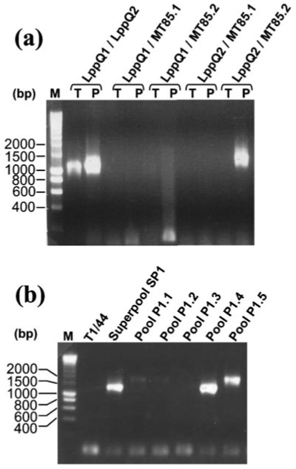
PCR-based screening for transposon insertions within the lppQ gene. (a) First round of PCR screening for lppQ mutants within superpool SP1 using different combinations of PCR primers (see Table 1) as indicated above the lanes. T, untransformed T1/44 control; P, superpool SP1; M, DNA Smart Ladder (Eurogentec). (b) Second-round PCR using primers LppQ2 and MT85.2 for the identification of the pools containing lppQ mutants. Pool P1.4 gave the strongest signal, of the same size as that detected in superpool SP1.
For each target, after identifying positive superpools, a second round of PCR was performed to determine the pool in which the mutant of interest could be found. In every case, PCR products were observed for at least one pool. For example, the superpool SP1 resulted from the mixing of genomic DNAs extracted from 10 pools of transformants and was PCR-positive for a lppQ mutant. During the second round of PCR, a fragment of about 1300 bp was amplified from pool P1.4, as in the superpool SP1 (Fig. 2b). Another pool, P1.5, was also found to be positive during this screening, with a PCR product of about 1500 bp (Fig. 2b), which suggested that the transposon was inserted within lppQ at a site different from that of the mutant in pool P1.4.
PCR products obtained from pools of transposon mutants were sequenced to identify the sites of insertion and to predict the lengths of the truncated proteins (Table 2). Sequencing of the amplification products confirmed integrations of the tranposon in each target locus. Two insertions were detected in the gtsABC locus, one in gtsB and one in gtsC. Three different mutants were characterized with transposon insertions within the glpO gene. Predicted GlpO proteins were 383, 37 and 79 aa in length, instead of 392 aa for the wild-type protein. Finally, two insertions were identified within the lppQ locus. The first one, detected in the P1.4 pool, was located 209 bp downstream of the ATG, leading to a truncated putative protein of 70 aa in length, instead of 445 aa for the wildtype protein. The second insertion, identified in the P1.5 pool, was found 205 bp upstream of the ATG, in a noncoding sequence (Table 2).
Table 2.
Transposon integration sites within specific target genes from M. mycoides subsp. mycoides SC
| Gene | Gene length (bp) | Wild-type protein length (aa) | Screening primers | Positive superpool | Positive pool | Insertion site* | Predicted length of truncated protein (aa) |
|---|---|---|---|---|---|---|---|
| glpK | 1518 | 505 | GlpO6′, GlpO7 | SP11 | P11.11 | +29 | 10 |
| glpO | 1179 | 392 | GlpO6, GlpO7 | SP10 | P10.9 | +1148 | 383 |
| GlpO6′, GlpO7 | SP10 | P10.11 | +109 | 37 | |||
| GlpO6′, GlpO7 | SP11 | P11.9 | +217 | 79 | |||
| gtsB | 1029 | 342 | GtsA3, GtsB3 | SP3 | P3.10 | +505 | 175 |
| gtsC | 810 | 269 | GtsB1, GtsC4 | SP2 | P2.1 | +588 | 200 |
| lppQ | 1338 | 445 | LppQ1, LppQ2 | SP1 | P1.4 | +209 | 70 |
| LppQ1, LppQ2 | SP1 | P1.5 | −285 | Untruncated |
From the ATG start codon.
Our results show that pMT85 can be used for efficient mutagenesis of M. mycoides subsp. mycoides SC and that insertion events in genes of interest can be detected by a PCR-based procedure.
Isolation and characterization of a lppQ mutant
Different methods can be used to isolate specific mutants from a pool identified as positive for a specific targeted gene. Taking into account the relatively low complexity of the pools generated by transformation of M. mycoides subsp. mycoides SC (about 10-1000 transformants), this could be achieved by a PCR-screening assay. Alternatively, mutants can be screened using antibodies directed against the encoded protein. Such a strategy was used for the isolation of a lppQ mutant by negative immunoscreening. After filtration through a 0.45 μm pore-size filter to eliminate cell aggregates, transformants from pool P1.4 were plated on solid medium supplemented with gentamicin. A colony-blot assay was then performed using anti-LppQ-N antibodies. Colonies that were revealed by a non-specific Ponceau red staining but not by the anti-LppQ-N antibodies were picked and grown in selective liquid medium. After a second round of filtration and negative immunodetection, a single mutant, named T1lppQ−MT1, was obtained.
Genomic DNA was extracted from the T1lppQ−MT1 mutant and the transposon integration site was verified by DNA-DNA hybridization (Fig. 3). Total DNA was EcoRI-digested and the restriction fragments were separated by gel electrophoresis. After transfer onto nylon membranes, the DNA was hybridized with probes complementary to inserted parts of the plasposon (colE1) or lppQ sequences. A unique EcoRI fragment of about 5500 bp was detected by both probes. This length was in agreement with the theoretical genomic organization inferred from the transposon integration site identified by sequencing. Moreover, only one band was detected using the colE1 probe, indicating that a single copy of the transposon had integrated the chromosome (Fig. 3). It should be noted that the genome of the T1/44 strain contains a single copy of the lppQ gene (Bischof et al., 2006), in contrast to that of the PG1 type strain, which contains two copies (Westberg et al., 2004). To evaluate its stability, the T1lppQ−MT1 mutant was cultured in the absence of gentamicin for 15 passages, and the lppQ locus was checked again by DNA hybridization. The same DNA pattern was observed, showing that the integrated transposon was maintained at the same locus (data not shown).
Fig. 3.
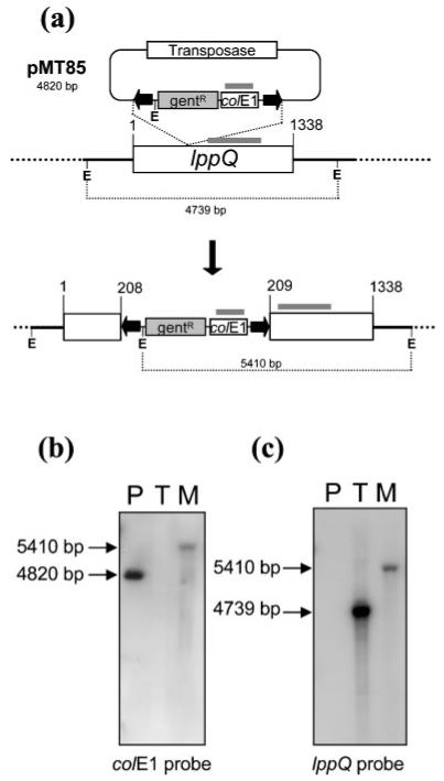
Location of the transposon within the lppQ gene of the T1lppQ−MT1 mutant. (a) Schematic integration of the transposon within the lppQ gene. Grey bars indicate the location of the probes colE1 and lppQ used in Southern blots. E, EcoRI restriction site. Elements are not drawn to scale. (b, c) Total genomic DNA from the T1lppQ−MT1 mutant was EcoRI-digested and fragments were separated by gel electrophoresis, transferred to nylon membranes and hybridized with a colE1 (b) or a lppQ probe (c). P, pMT85 control; T, T1/44; M, T1lppQ−MT1 mutant.
In order to confirm that the LppQ antigen was not expressed by the T1lppQ−MT1 mutant, total proteins extracted from both wild-type and mutant cells were separated by SDS-PAGE (Fig. 4). After transfer onto a nitrocellulose membrane, immunodetection of the LppQ protein was performed using anti-LppQ-N antibodies. Whereas the LppQ protein was detected in the T1/44 control extract as a band corresponding to a protein of about 48 kDa, it was not found in the extract from the mutant strain, which confirmed that the transposon integration prevents LppQ expression. It is still possible that a 70 aa truncated LppQ protein was produced that could not be detected under the experimental conditions used. The T1lppQ−MT1 mutant was then transformed with the pCJ13 plasmid, which is a derivative of the oriC-based plasmid pMYSO1 and carries the lppQ gene under the control of its own promoter. This transformation complemented the lppQ mutation, as the production of the LppQ protein was restored (Fig. 4b).
Fig. 4.
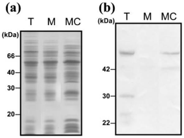
Lack of LppQ expression in the T1lppQ−MT1 mutant. (a) Total proteins were extracted using SDS from untransformed and mutant cells and separated by 12% PAGE. T, T1/44 (60 μg); M, T1lppQ−MT1 mutant (60 μg); MC, T1lppQ−MT1 mutant complemented with wild-type lppQ gene (120 μg). Proteins were stained with Coomassie Blue. Molecular mass markers LMW were used (Amersham Biosciences). (b) Immunodetection of LppQ after transfer onto a nitrocellulose membrane. A polyclonal serum directed against the N-terminal part of LppQ was used to detect the lipoprotein (dilution 1: 500). The two panels correspond to three independent strips that were parts of a single experiment and that were grouped for the purpose of this figure.
Improvement of the pMT85 plasposon to generate unmarked mutations
With the goal of being able to delete the antibiotic resistance marker from the mycoplasma genome to obtain unmarked mutations, the pMT85 plasposon was modified by cloning res sequences within the element surrounded by the IR sequences, yielding the pMT85/2res plasmid (Fig. 5). The res sequences are recognized specifically by the transposon γδ resolvase, which is encoded by the tnpR gene carried by the pMYSO1-derived pCJ15 plasmid.
Fig. 5.
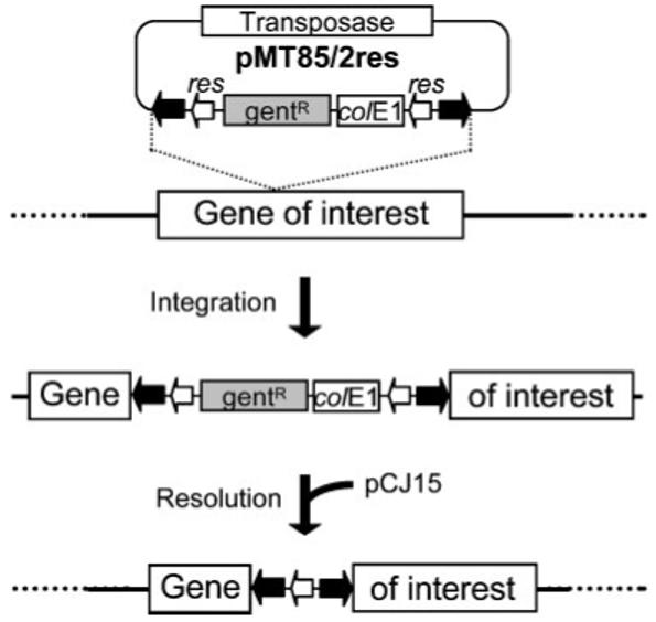
Schematic outline of the two-step procedure to obtain unmarked mutants. In the first step the transposon integrates into the chromosome. After selection of a mutant of interest, resolution of most of the transposed sequence is induced by the γδ resolvase that is encoded by a gene carried by the oriC plasmid pCJ15. Thick black arrows indicate the IRs of the transposon harboured by the plasposon pMT85/2res. Open arrows indicate the resolvase target sequences (res). colE1, replication origin in E. coli; gentR, gentamicin-resistance marker. The transposase-encoding gene is located outside the transposed region of the plasposon.
The pMT85/2res plasposon was used to create a new library of M. mycoides subsp. mycoides SC insertion mutants using conditions similar to those described above. However, instead of keeping the mutants as a pool in liquid medium, after 17-24 h culture with antibiotic, mycoplasmas were plated on selective medium containing gentamicin. A total of 984 clones were collected from a set of eight transformations, and were amplified in 1.5 ml cultures.
Isolation and characterization of an unmarked lppQ mutant
The new library of lppQ mutants was screened by PCR, using the same strategy as described in Fig. 1, which resulted in the selection of the clone T2A108. The sequencing of PCR products from this clone indicated that the transposed element from pMT85/2res had inserted between nucleotides 989 and 990 of the lppQ gene. Clone T2A108 was transformed with the pCJ15 plasmid in order to remove the aacA-aphD marker. After a single passage of the transformed clone in the presence of tetracycline, the antibiotic was removed to facilitate the loss of the pCJ15 plasmid, and a new clone, named T1lppQ−RSP1, was obtained after plating on a medium without antibiotic. We verified that the clone T1lppQ−RSP1 had the same sensitivity to tetracycline and gentamicin as the original T1/44 strain, which indicated that it had lost the aacA-aphD gene and the tet(M) gene carried by the pCJ15 plasmid. However, further sequencing analysis showed that the T1lppQ−RSP1 clone had retained the tnpA gene, which indicated that the initial transposition had not occurred exactly as expected (Fig. 6). Indeed, analysis of the junction between the 5′ end of the lppQ gene and the inserted element showed that an IR-like sequence was used during transposition. This sequence is only partially homologous to the genuine IR, with 17 conserved positions out of 26. This clone, which showed the required phenotype, was selected for further characterization.
Fig. 6.
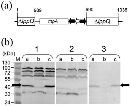
Molecular characterization of the mutant T1lppQ−RSP1. (a) Genomic structure of the disrupted lppQ gene in the T1lppQ−RSP1 mutant. The position of the transposon within the nucleotide sequence of lppQ is indicated. tnpA, transposase gene; black arrows, transposon IRs; open arrow, remaining res sequence. (b) Immunodetection of LppQ in T1/44, T1lppQ−RSP1 and Afadé. Total proteins were extracted from mycoplasma cells of T1/44 (lanes a), T1lppQ−RSP1(lanes b) and Afadé (lanes c) and separated by 10% PAGE. The blot in panel 1 was reacted with anti-T1/44 antibodies. The blot in panel 2 was reacted with serum from a mouse that was immunized with T1lppQ−RSP1. The blot in panel 3 was reacted with anti-LppQ-N antibodies. M, prestained molecular mass markers. The black arrows indicate the 48 kDa LppQ antigen.
Mice immunized with live M. mycoides subsp. mycoides SC strain T1/44 or T1lppQ−RSP1 showed no side-effects from immunization with either strain. Sera collected from these animals were used to detect mycoplasma antigens obtained from M. mycoides subsp. mycoides SC strains T1/44, T1lppQ−RSP1 and the virulent strain Afadé. The immunoblot results were compared with that of an identical immunoblot incubated with monospecific anti-LppQ-N antibodies (Fig. 6). Sera from mice infected with the vaccine strain T1/44 of M. mycoides subsp. mycoides SC showed a strong reaction to the 48 kDa protein LppQ in whole-cell preparations of strains T1/44 and Afadé, whereas no reaction to this antigen could be detected with a whole-cell preparation of strain T1lppQ−RSP1 (Fig. 6a). When sera from mice immunized with the T1lppQ−RSP1 clone were tested against the same bacterial preparations from the three strains, there was no reaction to LppQ (Fig. 6b). Anti-LppQ-N antibodies showed that the 48 kDa protein LppQ was present in strains T1/44 and Afadé. However, they reacted only weakly to a 37 kDa antigen from T1lppQ−RSP1 (Fig. 6c, lane b), which suggests that there was production of minor amounts of the residual N-terminal part of LppQ in strain T1lppQ−RSP1. The size of the detected antigen was consistent with the calculated mass of the truncated LppQ protein (38.36 kDa) in T1lppQ−RSP1. Interestingly, sera from all three mice infected with T1lppQ−RSP1 did not react to a protein of ∼85 kDa present in all three strains, T1/44, T1lppQ−RSP1 and Afadé, while serum from all four mice immunized with T1/44 reacted predominantly to this 85 kDa protein band in all strains. This 85/80 kDa antigen of M. mycoides subsp. mycoides SC is known as a major antigen of experimentally or naturally infected cattle (Abdo et al., 2000). The lack of antigenicity of the 85 kDa protein in strain T1lppQ−RSP1 could be due to a number of factors, such as the reduced immunogenicity of the cell antigens because the major LppQ lipoprotein is not produced. Another possibility is that there is altered production or membrane exposure of the 85 kDa protein, for reasons that are currently unknown.
DISCUSSION
Two transposons and various derivatives have already been used for gene inactivation in mollicutes. Transposon Tn916, originating from Enterococcus faecalis and carrying the tet(M) gene that confers resistance to tetracycline, has been used with success in Acholeplasma laidlawii, M. pulmonis, Mycoplasma hyorhinis (Dybvig & Cassell, 1987; Dybvig & Alderete, 1988), M. mycoides subsp. mycoides LC (King & Dybvig, 1991) and Mycoplasma gallisepticum (Whetzel et al., 2003). However, the instability of the integrated transposon has been identified as a potential drawback (Dybvig & Alderete, 1988). A second transposon, Tn4001, initially isolated from Staphylococcus aureus, has been used to create mutants in M. pneumoniae, Mycoplasma genitalium (Hedreyda et al., 1993; Reddy et al., 1996), M. gallisepticum (Mudahi-Orenstein et al., 2003) and Spiroplasma citri (Foissac et al., 1997). This transposon carries the gene aacA-aphD, which confers resistance to gentamicin, kanamycin and tobramycin. Because gentamicin resistance could not be used as a selective marker in M. pulmonis and M. hyorhinis, derivatives carrying the tet(M) gene (Tn4001T) or the cat gene (Tn4001C) were developed for use in these two species (Dybvig et al., 2000). Another derivative of Tn4001 with the transposase gene cloned outside the transposable region of the harbouring plasmid pMT85 was recently used for haystack mutagenesis in M. pneumoniae (Halbedel & Stulke, 2007; Zimmerman & Herrmann, 2005). In the present study, we extended this work by modifying this Tn4001 derivative to create unmarked mutations in the genome of M. mycoides subsp. mycoides SC, a major bovine pathogen.
In this study, we have shown that the mutant T1lppQ−MT1, obtained by using the pMT85 plasposon, is stable. Furthermore, we also have shown that the lppQ mutation can be complemented by using derivatives of the pMYSO1 oriC plasmid (Lartigue et al., 2003). The combination of a transposon that confers resistance to gentamicin and a replicative plasmid that harbours a marker for tetracycline resistance constitutes an efficient, and as yet unreported, panel of molecular tools for the identification of the genetic determinants of the pathogenicity of M. mycoides subsp. mycoides SC.
Three particular loci of the M. mycoides subsp. mycoides SC genome have been chosen as targets for this study because of their proposed involvement in mycoplasma pathogenicity (Pilo et al., 2007). Disruption of the genes lppQ, glpO, gtsB and gtsC indicates that these genes are not essential, at least for growth in rich medium. For both glpO and gtsABC, this conclusion is in agreement with previous studies reporting M. mycoides subsp. mycoides SC strains in which these genes were inactivated or missing (Vilei & Frey, 2001; Wadher et al., 1990). Our study indicates that the gene encoding the major LppQ antigen is not essential, as we were able to isolate and characterize two independent mutants, T1lppQ−MT1 and T1lppQ−RSP1. These mutants are of interest because LppQ is hypothesized to play a role in producing adverse reactions in vaccinated animals. Indeed, a recent study has shown that cattle immunized with purified recombinant LppQ, using different adjuvant methods, were significantly more susceptible to challenge with M. mycoides subsp. mycoides SC than cattle that were not vaccinated with LppQ (Nicholas et al., 2004). LppQ may therefore contribute to the immunopathology induced by M. mycoides subsp. mycoides SC, including the strong post-vaccine reactions observed after immunization of animals with the T1/44 vaccine strains. The lppQ mutants derived in this study from strain T1/44 provide a unique opportunity to test this hypothesis. The unmarked T1lppQ−RSP1 mutant is particularly suited for such studies because of the removal of the antibiotic-resistance marker, which may be regarded as a potential environmental hazard if left in a genetically modified vaccine. The development of a vaccine strain that does not produce LppQ, such as the T1lppQ−RSP1 mutant, follows the modern vaccinology concept of DIVA (differentiating infected from vaccinated animals); hence, existing serological assays could be employed to differentiate between infected and vaccinated animals (Bruderer et al., 2002; Le Goff & Thiaucourt, 1998).
The development of improved M. mycoides subsp. mycoides SC vaccine strains remains an important goal for the control of CBPP in countries where the disease is endemic. The inactivation of specific genes is now a practicable strategy for attenuating virulent strains, or existing vaccine strains for which side-effects indicative of residual virulence have been reported. One of the main challenges of vaccine strain development is to eliminate virulence while maintaining protective immunogenicity. This last constraint implies that gene-inactivation strategies should minimize the number of passages from the initial strain to the isolated mutant. The genetic tools that we have adapted for M. mycoides subsp. mycoides SC enable the isolation of specific mutants in fewer than 10 passages, including steps for the removal of the antibiotic-resistance marker.
ACKNOWLEDGEMENTS
This research was supported by grants from the INRA, the Région Aquitaine, the University Victor Segalen Bordeaux 2 and grant number 075804 of the Wellcome Trust, London, UK (to J. F.). We thank Richard Herrmann, Zentrum für Molekulare Biologie Heidelberg, for providing us with the pMT85 plasposon, Sybille Duret, UMR 1090 INRA, for the plasmids pSD6 and pSD25, François Poumarat, AFSSA, Lyon, France, for the strain T1/44, and Yvonne Schlatter for technical support. We also thank Joanna Cambray-Young for carefully reading the manuscript.
Abbreviations
- CBPP
contagious bovine pleuropneumonia
- IR
inverted repeat
REFERENCES
- Abdo EM, Nicolet J, Frey J. Antigenic and genetic characterization of lipoprotein LppQ from Mycoplasma mycoides subsp. mycoides SC. Clin Diagn Lab Immunol. 2000;7:588–595. doi: 10.1128/cdli.7.4.588-595.2000. [DOI] [PMC free article] [PubMed] [Google Scholar]
- Bischof DF, Vilei EM, Frey J. Genomic differences between type strain PG1 and field strains of Mycoplasma mycoides subsp. mycoides small-colony type. Genomics. 2006;88:633–641. doi: 10.1016/j.ygeno.2006.06.018. [DOI] [PMC free article] [PubMed] [Google Scholar]
- Bischof DF, Janis C, Vilei EM, Bertoni G, Frey J. Cytotoxicity of Mycoplasma mycoides subsp. mycoides SC toward bovine epithelial cells. Infect Immun. 2008;76:263–269. doi: 10.1128/IAI.00938-07. [DOI] [PMC free article] [PubMed] [Google Scholar]
- Bonvin-Klotz L, Vilei EM, Kuhni-Boghenbor K, Kapp N, Frey J, Stoffel MH. Domain analysis of lipoprotein LppQ in Mycoplasma mycoides subsp. mycoides SC. Antonie Van Leeuwenhoek. 2008;93:175–183. doi: 10.1007/s10482-007-9191-1. [DOI] [PMC free article] [PubMed] [Google Scholar]
- Boonstra E, Lindbaek M, Fidzani B, Bruusgaard D. Cattle eradication and malnutrition in under five’s: a natural experiment in Botswana. Public Health Nutr. 2001;4:877–882. doi: 10.1079/phn2001129. [DOI] [PubMed] [Google Scholar]
- Bruderer U, Regalla J, Abdo E-M, Huebschle OJ, Frey J. Serodiagnosis and monitoring of contagious bovine pleuropneumonia (CBPP) with an indirect ELISA based on the specific lipoprotein LppQ of Mycoplasma mycoides subsp. mycoides SC. Vet Microbiol. 2002;84:195–205. doi: 10.1016/s0378-1135(01)00466-7. [DOI] [PubMed] [Google Scholar]
- Cheng X, Nicolet J, Poumarat F, Regalla J, Thiaucourt F, Frey J. Insertion element IS1296 in Mycoplasma mycoides subsp. mycoides small colony identifies a European clonal line distinct from African and Australian strains. Microbiology. 1995;141:3221–3228. doi: 10.1099/13500872-141-12-3221. [DOI] [PubMed] [Google Scholar]
- Cordova CM, Lartigue C, Sirand-Pugnet P, Renaudin J, Cunha RA, Blanchard A. Identification of the origin of replication of the Mycoplasma pulmonis chromosome and its use in oriC replicative plasmids. J Bacteriol. 2002;184:5426–5435. doi: 10.1128/JB.184.19.5426-5435.2002. [DOI] [PMC free article] [PubMed] [Google Scholar]
- Duret S, Andre A, Renaudin J. Specific gene targeting in Spiroplasma citri: improved vectors and production of unmarked mutations using site-specific recombination. Microbiology. 2005;151:2793–2803. doi: 10.1099/mic.0.28123-0. [DOI] [PubMed] [Google Scholar]
- Dybvig K, Alderete J. Transformation of Mycoplasma pulmonis and Mycoplasma hyorhinis: transposition of Tn916 and formation of cointegrate structures. Plasmid. 1988;20:33–41. doi: 10.1016/0147-619x(88)90005-4. [DOI] [PubMed] [Google Scholar]
- Dybvig K, Cassell GH. Transposition of Gram-positive transposon Tn916 in Acholeplasma laidlawii and Mycoplasma pulmonis. Science. 1987;235:1392–1394. doi: 10.1126/science.3029869. [DOI] [PubMed] [Google Scholar]
- Dybvig K, French CT, Voelker LL. Construction and use of derivatives of transposon Tn4001 that function in Mycoplasma pulmonis and Mycoplasma arthritidis. J Bacteriol. 2000;182:4343–4347. doi: 10.1128/jb.182.15.4343-4347.2000. [DOI] [PMC free article] [PubMed] [Google Scholar]
- Foissac X, Saillard C, Bové JM. Random insertion of transposon Tn4001 in the genome of Spiroplasma citri strain GII3. Plasmid. 1997;37:80–86. doi: 10.1006/plas.1996.1271. [DOI] [PubMed] [Google Scholar]
- Freund EA. Culture media for classic mycoplasmas. In: Razin S, Tully JG, editors. Methods in Mycoplasmology, vol. 1, Mycoplasma Characterization. Academic Press; New York: 1983. pp. 127–135. [Google Scholar]
- Frey J. Biological safety concepts of genetically modified live bacterial vaccines. Vaccine. 2007;25:5598–5605. doi: 10.1016/j.vaccine.2006.11.058. [DOI] [PubMed] [Google Scholar]
- Halbedel S, Stulke J. Tools for the genetic analysis of Mycoplasma. Int J Med Microbiol. 2007;297:37–44. doi: 10.1016/j.ijmm.2006.11.001. [DOI] [PubMed] [Google Scholar]
- Hedreyda CT, Lee KK, Krause DC. Transformation of Mycoplasma pneumoniae with Tn4001 by electroporation. Plasmid. 1993;30:170–175. doi: 10.1006/plas.1993.1047. [DOI] [PubMed] [Google Scholar]
- Janis C, Lartigue C, Frey J, Wroblewski H, Thiaucourt F, Blanchard A, Sirand-Pugnet P. Versatile use of oriC plasmids for functional genomics of Mycoplasma capricolum subsp. capricolum. Appl Environ Microbiol. 2005;71:2888–2893. doi: 10.1128/AEM.71.6.2888-2893.2005. [DOI] [PMC free article] [PubMed] [Google Scholar]
- King KW, Dybvig K. Plasmid transformation of Mycoplasma mycoides subspecies mycoides is promoted by high concentrations of polyethylene glycol. Plasmid. 1991;26:108–115. doi: 10.1016/0147-619x(91)90050-7. [DOI] [PubMed] [Google Scholar]
- King KW, Dybvig K. Transformation of Mycoplasma capricolum and examination of DNA restriction modification in M. capricolum and Mycoplasma mycoides subsp. mycoides. Plasmid. 1994;31:308–311. doi: 10.1006/plas.1994.1033. [DOI] [PubMed] [Google Scholar]
- Lartigue C, Blanchard A, Renaudin J, Thiaucourt F, Sirand-Pugnet P. Host specificity of mollicutes oriC plasmids: functional analysis of replication origin. Nucleic Acids Res. 2003;31:6610–6618. doi: 10.1093/nar/gkg848. [DOI] [PMC free article] [PubMed] [Google Scholar]
- Le Goff C, Thiaucourt F. A competitive ELISA for the specific diagnosis of contagious bovine pleuropneumonia (CBPP) Vet Microbiol. 1998;60:179–191. doi: 10.1016/s0378-1135(98)00156-4. [DOI] [PubMed] [Google Scholar]
- Lorenzon S, Arzul I, Peyraud A, Hendrikx P, Thiaucourt F. Molecular epidemiology of contagious bovine pleuropneumonia by multilocus sequence analysis of Mycoplasma mycoides subspecies mycoides biotype SC strains. Vet Microbiol. 2003;93:319–333. doi: 10.1016/s0378-1135(03)00043-9. [DOI] [PubMed] [Google Scholar]
- Mbulu RS, Tjipura-Zaire G, Lelli R, Frey J, Pilo P, Vilei EM, Mettler F, Nicholas RA, Huebschle OJ. Contagious bovine pleuropneumonia (CBPP) caused by vaccine strain T1/44 of Mycoplasma mycoides subsp. mycoides SC. Vet Microbiol. 2004;98:229–234. doi: 10.1016/j.vetmic.2003.11.007. [DOI] [PubMed] [Google Scholar]
- Mudahi-Orenstein S, Levisohn S, Geary SJ, Yogev D. Cytadherence-deficient mutants of Mycoplasma gallisepticumgenerated by transposon mutagenesis. Infect Immun. 2003;71:3812–3820. doi: 10.1128/IAI.71.7.3812-3820.2003. [DOI] [PMC free article] [PubMed] [Google Scholar]
- Nicholas RAJ, Tjipura-Zaire G, Mbulu RS, Scacchia M, Mettler F, Frey J, Abusugra I, Huebschle OJB. An inactivated whole cell vaccine and LppQ subunit vaccine appear to exacerbate the effects of CBPP in adult cattle; Towards Sustainable CBPP Control Programmes for Africa, Proceedings of the FAO-OIE-AU/IBAR-IAEA Consultative Group on Contagious Bovine Pleuropneumonia Third Meeting; Rome: FAO. 2004.pp. 91–97. [Google Scholar]
- Pilo P, Vilei EM, Peterhans E, Bonvin-Klotz L, Stoffel MH, Dobbelaere D, Frey J. A metabolic enzyme as a primary virulence factor of Mycoplasma mycoides subsp. mycoides small colony. J Bacteriol. 2005;187:6824–6831. doi: 10.1128/JB.187.19.6824-6831.2005. [DOI] [PMC free article] [PubMed] [Google Scholar]
- Pilo P, Frey J, Vilei EM. Molecular mechanisms of pathogenicity of Mycoplasma mycoides subsp. mycoides SC. Vet J. 2007;174:513–521. doi: 10.1016/j.tvjl.2006.10.016. [DOI] [PMC free article] [PubMed] [Google Scholar]
- Provost A, Perreau P, Breard A, Le Goff C, Martel J, Cottew G. Contagious bovine pleuropneumonia. Rev Sci Tech. 1987;6:625–679. doi: 10.20506/rst.6.3.306. [DOI] [PubMed] [Google Scholar]
- Reddy SP, Rasmussen WG, Baseman JB. Isolation and characterization of transposon Tn4001-generated, cytadherence-deficient transformants of Mycoplasma pneumoniae and Mycoplasma genitalium. FEMS Immunol Med Microbiol. 1996;15:199–211. doi: 10.1111/j.1574-695X.1996.tb00086.x. [DOI] [PubMed] [Google Scholar]
- Thiaucourt F, Dedieu L, Maillard JC, Bonnet P, Lesnoff M, Laval G, Provost A. Contagious bovine pleuropneumonia vaccines, historic highlights, present situation and hopes. Dev Biol (Basel) 2003;114:147–160. [PubMed] [Google Scholar]
- Thiaucourt F, Aboubakar Y, Wesonga H, Manso-Silvan L, Blanchard A. Contagious bovine pleuropneumonia vaccines and control strategies: recent data. Dev Biol (Basel) 2004;119:99–111. [PubMed] [Google Scholar]
- Vilei EM, Frey J. Genetic and biochemical characterization of glycerol uptake in Mycoplasma mycoides subsp. mycoides SC: its impact on H2O2 production and virulence. Clin Diagn Lab Immunol. 2001;8:85–92. doi: 10.1128/CDLI.8.1.85-92.2001. [DOI] [PMC free article] [PubMed] [Google Scholar]
- Vilei EM, Abdo EM, Nicolet J, Botelho A, Goncalves R, Frey J. Genomic and antigenic differences between the European and African/Australian clusters of Mycoplasma mycoides subsp. mycoides SC. Microbiology. 2000;146:477–486. doi: 10.1099/00221287-146-2-477. [DOI] [PubMed] [Google Scholar]
- Wadher BJ, Henderson CL, Miles RJ, Varsani H. A mutant of Mycoplasma mycoides subsp. mycoides lacking the H2O2-producing enzyme l-α-glycerophosphate oxidase. FEMS Microbiol Lett. 1990;72:127–130. doi: 10.1016/0378-1097(90)90358-w. [DOI] [PubMed] [Google Scholar]
- Westberg J, Persson A, Holmberg A, Goesmann A, Lundeberg J, Johansson KE, Pettersson B, Uhlen M. The genome sequence of Mycoplasma mycoides subsp. mycoides SC type strain PG1T, the causative agent of contagious bovine pleuropneumonia (CBPP) Genome Res. 2004;14:221–227. doi: 10.1101/gr.1673304. [DOI] [PMC free article] [PubMed] [Google Scholar]
- Whetzel PL, Hnatow LL, Keeler CL, Jr, Dohms JE. Transposon mutagenesis of Mycoplasma gallisepticum. Plasmid. 2003;49:34–43. doi: 10.1016/s0147-619x(02)00114-2. [DOI] [PubMed] [Google Scholar]
- Zimmerman CU, Herrmann R. Synthesis of a small, cysteine-rich, 29 amino acids long peptide in Mycoplasma pneumoniae. FEMS Microbiol Lett. 2005;253:315–321. doi: 10.1016/j.femsle.2005.09.054. [DOI] [PubMed] [Google Scholar]


