Abstract
Context:
Excessive quadriceps contraction with insufficient hamstrings muscle cocontraction has been shown to be a possible contributing factor for noncontact anterior cruciate ligament (ACL) injuries. Assessing the relationships among lower extremity internal moments may provide some insight into avoiding muscle contraction patterns that increase ACL injury risk.
Objective:
To examine the relationships of knee-extensor moment with ankle plantar-flexor and hip-extensor moments and to examine the relationship between knee moment and center of pressure as a measure of neuromuscular response to center-of-mass position.
Design:
Cross-sectional study.
Setting:
Applied Neuromechanics Research Laboratory.
Patients or Other Participants:
Eighteen healthy, recreationally active women (age = 22.3 ± 2.8 years, height = 162.5 ± 8.1 cm, mass = 57.8 ± 9.3 kg).
Intervention(s):
Participants performed a single-leg landing from a 45-cm box onto a force plate. Kinetic and kinematic data were collected.
Main Outcome Measure(s):
Pearson product moment correlation coefficients were calculated among the net peak knee-extensor moment (KEMpk), sagittal-plane ankle (AM) and hip (HM) net internal moments, and anterior-posterior center of pressure relative to foot center of mass at KEMpk (COP).
Results:
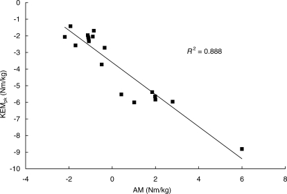
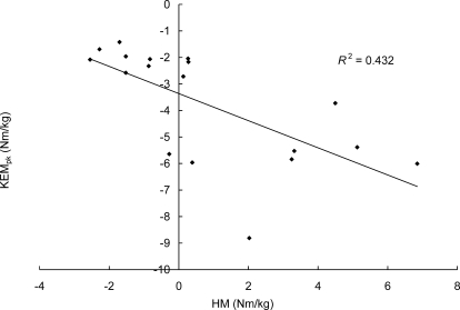
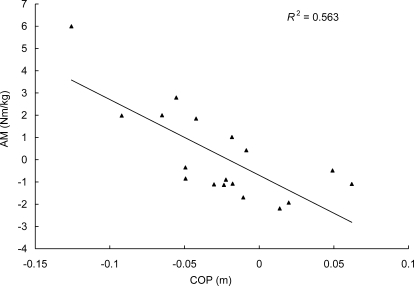
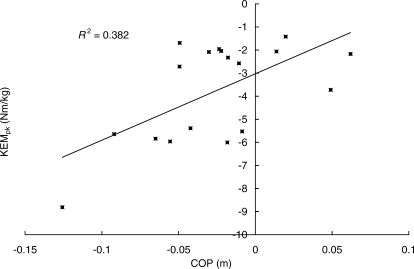
Lower KEMpk related to both greater AM (r = −0.942, P < .001) and HM (r = −0.657, P = .003). We also found that more anterior displacement of COP was related to greater AM (r = −0.750, P < .001) and lower KEMpk (r = 0.618, P = .006).
Conclusions:
Our results suggest that participants who lean the whole body forward during landing may produce more plantar-flexor moment and less knee-extensor moment, possibly increasing hip-extensor moment and decreasing knee-extensor moment production. These results suggest that leaning forward may be a technique to decrease quadriceps contraction demand while increasing hamstrings cocontraction demand during a single-leg landing.
Keywords: quadriceps muscle contraction, hamstrings muscle contraction
Key Points
We found that a lower knee-extensor moment related to higher ankle plantar-flexor and hip-extensor moments during a single-leg landing.
Greater anterior displacement of anterior-posterior center of pressure relative to foot center of mass at net peak knee-extensor moment was related to greater plantar-flexor and lower knee-extensor moments.
Leaning forward during landing may help to stabilize the knee by reducing the quadriceps contraction demand while increasing and/or maintaining the hamstrings muscle contraction demand.
The mechanisms of noncontact anterior cruciate ligament (ACL) injury have often been observed during sudden decelerating motions, such as landing.1–3 During such decelerating weight-bearing motions, excessive contraction force of the quadriceps muscles at shallow knee-flexion angles (ie, less than 40°–60° of knee flexion) may increase ACL tensile force,4–8 possibly rupturing the ACL. Conversely, cocontractions of the hamstrings muscles are thought to stabilize the knee9,10 because they have been shown to reduce ACL tensile force when quadriceps contraction force exists, even during shallow knee-flexion angles.6,7 Furthermore, authors5–7 have reported that ACL tensile forces due to quadriceps force decrease as the knee moves into deeper knee-flexion angle. Collectively, these findings suggest that reducing the quadriceps contraction demand while increasing hamstrings muscle cocontraction demand during sudden deceleration tasks, especially at or near full knee extension, is important to preventing noncontact ACL injuries.
The position of the body during decelerating motion has been implicated as a factor that may be associated with noncontact ACL injuries. Boden et al2 reported that ACL injuries often occur if the upper body is leaning backward with the leg positioned forward when decelerating from forward running. This body positioning may place the center of mass (COM) more posterior and could potentially increase the contraction demand for the knee-extensor muscles (ie, quadriceps muscles) while decreasing the contraction demands for the plantar flexors and hip extensors (ie, hamstrings muscles). Conversely, a more anterior COM placement may reduce knee-extensor muscle contraction demand, possibly increasing the hamstrings muscle contraction demand. Examining such relationships in the lower extremity is important when considering a landing strategy that may protect the ACL.
One way to understand these relationships is to examine net internal moments about each lower extremity joint. Moment (or torque) may be defined as the turning effect of a force that causes angular acceleration of an object about a specific axis.11 In biomechanics, the term internal moment to a body segment is used to describe the moment that is caused by the forces exerted within the body segment itself, such as muscle forces and tensile forces of the skin, joint capsules, or ligaments.11 We can approximate the amount of net internal moment about each joint using an inverse dynamics approach, which estimates the amount of forces and moments acting on each body segment from kinematics, anthropometric data, and all external forces applied on each segment. One of the limitations of this inverse dynamics approach is that only the net internal moment can be obtained, as individual tissue forces acting on the segment and their moment arms cannot be determined.
However, because muscle forces represent the major forces producing internal moments about a joint, examining the amount of lower extremity net internal moment may improve our understanding of how muscle contraction demands change across each joint during a given task. For example, increasing net internal knee-extensor moment may be associated with greater quadriceps muscle contraction demand.
We have found no studies that examined how the demands of lower extremity muscle contraction relate during decelerating motions, such as a single-leg landing. Thus, the primary purpose of our study was to examine the relationships among lower extremity sagittal-plane internal moment productions at the ankle, knee, and hip during a single-leg landing in order to consider a possible strategy to reduce quadriceps contraction demand while increasing or maintaining hamstrings muscle contraction demand. Our secondary purpose was to examine whether differences in landing strategies reduce knee-extensor contraction demand based on the center-of-pressure position of the body. In the inverse pendulum model, the ankle moment is responsible for changing the COM position. If the COM moves anteriorly (or posteriorly), one moves the center of pressure more anteriorly (or posteriorly) by producing greater plantar-flexor (or dorsiflexor) moment to maintain balance.12 Because the landing task required participants to balance for 2 seconds after landing, we analyzed the relationship of sagittal-plane ankle and knee moment with center-of-pressure position at peak knee-extensor moment (KEMpk) as an indicator of neuromuscular response to COM position. We hypothesized that lower knee-extensor moments would relate to greater plantar-flexor and hip-extensor moments and that anteriorly displaced center of pressure would relate to greater ankle plantar-flexor and lower knee-extensor moments.
Methods
Participants
Eighteen healthy, recreationally active women (age = 22.3 ± 2.8 years, height = 162.5 ± 8.1 cm, mass = 57.8 ± 9.3 kg) were recruited from the university and surrounding community. Female participants were chosen because this population has a higher incidence of ACL injuries than men13 and because we wanted to eliminate any confounding sex differences. No participant had lower extremity ligamentous injury or pain in either extremity at the time of participation. Before data collection, each participant gave informed consent, and the study was approved by the Institutional Review Board for the Protection of Human Participants at the University of North Carolina at Greensboro. Each participant stated her age and dominant leg (ie, the leg with which she preferred to kick a ball). The mass and height of each participant were measured and manually recorded.
Procedures
Participants performed all landing tasks with a bare foot to reduce data variability due to different shoe types. Before data collection, each participant practiced single-leg landings from a 45-cm box until she was comfortable performing them. The participant was instructed to stand on the box using the dominant leg. From this position, she was instructed to drop off the box without lowering the COM or jumping up, land on the center of the force plate using the dominant leg, and maintain balance on the dominant leg for at least 2 seconds. She was then told to place both thumbs on the iliac crests throughout the landing task to eliminate the potential effects of arm swing that could change the COM momentum.
We used a 3-dimensional electromagnetic tracking system (Motion Star hardware; Ascension Technology Corp, Burlington, VT) and nonconductive force plate (model 4060; Bertec Corp, Columbus, OH) to collect linear and angular position data of each segment at 140 Hz and ground reaction force at 1000 Hz. Four motion sensors were attached with double-sided tape on the dominant leg over the dorsum of the foot, the flattest part of the mid-anterior tibial shaft, the lateral aspect of the thigh over the iliotibial band, and the sacrum. To minimize sensor movement during the landing, elastic prewrap and regular white tape were used to further secure each motion sensor to each segment. The knee and ankle joint centers were defined as the midpoint between the lateral and medial epicondyles and between the lateral and medial malleoli, respectively.14 The center of the hip joint was determined as described by Leardini et al.15 All data were imported into the MotionMonitor software (Innovative Sports Training Inc, Chicago, IL), and the linked rigid-segment system and local coordinate system for each segment were constructed during the digitization procedure. The linked rigid-segment system was made based on the anthropometric data of Dempster.16
After the digitization procedure, participants performed the single-leg landing from a 45-cm box, as described. Kinetic and kinematic data were obtained from 5 successful landings. A trial during which the participant lost balance, lowered her COM before dropping off the box, and/or did not keep her thumbs on the iliac crests was considered a mistrial, and the trial was discarded and repeated.
Data Reductions and Analyses
All kinematic and kinetic calculations were performed using the MotionMonitor software. Kinetic and kinematic data were low-pass filtered at 60 Hz and 12 Hz, using a fourth-order, zero-lag, digital Butterworth filter. Kinetic data were calculated using Newtonian inverse dynamics. Sagittal-plane ankle and knee internal moments about the medial-lateral axis of the shank and sagittal-plane hip internal moments about the medial-lateral axis of the thigh were obtained. Net internal moments at the distal and proximal shank about the medial-lateral axis of the shank local coordinate system were defined as net internal ankle and knee moment, and net internal moment at the proximal thigh about the medial-lateral axis of the thigh local coordinate system was defined as net internal hip moment. From each trial, we identified KEMpk after touch down. At the time of KEMpk, corresponding sagittal-plane ankle moment (AM), sagittal-plane hip moments (HM), and anterior-posterior center of pressure relative to foot COM (COP) were also obtained. All kinetic variables were normalized to body mass (in kilograms).
We initially obtained all sagittal-plane kinetic and kinematic data using the right-hand rule. However, to match the direction of lower extremity sagittal-plane moment data with our hypotheses, the direction of moment data was changed as indicated in Table 1. For example, using the right-hand rule, knee-extensor moment would be directed to positive; however, in our convention, it was directed to negative. Table 1 summarizes the abbreviations, definitions, and directions for all variables. These values were obtained from each trial and averaged across the 5 trials.
Table 1.
Abbreviations and Definitions for Each Variable
Statistical Analyses
We used Pearson product moment correlations to examine the relationships among AM, KEMpk, HM, and COP. The α level was set at .05.
Results
The mean and SD for each variable are presented in Table 2, and the Pearson product moment correlations between the variables of interest are shown in Table 3. Scatterplots for the relationships between KEMpk and AM and between KEMpk and HM are presented in Figures 1 and 2, respectively. All KEMpk values were obtained shortly after the touch down (81.56 ± 24.11 milliseconds; range, 42–125 milliseconds). All moments were moderately to highly correlated at the time of KEMpk (Table 3). Specifically, greater plantar-flexor moment production was associated with lower knee-extensor moment production, and both greater plantar-flexor and lower knee-extensor moments were associated with greater hip-extensor moments.
Table 2.
Means and SDs for Each Variable at Peak Knee-Extensor Moment
Table 3.
Pearson Product Moment Correlation Matrix of Lower Extremity Internal Moments and Center of Pressure at Peak Knee-Extensor Moment
Figure 1. Scatterplot demonstrating the relationship between peak knee-extensor moment and ankle internal moment at peak knee-extensor moment. KEMpk, initial peak knee-extensor moment after foot contact; AM, sagittal-plane ankle internal moment at initial peak knee-extensor moment after foot contact.
Figure 2. Scatterplot demonstrating the relationship between peak knee-extensor moment and hip internal moment at peak knee-extensor moment. KEMpk, initial peak knee-extensor moment after foot contact; HM, sagittal-plane hip internal moment at initial peak knee-extensor moment after foot contact.
We found a relatively high negative correlation between AM and COP (r = −0.750) (Figure 3) and a moderately positive correlation between KEMpk and COP (r = 0.618) (Figure 4). These results indicated that anterior displacement of COP is related not only to greater plantar-flexor moment production but also to less knee-extensor moment production.
Figure 3. Scatterplot demonstrating the relationship between ankle internal moment and center of pressure at peak knee-extensor moment. AM, sagittal-plane ankle internal moment at initial peak knee-extensor moment after foot contact; COP, anterior-posterior center of pressure relative to the foot center of mass at initial peak knee-extensor moment after foot contact.
Figure 4. Scatterplot demonstrating the relationship between peak knee-extensor internal moment and center of pressure at peak knee-extensor moment. KEMpk, initial peak knee-extensor moment after foot contact; COP, anterior-posterior center of pressure relative to the foot center of mass at initial peak knee-extensor moment after foot contact.
Discussion
Our main finding was moderate to high correlations among sagittal-plane lower extremity internal moments at KEMpk. Specifically, less knee-extensor moment production was related to greater ankle plantar-flexor and hip-extensor moment productions during a single-leg landing.
Researchers4–8,17–19 widely accept that quadriceps contractions and weight bearing load the ACL, especially during shallow knee flexion. Anterior cruciate ligament loading with quadriceps force, especially near full knee extension, may be explained by the angle between the tibial shaft and infrapatellar tendon.20 During shallow knee flexion (ie, less than 40°–60° of knee flexion) when the quadriceps contraction produces force, the anterior component of the infrapatellar tendon force may pull the tibia anteriorly, resulting in ACL loading.4–8 DeMorat et al8 showed that violent quadriceps contraction might be a mechanism of ACL injury, and it is thought that preventing such violent quadriceps contraction during weight-bearing deceleration tasks may prevent ACL injury. Our data indicate that KEMpk occurred shortly after the touch down (81.56 ± 24.11 milliseconds) with relatively shallow knee-flexion angles (43.2 ± 7.5°). This finding is not surprising because during landing, the body generally receives much higher ground reaction force in the early phase of landing, and this higher ground reaction force may increase external knee-flexor moment, possibly increasing the demand for knee-extensor moment production. Thus, we believe that analyzing the relationship among KEMpk, AM, and HM is important to understanding how we may reduce the demand on quadriceps muscle contraction during landing.
Our results indicate that more plantar-flexor moment production was related to less knee-extensor moment production, which suggests the importance of using the plantar-flexor muscles to effectively absorb the shock of landing, so that quadriceps muscle contraction demand is reduced. Theoretically, by effectively using the ankle joint as the first shock absorber to reduce the linear momentum, the muscle contraction demands at the proximal joint (ie, knee joint) may be reduced. In our single-leg drop landing (toe-to-heel type of landing), the ground reaction force creates an external dorsiflexor moment. The plantar-flexor muscles create a countermoment (plantar-flexor moment) to resist excessive dorsiflexion. If the plantar-flexor muscles contract less to absorb the shock of landing, then the knee-extensor muscles aid in shock absorption, resulting in an increase in quadriceps muscle contraction demand. Our results support this theory because participants with greater ankle plantar-flexor moment production demonstrated less knee-extensor moment, which may be related to less quadriceps contraction demand.21
Our results also indicate that less knee-extensor moment and greater ankle plantar-flexor moment were related to greater hip-extensor moment production. As the hamstrings muscles act to flex the knee and extend the hip,22 greater hip-extensor moment would indicate an increase or maintenance of hamstrings muscle contraction demand. This is important because investigators4,6,7,19 have shown a decrease in ACL loading due to quadriceps force or knee-extensor exercise, such as squatting with hamstrings muscle cocontraction force. Hamstrings contractions also have been shown to provide stability to transverse-plane knee loading,23 which increases ACL loading.5,6,18,24,25 Thus, the reduction of quadriceps contraction demand and the increase or maintenance in hamstrings muscle contraction demand during high-risk activities, such as landing, may be important for increasing knee stability and possibly protecting the ACL.
Our correlation analyses also revealed that the relationships between anterior placement of COP with lower knee-extensor moment and greater plantar-flexor moment at KEMpk are relatively strong. Biomechanically, placement of the center of pressure in the anterior-posterior plane is controlled by plantar-flexor or dorsiflexor moment production at the ankle and is a result of neuromuscular responses to the location of the body's COM.12 Although the position of the center of pressure does not directly correspond with the actual COM position, we think that participants produced more plantar-flexor moment in response to anterior COM, resulting in a more anteriorly placed center of pressure. Therefore, a slight anterior lean when landing may facilitate more effective use of plantar-flexor muscles, thereby decreasing the quadriceps contraction demand and increasing the hip-extensor (eg, hamstrings muscle) contraction demand (through decreasing external knee-flexor and hip-flexor moments).
Collectively, these results suggest a possible landing strategy that may offer more protection for the ACL. Injury prevention programs for the ACL, such as plyometric or balance training, have been shown effective.26–28 However, the biomechanical rationale for these intervention programs is still unknown. Although our results are based on biomechanical data without actual ACL loading, they suggest that athletes who are trained to absorb shock during toe-to-heel landing by landing slightly forward and using more plantar-flexor muscles may experience less quadriceps and greater hip-extensor (which, in part, includes the hamstrings) muscle contraction demand to decelerate the body. Using such a landing strategy may increase the stability of the knee, possibly reducing loads on the ACL.
Limitations
Our study had several limitations. First, we do not know whether these relationships hold true in other landing styles, such as the landing phase of a drop jump or cutting or heel-to-toe landing. Furthermore, all landing tasks were performed with a bare foot by recreationally active participants. Thus, our findings may be limited to similar tasks and populations, and further work is needed to examine whether these findings may be generalized to other tasks and populations. Second, because we did not digitize the whole body, our discussions about the body COM position were based on the center-of-pressure data and basic biomechanical concepts and assumptions. To further understand how landing technique might protect the ACL, more comprehensive biomechanical studies are needed. Not obtaining surface electromyographic data or using a forward dynamics model to assess actual muscle activations or forces is also a limitation. Our discussions of muscle contraction demands were based only on net joint internal moment data and basic biomechanical concepts. Future work could validate whether greater hip-extensor moment is also associated with greater hamstrings muscle activation or forces using different research models. Finally, we did not measure the actual ACL loading during landing. Therefore, the safer landing strategy for the ACL implicated by our study is only based on the theory of what is known about the relationship between muscle contractions and knee stability.4,6,7,9,10,19,23 Further research using in vivo and in vitro models or computer simulation is needed to validate whether such a landing strategy actually reduces ACL loading.
Conclusions
Our results suggest that during a single-leg landing, an increase in plantar-flexor moment and an increase in hip-extensor moment are associated with a decrease in knee-extensor moment. Landing styles that place the COM of the whole body anteriorly may be an effective way to reduce quadriceps contraction demand and the resulting knee-extensor moment while increasing or maintaining hamstrings muscle and plantar-flexor muscle contraction demands. Increasing hamstrings muscle cocontractions during knee-extensor dominant activities, such as landing, may help to stabilize the knee joint and reduce ACL loading. This landing technique may be an important consideration in developing an ACL injury-prevention program for athletes who engage in jumping and landing activities, such as basketball or volleyball players, but future researchers must validate whether this landing technique actually reduces ACL loading in vivo.
Acknowledgments
We thank the University of North Carolina at Greensboro for allowing us to use the Applied Neuromechanics Research Laboratory.
Footnotes
Yohei Shimokochi, PhD, ATC, contributed to conception and design; acquisition and analysis and interpretation of the data; and drafting, critical revision, and final approval of the article. Sae Yong Lee, PhD, ATC; Sandra J. Shultz, PhD, ATC, FNATA, FACSM; and Randy J. Schmitz, PhD, ATC, contributed to conception and design, analysis and interpretation of the data, and critical revision and final approval of the article.
References
- 1.Olsen O.E, Myklebust G, Engebretsen L, Bahr R. Injury mechanisms for anterior cruciate ligament injuries in team handball: a systematic video analysis. Am J Sports Med. 2004;32(4):1002–1012. doi: 10.1177/0363546503261724. [DOI] [PubMed] [Google Scholar]
- 2.Boden B.P, Dean G.S, Feagin J.A, Jr, Garrett W.E., Jr Mechanisms of anterior cruciate ligament injury. Orthopedics. 2000;23(6):573–578. doi: 10.3928/0147-7447-20000601-15. [DOI] [PubMed] [Google Scholar]
- 3.McNair P.J, Marshall R.N, Matheson J.A. Important features associated with acute anterior cruciate ligament injury. N Z Med J. 1990;103(901):537–539. [PubMed] [Google Scholar]
- 4.Pandy M.G, Shelburne K.B. Dependence of cruciate-ligament loading on muscle forces and external load. J Biomech. 1997;30(10):1015–1024. doi: 10.1016/s0021-9290(97)00070-5. [DOI] [PubMed] [Google Scholar]
- 5.Arms S, Pope M.H, Johnson R.J, Fischer R.A, Arvidsson I, Eriksson E. The biomechanics of anterior cruciate ligament rehabilitation and reconstruction. Am J Sports Med. 1984;12(1):8–18. doi: 10.1177/036354658401200102. [DOI] [PubMed] [Google Scholar]
- 6.Markolf K.L, O'Neil G, Jackson S.R, McAllister D.R. Effects of applied quadriceps and hamstrings muscle loads on forces in the anterior and posterior cruciate ligaments. Am J Sports Med. 2004;32(5):1144–1149. doi: 10.1177/0363546503262198. [DOI] [PubMed] [Google Scholar]
- 7.Li G, Rudy T.W, Sakane M, Kanamori A, Ma C.B, Woo S.L.Y. The importance of quadriceps and hamstring muscle loading on knee kinematics and in-situ forces in the ACL. J Biomech. 1999;32(4):395–400. doi: 10.1016/s0021-9290(98)00181-x. [DOI] [PubMed] [Google Scholar]
- 8.DeMorat G, Weinhold P, Blackburn T, Chudik S, Garrett W. Aggressive quadriceps loading can induce noncontact anterior cruciate ligament injury. Am J Sports Med. 2004;32(2):477–483. doi: 10.1177/0363546503258928. [DOI] [PubMed] [Google Scholar]
- 9.Baratta R, Solomonow M, Zhou B.H, Letson D, Chuinard R, D'Ambrosia R. Muscular coactivation: the role of the antagonist musculature in maintaining knee stability. Am J Sports Med. 1988;16(2):113–122. doi: 10.1177/036354658801600205. [DOI] [PubMed] [Google Scholar]
- 10.Solomonow M, Baratta R, Zhou B.H, et al. The synergistic action of the anterior cruciate ligament and thigh muscles in maintaining joint stability. Am J Sports Med. 1987;15(3):207–213. doi: 10.1177/036354658701500302. [DOI] [PubMed] [Google Scholar]
- 11.Nigg B.M, MacIntosh B.R, Mester J, editors. Biomechanics and Biology of Movement. Champaign, IL: Human Kinetics; 2000. p. 258. [Google Scholar]
- 12.Winter D.A. Biomechanics and Motor Control of Human Movement. 2nd ed. New York, NY: John Wiley & Sons; 1990. pp. 93–94. [Google Scholar]
- 13.Powell J.W, Barber-Foss K.D. Sex-related injury patterns among selected high school sports. Am J Sports Med. 2000;28(3):385–391. doi: 10.1177/03635465000280031801. [DOI] [PubMed] [Google Scholar]
- 14.Madigan M.L, Pidcoe P.E. Changes in landing biomechanics during a fatiguing landing activity. J Electromyogr Kinesiol. 2003;13(5):491–498. doi: 10.1016/s1050-6411(03)00037-3. [DOI] [PubMed] [Google Scholar]
- 15.Leardini A, Cappozzo A, Catani F, et al. Validation of a functional method for the estimation of hip joint centre location. J Biomech. 1999;32(1):99–103. doi: 10.1016/s0021-9290(98)00148-1. [DOI] [PubMed] [Google Scholar]
- 16.Dempster W.T. Space Requirements of the Seated Operator: Geometrical Kinematic, and Mechanical Aspects of the Body With Special Reference to the Limbs. OH: Wright-Patterson Air Force Base; 1955. Wright Air Development Center Technical Report 55–159. [Google Scholar]
- 17.Cerulli G, Benoit D.L, Lamontagne M, Caraffa A, Liti A. In vivo anterior cruciate ligament strain behaviour during a rapid deceleration movement: case report. Knee Surg Sports Traumatol Arthrosc. 2003;11(5):307–311. doi: 10.1007/s00167-003-0403-6. [DOI] [PubMed] [Google Scholar]
- 18.Fleming B.C, Renstrom P.A, Beynnon B.D, et al. The effect of weightbearing and external loading on anterior cruciate ligament strain. J Biomech. 2001;34(2):163–170. doi: 10.1016/s0021-9290(00)00154-8. [DOI] [PubMed] [Google Scholar]
- 19.Shelburne K.B, Pandy M.G. Determinants of cruciate-ligament loading during rehabilitation exercise. Clin Biomech (Bristol, Avon) 1998;13(6):403–413. doi: 10.1016/s0268-0033(98)00094-1. [DOI] [PubMed] [Google Scholar]
- 20.Isaac D.L, Beard D.J, Price A.J, Rees J, Murray D.W, Dodd C.A.F. In-vivo sagittal plane knee kinematics: ACL intact, deficient and reconstructed knees. Knee. 2005;12(1):25–31. doi: 10.1016/j.knee.2004.01.002. [DOI] [PubMed] [Google Scholar]
- 21.Chappell J.D, Yu B, Kirkendall D.T, Garrett W.E. A comparison of knee kinetics between male and female recreational athletes in stop-jump tasks. Am J Sports Med. 2002;30(2):261–267. doi: 10.1177/03635465020300021901. [DOI] [PubMed] [Google Scholar]
- 22.Moore K.L. Clinically Oriented Anatomy. 3rd ed. Baltimore, MD: Williams & Wilkins; 1992. p. 593. [Google Scholar]
- 23.MacWilliams B.A, Wilson D.R, DesJardins J.D, Romero J, Chao E.Y. Hamstrings cocontraction reduces internal rotation, anterior translation, and anterior cruciate ligament load in weight-bearing flexion. J Orthop Res. 1999;17(6):817–822. doi: 10.1002/jor.1100170605. [DOI] [PubMed] [Google Scholar]
- 24.Markolf K.L, Gorek J.F, Kabo J.M, Shapiro M.S. Direct measurement of resultant forces in the anterior cruciate ligament: an in vivo study performed with a new experimental technique. J Bone Joint Surg Am. 1990;72(4):557–567. [PubMed] [Google Scholar]
- 25.Wascher D.C, Markolf K.L, Shapiro M.S, Finerman G.A.M. Direct in vitro measurement of forces in the cruciate ligaments, part I: the effect of multiplane loading in the intact knee. J Bone Joint Surg Am. 1993;75(3):377–386. doi: 10.2106/00004623-199303000-00009. [DOI] [PubMed] [Google Scholar]
- 26.Myklebust G, Engebretsen L, Braekken I.H, Skjolberg A, Olsen O.E, Bahr R. Prevention of anterior cruciate ligament injuries in female team handball players: a prospective intervention study over three seasons. Clin J Sport Med. 2003;13(2):71–78. doi: 10.1097/00042752-200303000-00002. [DOI] [PubMed] [Google Scholar]
- 27.Hewett T.E, Lindenfeld T.N, Riccobene J.V, Noyes F.R. The effect of neuromuscular training on the incidence of knee injury in female athletes: a prospective study. Am J Sports Med. 1999;27(6):699–706. doi: 10.1177/03635465990270060301. [DOI] [PubMed] [Google Scholar]
- 28.Hewett T.E, Stroupe A.L, Nance T.A, Noyes F.R. Plyometric training in female athletes: decreased impact forces and increased hamstring torques. Am J Sports Med. 1996;24(6):765–773. doi: 10.1177/036354659602400611. [DOI] [PubMed] [Google Scholar]


