Abstract
Context:
Individuals suffering from patellofemoral pain have previously been reported to have decreased isometric strength of the hip musculature; however, no researchers have investigated concentric and eccentric torque of the hip musculature in individuals with patellofemoral pain.
Objective:
To compare concentric and eccentric torque of the hip musculature in individuals with and without patellofemoral pain.
Design:
Case control.
Setting:
Research laboratory.
Patients or Other Participants:
Twenty participants with patellofemoral pain (age = 26.8 ± 4.5 years, height = 171.8 ± 8.4 cm, mass = 72.4 ± 16.8 kg) and 20 control participants (age = 25.6 ± 2.8 years, height = 169.5 ± 8.9 cm, mass = 70.0 ± 16.9 kg) were tested. Volunteers with patellofemoral pain met the following criteria: knee pain greater than or equal to 3 cm on a 10-cm visual analog scale, insidious onset of symptoms not related to trauma, pain with palpation of the patellar facets, and knee pain during 2 of the following activities: stair climbing, jumping or running, squatting, kneeling, or prolonged sitting. Control participants were excluded if they had a prior history of patellofemoral pain, knee surgery in the past 2 years, or current lower extremity injury that limited participation in physical activity.
Intervention(s):
Concentric and eccentric torque of the hip musculature was measured on an isokinetic dynamometer. All volunteers performed 5 repetitions of each strength test. Separate multivariate analyses of variance were performed to compare concentric and eccentric torque of the hip extensors, abductors, and external rotators between groups.
Main Outcome Measure(s):
Average and peak concentric and eccentric torque of the hip extensors, abductors, and external rotators. Torque measures were normalized to the participant's body weight multiplied by height.
Results:
The patellofemoral pain group was weaker than the control group for peak eccentric hip abduction torque (F1,38 = 6.630, P = .014), and average concentric (F1,38 = 4.156, P = .048) and eccentric (F1,38 = 4.963, P = .032) hip external rotation torque.
Conclusions:
The patellofemoral pain group displayed weakness in eccentric hip abduction and hip external rotation, which may allow for increased hip adduction and internal rotation during functional movements.
Keywords: anterior knee pain, lower extremity, muscle strength
Key Points
Peak eccentric hip abduction torque and average concentric and eccentric hip external rotation torque were lower in the group with patellofemoral pain.
Assessing hip muscle strength is a crucial step in the evaluation of patients with patellofemoral pain.
Patellofemoral pain syndrome is one of the most common knee conditions reported by males and females in the United States.1 Although this condition is common, its cause is not well understood. Many researchers2–4 speculate that abnormal alignment of the patella within the femoral trochlea may lead to the development of patellofemoral pain. One underlying cause of this malalignment is speculated to be abnormal transverse-plane or frontal-plane (or both) motion of the femur during functional movements.2
Hip musculature plays an important role in controlling transverse-plane and frontal-plane motions of the femur. More specifically, weakness of the gluteus medius muscle is believed to increase hip adduction and knee valgus angles.2,5–7 Additionally, weakness of the “deep 6” hip external rotators (piriformis, obturator internus and externus, gemellus superior and inferior, and quadratus femoris) is also proposed to increase hip internal rotation and knee valgus angles.2,5,8 Although the gluteus maximus is most commonly thought to control sagittal-plane motion at the hip and trunk,9 researchers10 have reported that the upper portion of the gluteus maximus functions like the gluteus medius during walking and stair ambulation; therefore, the gluteus maximus may play a role in controlling frontal-plane and transverse-plane motions of the hip during functional tasks. Based on the functions of these muscles, weakness of the hip muscles may lead to malalignment of the patella within the femoral trochlea due to excessive movements of the femur in hip adduction and internal rotation.
Previous investigators5,11–13 have reported that individuals with patellofemoral pain have decreased isometric strength of the hip abductors, external rotators, and extensors. Females with patellofemoral pain have been reported to be 26% and 24% to 36% weaker than healthy females on measures of isometric hip abduction and hip external rotation strength, respectively.5,11 In a separate study,13 females with patellofemoral pain had 27% less hip abduction strength, 30% less hip external rotation strength, and 52% less hip extension strength than healthy females. Although difference have been reported among females, when males and females with patellofemoral pain were compared with healthy males and females, no differences in hip abductor and external rotation isometric strength were noted.12
To our knowledge, no authors have determined if concentric and eccentric torque of the hip musculature differ in individuals with and without patellofemoral pain. Research investigating concentric and eccentric muscle torque is needed because of the importance of concentric and eccentric muscle contraction during functional tasks. Concentric muscle contraction produces the joint torque necessary to accelerate the limb through space and facilitate joint motion. In contrast, eccentric muscle contraction decelerates the limb during motion.14 Therefore, decreased muscle torque may have a significant effect on the ability to accelerate and decelerate joint motion. If patients with patellofemoral pain are weaker than healthy controls on measures of concentric and eccentric hip abduction and hip external rotation torque, frontal-plane and transverse-plane motions of the femur during functional movements may be less controlled and, in turn, lead to patellar malalignment.
The literature offers conflicting results on the relationship between isometric strength and isokinetic torque. Some researchers15,16 have observed strong relationships between these measures, whereas others17 have reported weaker relationships. Based on the lack of a clear relationship between isometric and isokinetic torque, we cannot conclude that decreased isometric strength of hip muscles in patients with patellofemoral pain suggests that they have decreased concentric and eccentric torque of the hip muscles. Therefore, the purpose of our investigation was to determine if differences in concentric and eccentric torque of the hip musculature (hip extensors, hip external rotators, and hip abductors) exist between individuals with and without patellofemoral pain. We hypothesized that both concentric and eccentric torque of the hip extensors, external rotators, and abductors would be decreased in individuals with patellofemoral pain compared with those without patellofemoral pain.
Methods
Participants
Forty volunteers (20 with patellofemoral pain, 20 controls) between the ages of 18 and 40 years were recruited from the University of North Carolina student, staff, and faculty populations. The patellofemoral pain group consisted of 7 male and 13 female participants (age = 26.8 ± 4.5 years, height = 171.8 ± 8.4 cm, mass = 72.4 ± 16.8 kg). Inclusion criteria for the patellofemoral pain group included 1) anterior or retropatellar knee pain present during at least 2 of the following activities: ascending or descending stairs, hopping or running, squatting, kneeling, and prolonged sitting; 2) insidious onset of symptoms not related to trauma; 3) pain on palpation of the patellar facets; and 4) worst pain in the past week greater than or equal to 3 cm on a 10-cm visual analog scale. Inclusion criteria were adapted from Cowan et al.18 The control group consisted of 7 males and 13 females (age = 25.6 ± 2.8 years, height = 169.5 ± 8.9 cm, mass = 70.0 ± 16.9 kg) who were matched on age, height, and mass to the patellofemoral pain volunteers. Exclusion criteria for the patellofemoral pain and control groups included 1) history of knee surgery, 2) clinical evidence of other knee injury, or 3) current significant injury affecting other lower extremity joints.
Before data collection, all participants signed an informed consent approved by the University's Biomedical Institutional Review Board, which also approved the study. Additionally, all volunteers completed a 10-cm visual analog scale (VAS) for “worst knee pain during the previous week” and the anterior knee pain scale (AKPS). The AKPS is a 13-item, self-administered questionnaire that assesses knee function. A higher score on the AKPS suggests better perceived function, with a maximum score of 100. The AKPS has previously been reported to be reliable and valid in a population with patellofemoral pain.19,20
Procedures
Concentric and eccentric torque of the hip extensors, hip external rotators, and hip abductors were assessed using an isokinetic dynamometer (Biodex Medical Systems Inc, Shirley, NY). Before testing, participants were provided with detailed instructions for the strength testing procedures. They were allowed 3 submaximal practice repetitions before testing. No volunteers needed more than 3 practice repetitions to perform the tests in a smooth manner. Five maximal repetitions for hip extension, external rotation, and abduction were collected for each strength test. All strength testing was performed at 60°/s. We chose 60°/s because a muscle produces greater concentric force at slower isokinetic testing velocities.21 Furthermore, as velocity increases during eccentric contractions, the force-producing capability stays the same or slightly increases.21 Therefore, the testing velocity of 60°/s is a good representation of both the concentric and eccentric force-producing capabilities of each muscle group we assessed.
Each participant was provided 2 minutes of rest between strength tests. Order of strength testing was randomized. The leg used for testing in each control volunteer corresponded with the injured limb tested in the matched patellofemoral pain volunteer. If the patellofemoral pain volunteer suffered from bilateral patellofemoral pain, the leg with the worst pain in the past week was used for testing.
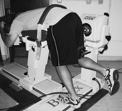
For the hip extension test, participants were positioned so that the trunk was flexed to 90° and their arms were wrapped around the chair of the dynamometer to stabilize the trunk (Figure 1). The axis of rotation for the dynamometer was aligned with the greater trochanter of the femur on the test leg. The lever arm applied resistance on the posterior thigh just superior to the knee. The nontest leg supported the body of the participant. The participant was instructed to exert maximal strength against the dynamometer in the direction of hip extension, keeping the knee flexed at approximately 90°. The torque of the hip extensors was tested concentrically and eccentrically from 90° of hip flexion to 60° of hip flexion.
Figure 1. Hip extension strength test.
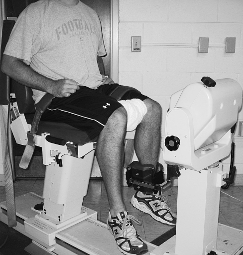
Hip external rotation torque was assessed with the participant in a seated position with the hip and knee flexed to 90° (Figure 2). The upper thigh of the test leg and the trunk were stabilized with straps. A bolster was placed between the participant's knees in order to control for hip adduction. The axis of rotation for the dynamometer was aligned with the knee joint line. Concentric and eccentric strength testing was performed through a range of 5° of hip internal rotation (neutral) to 20° of hip external rotation, allowing for 25° of total hip internal and external rotation range of motion.
Figure 2. Hip external rotation strength test.
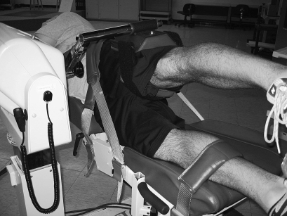
Hip abduction torque was assessed with the test leg on top of the nontest leg and the participant in a side-lying position (Figure 3). The nontest leg and trunk were stabilized with straps. The dynamometer axis of rotation was aligned medial to the anterior superior iliac spine at the level of the greater trochanter on the test leg. The lever arm applied resistance to the lateral aspect of the distal thigh superior to the lateral femoral condyle. The participant was instructed to exert maximal strength against the dynamometer in the direction of hip abduction during the entire strength test. Concentric and eccentric torque of the hip abductors was tested from 0° to 20° of hip abduction.
Figure 3. Hip abduction strength test.
Intrasession reliability (intraclass correlation coefficient [2,1]) for the testing protocol used in this investigation has previously been reported from our laboratory22 and is presented in Table 1. For the reliability investigation, concentric and eccentric torque of the hip muscles was assessed in 20 healthy, physically active volunteers. Each volunteer performed 3 repetitions of hip extension, abduction, and external rotation using the same positioning and testing procedures described above.22
Table 1.
Intrasession Reliability Values for Isokinetic Strength Testsa
Data Reduction and Statistical Analysis
Peak and average torque data for the concentric and eccentric phases of isokinetic strength testing were determined using custom software (MATLAB; The MathWorks, Inc, Natick, MA). Concentric and eccentric phases were determined by the direction of motion. For example, during hip abduction, the concentric phase occurred when the dynamometer arm moved from 0° to 20° of hip abduction; the eccentric phase occurred when the dynamometer arm moved from 20° to 0° of hip abduction. The average of the middle 3 trials for each strength test was used for data analysis to eliminate the possibility of learning (trial 1) and fatigue effects (trial 5). All torque data (Nm) were normalized to body weight multiplied by height.23
We performed independent-samples t tests to determine differences in age, height, mass, visual analog scale values, and AKPS values between the patellofemoral pain and control groups. Three separate, 1-way multivariate analyses of variance (MANOVAs) were conducted to determine the effect of group (patellofemoral pain and control) on peak and average concentric and eccentric torque for the hip extensors, abductors, and external rotators. Analyses of variance (ANOVAs) on each dependent variable were conducted as follow-up tests to the MANOVA. We used SPSS (version 14.0; SPSS Inc, Chicago, IL) to perform all analyses. An a priori α level was set at .05.
Results
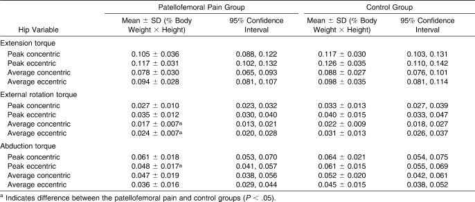
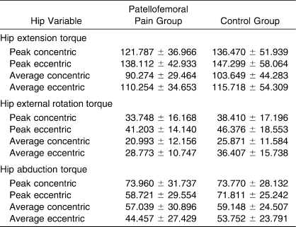
The patellofemoral pain group did not differ from the control group on measures of age, height, or mass (P > .05); however, both the visual analog scale (5.06 ± 2.27 cm versus 0.14 ± 0.28 cm, t38 = 9.57, P = .001) and AKPS (68.90 ± 9.15 versus 99.10 ± 2.73, t38 = −14.15, P = .001) scores were different, indicating that knee pain and function were worse in the patellofemoral pain participants. A significant MANOVA was found for both hip external rotation torque (Pillai trace = 0.279, F4,35 = 3.386, P = .019) and hip abduction torque (Pillai trace = 0.250, F4,35 = 2.919, P = .035). Follow-up, 1-way ANOVA tests for hip abduction torque revealed weaker peak eccentric torque in the patellofemoral pain group than the control group (F1,38 = 6.630, P = .014). Similarly, follow-up analyses of hip external rotation torque demonstrated decreased average concentric torque (F1,38 = 4.156, P = .048) and average eccentric torque (F1,38 = 4.963, P = .032) in the patellofemoral pain group compared with the control group. The MANOVA for hip extension torque was not significant (Pillai trace = 0.093, F4,35 = 0.892, P = .479, 1-β = .254). Means, SDs, and 95% confidence intervals for normalized data and means and SDs for raw (Nm) data for each dependent variable are presented in Tables 2 and 3.
Table 2.
Normalized Peak and Average Hip Concentric and Eccentric Torques
Table 3.
Peak and Average Hip Concentric and Eccentric Torques (Raw Data) (Nm, Mean ± SD)
Discussion
Recently, researchers5,11–13 have reported that patients with patellofemoral pain have decreased isometric strength of the hip extensors, external rotators, and abductors; however, no authors have investigated the concentric and eccentric torque of these muscles. The findings of this study indicate that patients with patellofemoral pain are weaker than healthy volunteers on measures of peak eccentric hip abduction torque and average concentric and eccentric hip external rotation torque. More specifically, the patellofemoral pain group was 21% weaker than the control group for peak eccentric hip abduction torque and 23% weaker than the control group for both average concentric and eccentric hip external rotation torque.
Our findings are similar to those of previous researchers5,11,13 who reported decreased isometric strength of the hip external rotators and abductors in females with patellofemoral pain. Direct comparisons with previous studies are difficult because no other researchers measured concentric and eccentric torque of the hip musculature in patients with patellofemoral pain. One finding in disagreement with previous research is hip extension strength. Robinson and Nee13 reported that patients with patellofemoral pain were 52% weaker on measures of isometric hip extension strength than controls. In our investigation, patients with patellofemoral pain were, on average, 8% weaker than the healthy volunteers. A possible reason for the conflicting results is differences in testing procedures. First, Robinson and Nee13 only assessed isometric strength, whereas we assessed isokinetic torque. Additionally, they positioned participants at 0° of hip flexion when testing isometric hip extension strength. In our investigation, volunteers started at approximately 90° of hip flexion and extended at the hip to approximately 60° of hip flexion (30° range of motion). The differences in the length-tension relationship due to participant positioning between these studies may explain the different results and should be further investigated. We performed a post hoc power analysis for hip extension torque and determined that we would need more than 140 volunteers to achieve a power of .80. Therefore, based on these findings and the large amount of overlap in the 95% confidence intervals for hip extension torque, increasing participant numbers to increase statistical power would not produce a clinically meaningful difference between individuals with and without patellofemoral pain.
The findings for hip external rotation and abduction strength are similar when our findings are compared with those of previous investigators who assessed isometric strength. We noted that patients with patellofemoral pain were approximately 8% weaker on measures of concentric hip abduction torque and 20% weaker on measures of eccentric hip abduction torque. The eccentric hip abduction torque deficit is similar to the deficit of approximately 25% reported by previous authors for isometric hip abduction strength; however, our findings for concentric hip abduction torque are much smaller than those previously reported for isometric hip abduction strength.5,11,13 Additionally, patients with patellofemoral pain in our investigation were approximately 21% weaker than the control group on measures of concentric hip external rotation torque and 18% weaker on measures of eccentric hip external rotation torque. These findings are also much smaller than the deficit of approximately 30% reported by previous authors5,11,13 assessing isometric strength of the hip external rotators. The lack of consistency in strength deficits among the concentric, eccentric, and isometric measures lends support to the need for future investigators to assess concentric and eccentric torque and not focus solely on isometric strength in the population with patellofemoral pain. Although investigators assessing isometric strength provide good information to clinicians for rehabilitation and prevention measures, concentric and eccentric torque is also an important piece of the puzzle that needs to be evaluated in patients with patellofemoral pain. To our knowledge, researchers have reported that no single strength assessment (isometric, isokinetic, or isotonic) is a better predictor of functional performance than another,24 but have stated that each strength assessment by itself is highly correlated with functional performance.25,26 Based on these findings, measurements of isometric, isokinetic and isotonic strength each provide useful information to clinicians and should all be assessed for use in prevention and rehabilitation programs.
Measures of concentric and eccentric torque are clinically important because during functional movements, muscles contract concentrically and eccentrically. If joints and body segments cannot withstand the external moments applied to the body by concentric and eccentric muscle torques, faulty movement patterns may occur. During running, the hip abductor muscles fire during midstance to stabilize the pelvis.27 If the hip abductors cannot stabilize the pelvis, frontal-plane motion at the hip and knee may increase.28 As stated previously, faulty movement patterns, such as increased hip adduction and internal rotation, are theorized risk factors for patellofemoral pain.2 Increased hip adduction and internal rotation may be prevented through concentric and eccentric contractions of the hip external rotators and abductors; however, if the hip abductors and external rotators are weak, excessive or abnormal motions of the femur are more difficult to control.
Many researchers18,29–31 have reported decreased pain and increased function in patients with patellofemoral pain after an intervention that included strengthening exercises for the hip musculature. Based on our findings of decreased hip muscle strength and previous findings of decreased pain and increased function after strengthening of the hip musculature, strengthening exercises for the hip muscles are warranted in the treatment of patients with patellofemoral pain. No investigators have determined if strength gains in the hip musculature lead to decreased frontal-plane and transverse-plane motions at the hip and knee during functional movements; therefore, future authors should investigate changes in hip and knee kinematics after an intervention that includes hip strengthening exercises.
Future researchers should also determine if patients with patellofemoral pain display sex differences on measures of hip muscle strength. Because of the sample size in this investigation, we did not have sufficient power to make this comparison. Additionally, future authors should determine if hip muscle weakness and lower extremity kinematics during dynamic activities are related in patients with patellofemoral pain. This information will provide additional results that may be used to develop effective intervention programs for patients with patellofemoral pain.
Limitations
A limitation of this investigation is the range of motion we used to assess the strength of the hip extensors, external rotators, and abductors. We decided on this range of motion based upon the level of comfort of the participants during pilot testing. Although we are limited in the ability to generalize these results to functional tasks because of the small range of motion used to assess strength, we feel the internal validity of our results is high, given that all participants performed the tests through the same arcs of motion. Future researchers should assess the concentric and eccentric strength of the hip extensors, external rotators, and abductors through a larger range of motion.
An additional limitation is that we did not assess participants' pain during the strength tests, which may have had an influence on the results of this investigation. However, we did not assess pain during the testing because it is not common for individuals with patellofemoral pain to complain of pain in the hip region. Another limitation of this study is the decreased ability to determine a cause-and-effect relationship within a case-control design. We cannot determine if hip muscle weakness was present before or after the patellofemoral pain developed. Future authors should use a prospective cohort design to determine if hip muscle weakness is a risk factor for patellofemoral pain.
Clinical Implications
Based on the findings of this investigation, assessing hip muscle strength is a crucial step in the evaluation of patients with patellofemoral pain. As denoted by the 95% confidence intervals, our measures of hip muscle torque were precise because of the small ratio between the lower and upper confidence intervals (ratios ranged from .61 to .79).32 Additionally, with the significant results, the confidence intervals displayed a very small amount of overlap. With the use of 95% confidence intervals, the clinical meaningfulness of our results is enhanced due to the divergence in confidence intervals and small confidence limit ratios for the significant findings. Therefore, including exercises that not only increase concentric but also eccentric strength of the hip muscles should be part of rehabilitation programs developed for patients with patellofemoral pain. The muscles targeted through strengthening exercises should include the gluteus medius and the “deep 6” hip external rotators (piriformis, obturator internus and externus, gemellus superior and inferior, and quadratus femoris).
Acknowledgments
We thank the University of North Carolina Injury Prevention Research Center for funding this project through its Student Small Grant program.
Footnotes
Michelle C. Boling, PhD, ATC, contributed to conception and design; acquisition and analysis and interpretation of the data; and drafting, critical revision, and final approval of the article. Darin A. Padua, PhD, ATC, contributed to conception and design, analysis and interpretation of the data, and drafting, critical revision, and final approval of the article. R. Alexander Creighton, MD, contributed to conception and design and drafting, critical revision, and final approval of the article.
References
- 1.DeHaven K.E, Lintner D.M. Athletic injuries: comparison by age, sport, and gender. Am J Sports Med. 1986;14(3):218–224. doi: 10.1177/036354658601400307. [DOI] [PubMed] [Google Scholar]
- 2.Powers C. The influence of altered lower-extremity kinematics on patellofemoral joint dysfunction: a theoretical perspective. J Orthop Sports Phys Ther. 2003;33(11):639–646. doi: 10.2519/jospt.2003.33.11.639. [DOI] [PubMed] [Google Scholar]
- 3.Fulkerson J.P. Diagnosis and treatment of patients with patellofemoral pain. Am J Sports Med. 2002;30(3):447–456. doi: 10.1177/03635465020300032501. [DOI] [PubMed] [Google Scholar]
- 4.Thomee R, Augustsson J, Karlsson J. Patellofemoral pain syndrome: a review of current issues. Sports Med. 1999;28(4):245–262. doi: 10.2165/00007256-199928040-00003. [DOI] [PubMed] [Google Scholar]
- 5.Ireland M.L, Willson J.D, Ballantyne B.T, Davis I.M. Hip strength in females with and without patellofemoral pain. J Orthop Sports Phys Ther. 2003;33(11):671–676. doi: 10.2519/jospt.2003.33.11.671. [DOI] [PubMed] [Google Scholar]
- 6.Fredericson M, Cookingham C.L, Chaudhari A.M, Dowdell B.C, Oestreicher N, Sahrmann S.A. Hip abductor weakness in distance runners with iliotibial band syndrome. Clin J Sport Med. 2000;10(3):169–175. doi: 10.1097/00042752-200007000-00004. [DOI] [PubMed] [Google Scholar]
- 7.Mizuno Y, Kumagai M, Mattessich S.M, et al. Q-angle influences tibiofemoral and patellofemoral kinematics. J Orthop Res. 2001;19(5):834–840. doi: 10.1016/S0736-0266(01)00008-0. [DOI] [PubMed] [Google Scholar]
- 8.Lee T.Q, Anzel S.H, Bennett K.A, Pang D, Kim W.C. The influence of fixed rotational deformities of the femur on the patellofemoral contact pressures in human cadaver knees. Clin Orthop Rel Res. 1994;302:69–74. [PubMed] [Google Scholar]
- 9.Lieberman D.E, Raichlen D.A, Pontzer H, Bramble D.M, Cutright-Smith E. The human gluteus maximus and its role in running. J Exp Biol. 2006;209(pt 11):2143–2155. doi: 10.1242/jeb.02255. [DOI] [PubMed] [Google Scholar]
- 10.Lyons K, Perry J, Gronley J.K, Barnes L, Antonelli D. Timing and relative intensity of hip extensor and abductor muscle action during level and stair ambulation: an EMG study. Phys Ther. 1983;63(10):1597–1605. doi: 10.1093/ptj/63.10.1597. [DOI] [PubMed] [Google Scholar]
- 11.Bolgla L.A, Malone T.R, Umberger B.R, Uhl T.L. Hip strength and hip and knee kinematics during stair descent in females with and without patellofemoral pain syndrome. J Orthop Sports Phys Ther. 2008;38(1):12–18. doi: 10.2519/jospt.2008.2462. [DOI] [PubMed] [Google Scholar]
- 12.Piva S.R, Goodnite E.A, Childs J.D. Strength around the hip and flexibility of soft tissues in individuals with and without patellofemoral pain syndrome. J Orthop Sports Phys Ther. 2005;35(12):793–801. doi: 10.2519/jospt.2005.35.12.793. [DOI] [PubMed] [Google Scholar]
- 13.Robinson R.L, Nee R.J. Analysis of hip strength in females seeking physical therapy treatment for unilateral patellofemoral pain syndrome. J Orthop Sports Phys Ther. 2007;37(5):232–238. doi: 10.2519/jospt.2007.2439. [DOI] [PubMed] [Google Scholar]
- 14.Perrin D.H, Gieck J.H. Principles of therapeutic exercise. In: Perrin D.H, editor. The Injured Athlete. Vol. 1999. Philadelphia, PA: Lippincott-Raven; pp. 123–139. 3rd ed. [Google Scholar]
- 15.Knapik J.J, Ramos M.U. Isokinetic and isometric torque relationships in the human body. Arch Phys Med Rehabil. 1980;61(2):64–67. [PubMed] [Google Scholar]
- 16.Lord J.P, Aitkens S.G, McCrory M.A, Bernauer E.M. Isometric and isokinetic measurement of hamstring and quadriceps strength. Arch Phys Med Rehabil. 1992;73(4):324–330. doi: 10.1016/0003-9993(92)90004-g. [DOI] [PubMed] [Google Scholar]
- 17.Osternig L.R, Bates B.T, James S.L. Isokinetic and isometric torque force relationships. Arch Phys Med Rehabil. 1977;58(6):254–257. [PubMed] [Google Scholar]
- 30.Cowan S.M, Bennell K.L, Crossley K.M, Hodges P.W, McConnell J. Physical therapy alters recruitment of the vasti in patellofemoral pain syndrome. Med Sci Sports Exerc. 2002;34(12):1879–1885. doi: 10.1097/00005768-200212000-00004. [DOI] [PubMed] [Google Scholar]
- 18.Crossley K.M, Bennell K.L, Cowan S.M, Green S. Analysis of outcome measures for persons with patellofemoral pain: which are reliable and valid. Arch Phys Med Rehabil. 2004;85(5):815–822. doi: 10.1016/s0003-9993(03)00613-0. [DOI] [PubMed] [Google Scholar]
- 19.Watson C.J, Propps M, Ratner J, Zeigler D.L, Horton P, Smith S.S. Reliability and responsiveness of the lower extremity functional scale and the anterior knee pain scale in patients with anterior knee pain. J Orthop Sports Phys Ther. 2005;35(3):136–146. doi: 10.2519/jospt.2005.35.3.136. [DOI] [PubMed] [Google Scholar]
- 20.Perrin D.H. Isokinetic Exercise and Assessment. Champaign, IL: Human Kinetics; p. 1993. [Google Scholar]
- 21.Hawkey T.J, Halverson S.D, Padua D.A, Guskiewicz K.M. Reliability of three different strength testing protocols for hip abduction, extension, and external rotation strength [abstract] J Athl Train. 2004;39(2 suppl):S–116. [Google Scholar]
- 22.Patel R.R, Hurwitz D.E, Bush-Joseph C.A, Bach BR, Andriacchi T.P. Comparison of clinical and dynamic knee function in patients with anterior cruciate ligament deficiency. Am J Sports Med. 2003;31(1):68–74. doi: 10.1177/03635465030310012301. [DOI] [PubMed] [Google Scholar]
- 23.Anderson M.A, Gieck J.H, Perrin D.H, Weltman A, Rutt R, Denegar C. The relationship among isometric, isotonic, and isokinetic concentric and eccentric quadriceps and hamstring force and three components of athletic performance. J Orthop Sports Phys Ther. 1991;14(3):114–120. doi: 10.2519/jospt.1991.14.3.114. [DOI] [PubMed] [Google Scholar]
- 24.McClements L.E. Power relative to strength of leg and thigh muscles. Res Q. 1966;37(1):71–78. [PubMed] [Google Scholar]
- 25.Miyashita M, Kanehisa H. Dynamic peak torque related to age, sex, and performance. Res Q. 1979;50(2):249–255. [PubMed] [Google Scholar]
- 26.Montgomery W.H, III, Pink M, Perry J. Electromyographic analysis of hip and knee musculature during running. Am J Sports Med. 1994;22(2):272–278. doi: 10.1177/036354659402200220. [DOI] [PubMed] [Google Scholar]
- 27.Niemuth P.E, Johnson R.J, Myers M.J, Thieman T.J. Hip muscle weakness and overuse injuries in recreational runners. Clin J Sport Med. 2005;15(1):14–21. doi: 10.1097/00042752-200501000-00004. [DOI] [PubMed] [Google Scholar]
- 28.Tyler T.F, Nicholas S.J, Mullaney M.J, McHugh M.P. The role of hip muscle function in the treatment of patellofemoral pain syndrome. Am J Sports Med. 2006;34(4):630–636. doi: 10.1177/0363546505281808. [DOI] [PubMed] [Google Scholar]
- 29.Mascal C.L, Landel R, Powers C. Management of patellofemoral pain targeting hip, pelvis, and trunk muscle function: 2 case reports. J Orthop Sports Phys Ther. 2003;33(11):647–660. doi: 10.2519/jospt.2003.33.11.647. [DOI] [PubMed] [Google Scholar]
- 31.Boling M.C, Bolgla L.A, Mattacola C.G, Uhl T.L, Hosey R.G. Outcomes of a weight-bearing rehabilitation program for patients diagnosed with patellofemoral pain syndrome. Arch Phys Med Rehabil. 2006;87(11):28–35. doi: 10.1016/j.apmr.2006.07.264. [DOI] [PubMed] [Google Scholar]
- 32.Poole C. Low P-values or narrow confidence intervals: which are more durable. Epidemiology. 2001;12(3):291–294. doi: 10.1097/00001648-200105000-00005. [DOI] [PubMed] [Google Scholar]


