Abstract
Purpose
To determine the sustaining effects of orthokeratology.
Methods
This study enrolled 58 eyes with moderate myopia. LK-DM lenses (Lucid Korea Dream Lens™) were fitted daily for at least eight hours on an overnight regimen. The effects of orthokeratology and it's sustainability throughout the day were recorded twice; immediately after removal in the morning and eight hours later. Sustainability was measured by comparing the changes from morning to afternoon for best uncorrected visual acuity, apical corneal power, keratometric values, spherical equivalent and induced astigmatism.
Results
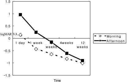
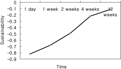
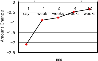
UCVA demonstrated improved values at all follow up periods. Fluctuations during the day stabilized after 4 weeks of lens wear. K values averaged a mean of 42.4 mm at baseline, and reduced to 40.9 mm by week 12. Unaided logMAR visual acuity changed from 0.94±0.14 at baseline to -0.11±0.17 by week 12. The sustainability of orthokeratology, defined as the difference between morning and afternoon values of unaided logMAR visual acuity, increased from -0.82 on day 1 to -0.11 on week 12.
Conclusions
UCVA and spherical refractive error did not change to a significant degree after 4 weeks. Although statistically insignificant minute fluctuations during the day were observed up to week 12, these fluctuations decreased to a statistically significant level after week 4.
Keywords: Corneal asphericity, Corneal topography, Myopia, Orthokeratology, Rigid gas-permeable contact lenses
There has been a renewal of clinical and research interest in orthokeratology (OK) since the development of reverse-geometry contact lens designs by Wlodyga and Stoyan in the late 1980s.1 Traditional OK involved the use of conventionally designed, usually flat-fitting, rigid contact lenses. To differentiate this traditional form from the current incarnation of OK the term 'accelerated orthokeratology' was developed. This term refers to the rapid onset of refractive and corneal topographic changes that occur with reverse-geometry lenses. Nichols et al2 and Swarbrick and Alharbi3 have reported that the refractive endpoint is typically reached with these lenses after 7 to 10 days of wear with the use of an overnight lens-wearing protocol. This was confirmed more recently by Chang et al4 showing stabilization of vision by 1 week and by Shin et al5 showing stable refractive endpoints by 14 days. This compares with traditional OK, where the refractive endpoint may take weeks to months of open-eye rigid lens wear to achieve.4-7
As early as 1968, Mandell and St. Helen10 reported that significant transient changes in corneal curvature could be induced by lid forces, digital pressure, and eye rubbing. Carney and Clark11 subsequently described short-term corneal topographic changes resulting from the pressure associated with applanation tonometry. More recently, Horner et al12 reported significant corneal flattening after 1 hour of reverse-geometry contact lens wear in the open eye. Swarbrick et al13 have also documented rapid changes in corneal curvature within the first few hours wearing reverse-geometry rigid contact lenses.
In clinical practice, many OK practitioners utilize a short lens-wearing trial as a predictive test of future success for subsequent OK lens wear. The usefulness of such trials and the appropriate period of lens wear for reliable prediction of a refractive endpoint have not been investigated. Also, initial fluctuations of the induced orthokeratologic effects are clinically observed. Refractive correction that results in the morning has a tendency not to persist throughout the day in the initial period of lens wear. Yoon et al14 have published reports on the reversibility of corneal parameters after discontinuation of lens wear, which occurs at 2 weeks for low myopes (less than -3.5D) and at 4 weeks for moderate myopes (more than -3.5D). However, there have not been any studies on short term reversibility occurring during the day to the naked eye.
In this article we report in detail the changes in visual acuity, refractive error and corneal topography immediately after lens removal in the morning and compare these changes with those induced eight hours later. Such a comparison should provide useful insight into the sustainability of OK lens wear on refractive effects during the initial periods of orthokeratology.
Materials and Methods
Fifty-eight eyes of 29 subjects were recruited from the employee population of our medical institution. After approval for this study by the institutional human research ethics committee, written informed consent was obtained from all subjects after the risks and benefits of OK lens wear and study procedures had been fully explained. Subjects were required to be 18 to 35 years of age, free of ocular disease, have no prior history of ocular surgery or contraindications to rigid contact lens wear. All subjects needed to have with-the-rule corneal astigmatism of less than 1.50D, with less than moderate myopia of 4.0D to enroll in the study. None of the subjects were current rigid gas-permeable or full-time soft lens wearers. Baseline parameters are summarized in Table 1.
Table 1.
Subject characteristics at baseline
*logMAR VA: logarithm of the minimum angle of resolution visual acuity.
The reverse-geometry rigid contact lenses used in this study were of XQ material (nominal Dk 145×10-11 [cm2.mL O2]/[sec.mL.mmHg]), with a nominal center thickness of 0.22 mm and a quadracurve design (Lucid Korea Dream Lens™) .The lenses were 11.0 mm in overall diameter with an optic zone of 6.0 mm.
Accelerated orthokeratology was performed on an overnight protocol of eight hours according to manufacturer's guidelines. Sustainability of the orthokeratologic effects during the day was clinically observed by comparing the refractive corrective effects observed right after lens removal in the morning to eight hours later. Changes in the sustainability of accelerated orthokeratology as subjects continued with lens wear was monitored over time.
Reverse-geometry lenses give a bull's eye fluorescein fitting pattern, with a central zone of light touch 3.5 to 4.0 mm in diameter, a midperipheral ring of fluorescein pooling, or 'tear reservoir', under the steeper secondary curve and a peripheral circular band of alignment tapering to the edge lift. A slit l-amp lens-fitting assessment of the recommended trial lens was carried out on all subjects to ensure good centration, lens movement of 1 to 2 mm on a blink, active tear exchange without the presence of bubbles in the tear reservoir and a clinically acceptable fluorescein fitting pattern.
Unaided visual acuity for distance was measured at six meters. This was performed using the same internally illuminated logarithm of the minimum angle of resolution (log MAR) type chart, with a luminance of 120 cd/m2 to monitor change in vision with OK lens wear. All visual acuity measurements were obtained under normal room illumination.
Subjective refraction was performed during each visit to determine the best-vision sphere (sphere+half cylinder) in each eye. Subjective refraction was obtained using the retinoscope by the same examiner (S.Y. Kang) for all patients during the entire period of study. A detailed biomicroscopic examination of the anterior segment, including fluorescein staining assessment, was performed before and after each session of lens wear to ensure good ocular health.
The corneal radius of curvature was measured using a Magnon keratometer (H. Ogino & Co, Yokohama, Japan). Three readings were taken and averaged on each measurement occasion. The same keratometer was used throughout the study, and was calibrated before the study began and periodically during the study using steel balls of known radius.
Corneal topography was measured using the Oculus corneal topographer. Three images were captured, and data from the associated map displays were averaged on each measurement occasion. The instrument was adjusted and refocused after every image capture. Parameters obtained from the topographic data display included apical corneal power and radius of curvature, simulated keratometry (SimK) readings, and the difference in apical corneal power between pre- and post-lens wear sessions (from the difference map). On the topography map, orthokeratology treatment creates a central circular zone of corneal flattening, termed the treatment zone, surrounded by a ring of midperipheral corneal steepening. The treatment zone diameter was measured manually from the topograph screen displaying the difference map, which demonstrates change in corneal topography relative to baseline. The criterion for determining treatment zone diameter was horizontal distance from inner edge to inner edge of the zero diopter change zone inside the ring of midperipheral steepening on the difference map.
Baseline data were collected before commencing lens wear. Subjects wore the same OK lens daily in both eyes for the full overnight protocol, a minimum of 8 hours. Subjects inserted their own lenses in the evening at home, and slept approximately eight hours while wearing the lenses. Lenses were removed by one of the authors the following morning at the hospital when the patients came in for work. Study variables were measured immediately after lens removal, and again when patients returned eight hours later before leaving work. Measurements were taken at 1 hour, 1 day, 1, 2, 4 and 12 weeks.
We combined analysis of variance (ANOVA) with post hoc paired t-tests with Bonferroni protection to compare parameters measured in the morning and afternoon. A critical p value of 0.05 was chosen to denote statistical significance.
Results
At the time of lens removal, statistically significant improvement in unaided visual acuity (p<0.001, ANOVA) relative to baseline was found for all periods of lens wear in all subjects. As expected, there was a greater improvement in visual acuity in the morning immediately after lens removal. Fig. 1 shows the change in unaided visual acuity for both morning and afternoon after different durations of OK lens wear. This difference between morning and afternoon was designated as the sustainability of orthokeratology, and is demonstrated in Fig. 2. Sustainability increased with increasing durations of lens wear, finally stabilizing after week 4 (p<0.05). These results and the outcome of statistical analyses are summarized in Table 2.
Fig. 1.
Changes in measurements for morning and afternoon for each respective period of lens wear in unaided logMAR visual acuity (UCVA).
Fig. 2.
The difference between morning and afternoon unaided logMAR visual acuity (UCVA), demonstrating increasing sustainability of orthokeratologic effects with increasing periods of lens wear.
Table 2.
Changes in measurements between morning and afternoon for each respective period of lens wear (mean±SD) in unaided logMAR visual acuity (UCVA), apical corneal power (ACP), average keratometry readings (Ka), corneal toricity (Cyl) and treatment zone diameter (TZD) after different durations of orthokeratology lens wear.
NS, not statistically significant (p>0.1).
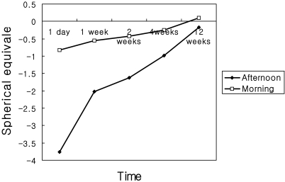
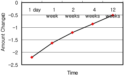
Apical corneal powers were obtained from the central 3.0 mm of optic zone cornea. Mean K value was calculated by averaging vertical and horizontal K values. Fluctuations during the day continuously changed up to week 12, although amplitude of change decreased to a statistically significant level after 4 weeks (p<0.05). Fig. 3 shows the change in best measured sphere for both morning and afternoon after different durations of OK lens wear. These results and the outcome of statistical analyses performed on these data are summarized in Table 2. Figs. 4 and 5 each demonstrate change in apical corneal power and K readings for both morning and afternoon after different durations of lens wear.
Fig. 3.
Change in best measured sphere for morning and afternoon after different durations of OK lens wear.
Fig. 4.
Change in apical corneal power between morning and afternoon after different durations of lens wear.
Fig. 5.
Change in keratometry readings between morning and afternoon after different durations of lens wear.
The treatment zone averaged 3.86±0.88 mm in diameter after 1 day of wear. This value became larger with increasing duration of lens wear, reaching 5.59±0.83 mm in the morning after 1 week. In the afternoon it decreased to an average of 0.84±0.23 mm after 1 day, but this value stabilized to 4.52±0.24 mm after 4 weeks of overnight protocol. Treatment zone diameters after different periods of lens wear are also presented in Table 2.
Discussion
Orthokeratology was defined by Mountford15 in 1997 as a clinical technique utilizing contact lenses to "reduce, modify, or eliminate refractive error". Although primitive forms of orthokeratology were practiced as early as 1968, there has been a resurgence of interest in its applicability and efficacy. This resurgence is secondary to the development of reverse-geometry lens designs, which incorporate a secondary curve steeper than the lens base curve to aid in centration. Reverse-geometry lenses are fitted with a base curve flatter than the central corneal curvature. This applies pressure to the central treatment zone, achieving sphericalization of the prolate cornea and resulting in reduction of myopic refractive error.
Swarbrick13 demonstrated reductions in central corneal asphericity, reporting that final corneal shape changed from prolate to oblate after an overnight session. One of the main advantages of reverse-geometry OK lenses is the speed with which significant corneal curvature and visual acuity changes can be achieved. A procedure that took months to induce significant reductions in myopia with conventional flat-fitting OK lenses6-9 has proven effective within days or even hours with reverse-geometry lenses. However, the results of this study clearly indicate that corneas experiencing orthokeratology regress in their refractive correcting effects in the initial periods of lens wear. This regression exposes patients to blurry vision in the afternoon as corneal asphericity returns, resulting in a decline of visual acuity. However the results presented here demonstrate that this morning to afternoon change stabilizes by week 4, when variability of unaided vision and refractive error diminish.
Swarbrick et al16 have suggested that changes in anterior corneal topography in OK are achieved through central corneal thinning and midperipheral thickening. The central thinning was mainly epithelial in origin, whereas the midperipheral thickening appeared to include a stromal contribution. The topographic thickness changes found in that study were able to explain almost all of the refractive effects of OK based on changes in corneal sagittal height. Based on this mechanism, our results suggest that the corneal epithelium is remodeled very rapidly in response to tear film forces generated behind reverse-geometry lenses. However, this initial discrepancy between morning and afternoon measurements may be due to a lack of epithelial compression by the lenses in the initial periods of lens wear. Thereby inherent forces in the cornea enabled it to return to its original configuration when the lens was not in place.
The overall mean changes in corneal toricity, measured using keratometry, were less than 0.05 D for all lens-wearing periods (see Table 2) and did not reach statistical significance at any point. SimK (simulated keratometry) readings obtained from the Oculus topographer were also analyzed with a similar outcome. Traditional OK is known to induce significant with-the-rule corneal toricity due to decentration of the flat-fitting lenses,6,7 and this undesirable effect is probably one of the reasons for the demise of traditional orthokeratology. Our study did not reveal any induction of corneal toricity in the short lens-wearing periods. Longer-term clinical studies will confirm whether this is also true for prolonged wear of these lenses. To date, studies of OK using reverse-geometry lenses have not reported significant increases in corneal toricity,2,3,15,17 presumably because lens centration is maintained more reliably due to the steeper secondary curve of these lenses. Indeed, some authors have claimed that modern OK with reverse-geometry lenses can reduce with-the-rule astigmatism by up to 60%.18 Mountford and Pesudovs19 report an average reduction in corneal toricity of 50% with accelerated OK using reverse-geometry lenses. Some researchers have suggested that the endpoint of OK treatment is reached when the cornea, which is typically prolate in shape (Q<0), has been sphericalized (Q=0).6,9,15,20,21 To better understand the effects of short-term OK lens wear on corneal shape, further studies are needed using a topographic instrument; which can provide accurate and repeatable values for corneal shape descriptors (e, p, or Q) over variable chord diameters.
This study has demonstrated that corneas experiencing orthokeratology regress in their refractive correcting effects in the initial periods of lens wear. This regression exposes the patients to blurry afternoon vision as corneal asphericity returns, resulting in a decline of visual acuity. However the results presented here show that this change between morning and afternoon stabilizes by week 4, when variability of unaided vision and refractive error diminish.
Footnotes
* Data was previously presented in Seoul Oct. 2003 as a poster at the 90th Annual Meeting of the Korean Ophthalmological Society
References
- 1.Wlodyga RJ, Bryla C. Corneal molding: the easy way. Contact Lens Spectrum. 1989;4:58–65. [Google Scholar]
- 2.Nichols JJ, Marsich MM, Nguyen M, et al. Overnight orthokeratology. Optom Vis Sci. 2000;77:252–259. doi: 10.1097/00006324-200005000-00012. [DOI] [PubMed] [Google Scholar]
- 3.Swarbrick HA, Alharbi A. Overnight orthokeratology induces central corneal epithelial thinning. Invest Ophthalmol Vis Sci. 2001;42:S597. [Google Scholar]
- 4.Chang JW, Choi TH, Lee HB. J Korean Ophthalmol Soc. 2004;45:908–912. [Google Scholar]
- 5.Shin DB, Yang KM, Lee SB, et al. J Korean Ophthalmol Soc. 2003;44:1748–1756. [Google Scholar]
- 6.Kerns RL. Research in orthokeratology. Part VIII: results, conclusions and discussion of techniques. J Am Optom Assoc. 1978;49:308–314. [PubMed] [Google Scholar]
- 7.Binder PS, May CH, Grant SC. An evaluation of orthokeratology. Ophthalmology. 1980;87:729–744. doi: 10.1016/s0161-6420(80)35171-3. [DOI] [PubMed] [Google Scholar]
- 8.Polse KA, Brand RJ, Schwalbe JS, et al. The Berkeley Orthokeratology Study. Part II: efficacy and duration. Am J Optom Physiol Opt. 1983;60:187–198. doi: 10.1097/00006324-198303000-00006. [DOI] [PubMed] [Google Scholar]
- 9.Coon LJ. Orthokeratology. Part II: evaluating the Tabb method. J Am Optom Assoc. 1984;55:409–418. [PubMed] [Google Scholar]
- 10.Mandell RB, Helen RS. Stability of the corneal contour. Am Acad Optom. 1968;45:797–806. doi: 10.1097/00006324-196812000-00002. [DOI] [PubMed] [Google Scholar]
- 11.Carney LG, Clark BA. Experimental deformation of the in vivo cornea. Am Acad Optom. 1972;49:28–34. doi: 10.1097/00006324-197201000-00005. [DOI] [PubMed] [Google Scholar]
- 12.Horner DG, Armitage KS, Wormsley KA, Mandell RB. Corneal molding recovery after contact lens wear. Optom Vis Sci. 1992;69:S156–S157. [Google Scholar]
- 13.Swarbrick HA, Wong G, O'Leary DJ. Corneal response to orthokeratology. Optom Vis Sci. 1998;75:791–799. doi: 10.1097/00006324-199811000-00019. [DOI] [PubMed] [Google Scholar]
- 14.Yoon YM, Kim MK, Lee JL. J Korean Ophthalmol Soc. 2005;46:1478–1485. [Google Scholar]
- 15.Mountford J. An analysis of the changes in corneal shape and refractive error induced by accelerated orthokeratology. ICLC. 1997;24:128–144. [Google Scholar]
- 16.Swarbrick HA, Wong G, O'Leary DJ. Corneal response to orthokeratology. Optom Vis Sci. 1998;75:791–799. doi: 10.1097/00006324-199811000-00019. [DOI] [PubMed] [Google Scholar]
- 17.Lui W-O, Edwards MH. Orthokeratology in low myopia. Part 1: efficacy and predictability. Cont Lens Anterior Eye. 2000;23:77–89. doi: 10.1016/s1367-0484(00)80016-8. [DOI] [PubMed] [Google Scholar]
- 18.Soni PS, Horner DG. Orthokeratology. In: Bennett ES, Weissman BA, editors. Clinical Contact Lens Practice. Philadelphia: Lippincott; 1994. pp. 1–7. [Google Scholar]
- 19.Mountford J, Pesudovs K. An analysis of the astigmatic changes induced by accelerated orthokeratology. Clin Exp Optom. 2002;85:284–293. doi: 10.1111/j.1444-0938.2002.tb03084.x. [DOI] [PubMed] [Google Scholar]
- 20.Freeman RA. Predicting stable changes in orthokeratology. Contact Lens Forum. 1978;3:21–31. [Google Scholar]
- 21.Lui W-O, Edwards MH. Orthokeratology in low myopia. Part 2: corneal topographic changes and safety over 100 days. Cont Lens Anterior Eye. 2000;23:90–99. doi: 10.1016/s1367-0484(00)80017-x. [DOI] [PubMed] [Google Scholar]


