Abstract
Purpose
To assess the reproducibility and agreement of anterior chamber measurements between the Pentacam (PTC) and the Anterior segment optical coherence tomography (AOCT) in normal healthy eyes with open angle.
Methods
Prospective cross-sectional comparative case series. A total of 162 eyes of 81 healthy volunteers with normal open angle were included in this study. Anterior chamber angle (ACA) and anterior chamber depth (ACD) were measured with PTC and AOCT. Intra-observer variability and inter-methods agreement of both instruments for ACA and ACD were evaluated.
Results
Values of temporal and nasal ACA measured by two instruments were similar, and the results of ACD were also not significantly different between modalities (p>0.01). ACA and ACD measurements by PTC and AOCT showed good intra-observer and inter-method agreements (all >0.9).
Conclusions
PTC and AOCT are presumed to be very useful for the anterior chamber angle examination. They may provide good images and quantitative data about the angle structures including ACA and ACD.
Keywords: Anterior chamber angle, Anterior chamber depth, Anterior segment optical coherence tomogaphy, Pentacam
Primary angle closure glaucoma (PACG) is highly prevalent in some regions, especially the Mongolian-type Asian populations.1,2-5 For example, Foster and Johnson2 have estimated that 1.7 million people in China are bilaterally blind from glaucoma and that 91% of glaucoma blindness is attributable to primary angle closure. The number of people with the predisposing risk of "occludable angles" is estimated to be 28.2 million, and the number of people with significant angle closure is 9.1 million.3 Although prospective data are lacking, it is believed that treating anatomically occludable angles with a laser peripheral iridectomy may prevent development of angle closure.6,7 Therefore, early screening of anatomically occludable angles is important for the high prevalence of PACG in certain regions. Assessing of the anterior chamber angle (ACA) and anterior chamber depth (ACD) are determinant components in the assessment of angle closure patient. PACG is diagnosed in cases with an occludable angle combined with glaucomatous optic neuropathy and consistent visual morbidity. An "occludable" angle is one in which the trabecular meshwork is seen in less than 90° of the angle circumference by gonioscopy.8 Currently, gonioscopy is the gold standard for identifying occludable angles by assessing the anatomy and morphology of anterior chamber. However, it is a subjective technique that requires the expertise of a highly skilled examiner (intraobserver and interobserver reproducibilities are generally poor) and can be uncomfortable or poorly tolerated by patients for contacting lens to their eyes. There are also no uniform gonioscopic criteria for identifying angles that require treatment.9,10
New modalities such as the Pentacam (PTC, Oculus Inc, Lynnwood, WA, USA) using the rotating Scheimpflug imaging and the Anterior Segment Optical Coherence Tomography (AOCT; SL-OCT, Heidelberg Engineering GmbH, Heidelberg, Germany) have recently become available. They promise to overcome some limitations of the conventional gonioscopy, and may be excellent candidates for assessment of angle structure.
In the present study, we evaluated the reproducibility and agreement of PTC and AOCT in normal healthy eyes with open angle. Our results provide strong backing to use these new imaging modalities for anterior segment examination.
Subjects and Methods
After obtaining approval of the Institutional Review Board, 81 healthy volunteers with gonioscopically confirmed normal open angle were enrolled to this study. Informed consent was obtained from each subject, then ACA and ACD were measured by PTC and AOCT under the uniform dim illumination by a single investigator. Images of the temporal and nasal angles can be taken more easily than those of superior and inferior angles, and they need no eyelid manipulation to expose the limbus. So, the angle images were captured using the horizontal linear scan protocol (from 3-o'clock to 9-o'clock direction). ACA was measured automatically by PTC and AOCT. ACD was defined as the distance from the posterior vertex of the corneal endothelium to the anterior surface of the crystalline lens along the optical axis. All measurements were repeated 3 times, and their average was used to further analyses.
All statistical analysis were performed using SPSS for Windows, version 11.0 (SPSS Inc, Chicago, Illinois, USA) except intraclass correlation coefficient (ICC) test calculated by using Statistical Analysis Software, version 8.2 (SAS Institute, Cary, NC, USA). p<0.01 was considered as statistically significant.
Results
Among 81 healthy volunteers, 51 (63.0%) were men. Mean age was 22.3±3.5 years (range, 18 to 33 years). And mean refractive error was -3.70±2.68 diopters for right eye and -3.62±2.81 diopters for left eye.
Data about ACA and ACD by two modalities are shown in Table 1. ACA taken with PTC were 45.41±5.30° (31.47-59.47) (temporal side), 43.58±5.04° (33.97-56.67) (nasal side) on right eyes and 47.32±5.66° (33.50-60.40) (temporal side), 44.80±5.38° (31.50-56.90) (nasal side) on left eyes. ACA taken with AOCT were 46.18±5.50° (31.67-59.67) (temporal side), 45.13±5.89° (31.00-57.33) (nasal side) on right eyes and 46.67±5.98° (34.67-61.00) (temporal side), 44.90±5.94° (30.33-59.67) (nasal side) on left eyes. ACD taken with PTC were 3.33±0.27 mm (2.57-3.96) on right eyes and 3.34±0.28 mm (2.59-4.04) on left eyes. ACD taken with AOCT were 3.32±0.26 mm (2.56-3.95) on right eyes and 3.31±0.28 mm (2.54-4.06) on left eyes.
Table 1.
Anterior chamber angle and anterior chamber depth measurements by Pentacam and anterior segment optical coherent tomography
ACA=anterior chamber angle; ACD=anterior chamber depth; AOCT=Anterior segment optical coherent tomography; PTC=Pentacam; °=degrees; Values given as means±standard deviation; If p<0.01: statistically significant; * p-value when compared temporal and nasal anterior chamber angle measured by each methods
Temporal and nasal ACA did not show significant difference between PTC and AOCT (p>0.01). And ACD measured by two instruments were also similar (p>0.01).
ICC for evaluating intra-observer variability of each instrument are shown in Table 2. Each parameter was measured 3 times. ACD and ACD measurements using two study modalities had good intraobserver agreements.
Table 2.
Intraobserver agreement between the Pentacam and anterior segment optical coherence tomography based on intraclass correlation coefficients for measuring anterior chamber angles and anterior chamber depth
ACA=anterior chamber angle; ACD=anterior chamber depth; AOCT=Anterior segment optical coherent tomography; PTC=Pentacam.
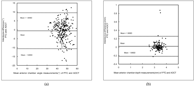
Inter-methods agreement was analyzed using the Bland-Altman analysis (Fig. 1). ACA and ACD measurements by two study modalities showed a good agreement each other.
Fig. 1.
Bland-Altman analysis for the intrer-methods agreement for Pentacam (PTC) and Anterior segment optical coherence tomography (AOCT). (a) Anterior chamber angle; (b) Anterior chamber depth.
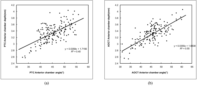
At last, the linear regression analysis was performed to seek the relationship between ACD and ACA (Fig. 2). Average of measurements on each eye were used for analysis, and the best-fit line is also shown in the figures (PTC, R2=0.40, p<0.001; AOCT, R2=0.55, p<0.001).
Fig. 2.
Linear regression analysis between anterior chamber angle and anterior chamber depth for the Pentacam (PTC) (a) and the Anterior segment optical coherence tomography (AOCT) (b).
Discussion
In the present study, the measurements of ACA and ACD were evaluated by two new image modalities; PTC and AOCT. Their data were similar each other, and they showed good intra-observer reproducibility and inter-method agreement.
To judge the intra-observer reproducibility, the ICCs were calculated. It is one of the most popularly used in order to find an intra-observer agreement for various tasks in other area of medical imaging.11 To our knowledge, this is the first article to report the intra-observer agreement of ACA and ACD by PTC and AOCT. They showed the excellent reproducibility of ACA and ACD using PTC and AOCT by a single observer.
Previously, Bland and Altman proposed an informative method to evaluate actual interdevice agreement that allows clinicians to determine for any given use whether the measurements provided by two devices are interchangeable.12 Numerically, the 95% Limit of agreement (LoA) gives the clinician an indication of how much the devices may differ in 95% of cases- that is, in most of their patients. In this report, the mean differences in ACA and ACD as measured with PTC and AOCT were not statistically significant and clinically negligible-approximately 0.543°, 0.021 mm (approximately 1%, 0.6%). The 95% LoA and Brand-Altman plots show a relatively large range of interdevice differences for all comparison, especially ACA (approximately 20° (45% of mean ACA); 0.47 mm (15% of mean ACD)), and this may be too broad for use interchangeably. Other studies have assessed agreement of ACD with Orbscan and PTC.11 Measurements with Orbscan were on the average 0.046 mm longer than with Pentacam. The 95% LoA corresponds to +5.6~-2.5% of the mean ACD. Reddy and associates13 compared ACD measurements by three methods: Orbscan II, IOL Master, and contact A-scan ultrasound. They found that ultrasound measured ACD 13% shorter, whereas the other two modalities showed good correlation. The authors, however, did not assess interdevice agreement by Bland-Altman plots or 95% LoA. Koranyi et al. also showed excellent correlations between three optical modalities, Orbscan, conventional noncontacting Scheimpflug camera, and optical pachymetry, to determine ACD with mean differences of approximately 1.5%, whereas ultrasound measured ACD shorter.14 In a recent study by Buehl et al., 95% LoA of approximately 0.4 mm were reported when ACD measurments by PTC, Orbscan I, and ACMaster were compared.15 It is similar to the result of this study.
In addition, in this study, it is also noticed that the temporal ACA was significantly larger than the nasal ACA by both instruments (all p<0.001, Table 1). And ACA and ACD showed moderate correlation, however they did not have an excellent relationship (Fig. 2).
Even though this study included only normal subjects with open angle, our methodology could be applied to other patients who had closed or occludable angle. PTC and AOCT are presumed to be very useful for angle examination. They may provide good images and quantitative data about the angle structures including ACA and ACD.
References
- 1.Bonomi L, Varotto A. Epidemiology of Angle-closure Glaucoma; Prevalence, Clinical Types, and Association with Peripheral Anterior Chamber Depth in the Egna-Neumarkt Galucoma Study. Opthalmology. 2000;107:998–1003. doi: 10.1016/s0161-6420(00)00022-1. [DOI] [PubMed] [Google Scholar]
- 2.Foster PJ, Johson GJ. Glaucoma in China-how big is the problem? Br J Ophthalmol. 2001;85:1271–1272. doi: 10.1136/bjo.85.11.1277. [DOI] [PMC free article] [PubMed] [Google Scholar]
- 3.Alsbrik PH. Anterior chamber depth and primary angle-closure glaucoma. I. An epidemiologic study in Greenland Eskimos. Acta Ophthalmol (Copenh) 1975;53:89–104. doi: 10.1111/j.1755-3768.1975.tb01142.x. [DOI] [PubMed] [Google Scholar]
- 4.Chew PT, Sung T. Primary angle-closure glaucoma in Asia. J Glaucoma. 2001;10(Suppl):S7–S8. doi: 10.1097/00061198-200110001-00004. [DOI] [PubMed] [Google Scholar]
- 5.Dandona L, Dandona R, Mandal P, et al. Angle-closure glaucoma in an urban population in southern India. The Andhra Pradesh eye disease study. Ophthalmology. 2000;107:1710–1716. doi: 10.1016/s0161-6420(00)00274-8. [DOI] [PubMed] [Google Scholar]
- 6.Radhakrishnan S, Goldsmith J, Huang D, et al. Comparison of Optical Coherence Tomography and Ultrasound Biomicroscopy for Detection of Narrow Anterior Chamber Angles. Arch Ophthalmol. 2005;123:1053–1059. doi: 10.1001/archopht.123.8.1053. [DOI] [PubMed] [Google Scholar]
- 7.Radhakrishnan S, Goldsmith J, Huang D, et al. Optical coherence tomography imaging of the anterior chamber angle. Ophthalmol Clin North Am. 2005;18:375–381. doi: 10.1016/j.ohc.2005.05.007. [DOI] [PubMed] [Google Scholar]
- 8.Foster PJ, Devereux JG, Alsbirk PH, et al. Detection of gonioscopically occludable angles and primary angle closure glaucoma by estimation of limbal chamber depth in Asians: modified grading scheme. Br J Ophthalmol. 2000;84:186–192. doi: 10.1136/bjo.84.2.186. [DOI] [PMC free article] [PubMed] [Google Scholar]
- 9.Foster PJ, Buhrmann R, Quigley HA, Johnson GJ. The definition and classification of glaucoma in prevalence surveys. Br J Ophthalmol. 2002;86:238–242. doi: 10.1136/bjo.86.2.238. [DOI] [PMC free article] [PubMed] [Google Scholar]
- 10.Friedman DS. Who needs an iridotomy? Br J Ophthalmol. 2001;85:1019–1021. doi: 10.1136/bjo.85.9.1019. [DOI] [PMC free article] [PubMed] [Google Scholar]
- 11.Lackner B, Schimidger G, Skorpik C. Validity and Repeatability of Anterior Chamber Depth Measurements With Pentacam and Orbscan. Optom Vis Sci. 2005;82:858–861. doi: 10.1097/01.opx.0000177804.53192.15. [DOI] [PubMed] [Google Scholar]
- 12.Bland JM, Altman DG. Statistical methods for assessing agreement between two methods of clinical measurement. Lancet. 1986;1:307–310. [PubMed] [Google Scholar]
- 13.Reddy AR, Pande MV, Finn P, El-Gogary H. Comparative estimation of anterior chamber depth by ultrasonography, Orbscan II, and IOLMaster. J Cataract Refract Surg. 2004;30:1268–1271. doi: 10.1016/j.jcrs.2003.11.053. [DOI] [PubMed] [Google Scholar]
- 14.Koranyi G, Lydahl E, Norrby S, Taube M. Anterior chamber depth measurement: a-scan versus optical methods. J Cataract Refract Surg. 2002;28:243–247. doi: 10.1016/s0886-3350(01)01039-2. [DOI] [PubMed] [Google Scholar]
- 15.Buehl W, Stojanac D, Sacu S, et al. Comparison of three methods of measuring corneal thickness and anterior chamber depth. Am J Ophthalmol. 2006;141:7–12. doi: 10.1016/j.ajo.2005.08.048. [DOI] [PubMed] [Google Scholar]


