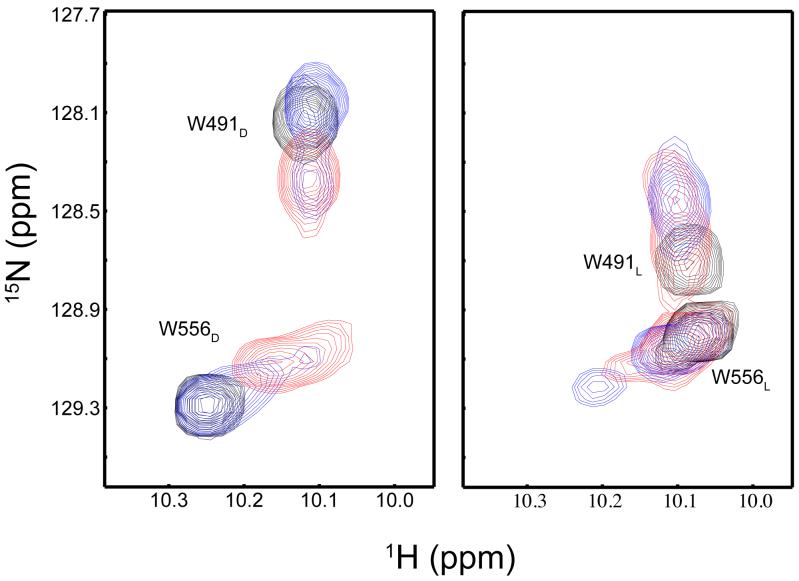
Figure 5. 15N/1H HSQC spectra of the tryptophan Hε1-Nε1 region.
The overlaid spectra of the dark (a) and lit (b) states of the wildtype AsLOV2 (black), Q513L (blue), and Q513N (red) are shown. The tryptophan indole assignments are indicated in both the dark and lit state panels with subscript D or L respectively. In the lit state, there is a significant shift in this region in wildtype protein (b).

