Abstract
The case of a 78-year-old African American woman who presented at the Mount Sinai Medical Center (Chicago, USA) with excruciating backache is presented. Computed tomography of the chest at the time of admission showed dissection of the aortic arch, descending aorta and dissection of an aberrant right subclavian artery. She was managed medically for Stanford type B acute aortic dissection. The patient was asymptomatic at presentation, but started complaining of new-onset dysphagia during her stay in the hospital. An esophagogram was performed and suggested posterior impingement of the esophagus, a classic sign of an aberrant right subclavian artery. Because the patient had multiple underlying comorbidities and the dysphagia was mild and intermittent, surgery was deferred. The patient was discharged home after complete stabilization and was scheduled for a follow-up appointment.
Keywords: Aortic arch syndromes, Dysphagia, Subclavian artery
Abstract
On présente ici le cas d’une patiente d’origine afro-américaine de 78 ans qui a consulté au Mount Sinai Medical Center (Chicago, USA) pour d’intenses douleurs au dos. La tomographie du thorax au moment de son admission a révélé une dissection de la crosse de l’aorte, de l’aorte descendante et de l’artère sous-clavière droite aberrante. Elle a été traitée pour une dissection de l’aorte aiguë de type Stanford B. La patiente était asymptomatique au moment de son arrivée, mais elle a commencé à se plaindre de dysphagie de novo durant son séjour hospitalier. Un œsophagogramme a été réalisé et a révélé une possible empreinte rétroœsophagienne, typique de l’artère sous-clavière droite aberrante. Étant donné que la patiente présentait de nombreuses comorbidités sous-jacentes et que sa dysphagie s’est révélée légère et transitoire, la chirurgie n’a pas été offerte. La patiente a reçu son congé après stabilisation complète et on a prévu chez elle un rendez-vous de suivi.
The most common embryological abnormality of the aortic arch is an aberrant right subclavian artery, which occurs in 0.5% to 1.8% of the population (1,2).
We present our experience with a rare case of adult onset dysphagia lusoria secondary to a dissecting aberrant right subclavian artery associated with Stanford type B acute aortic dissection.
CASE PRESENTATION
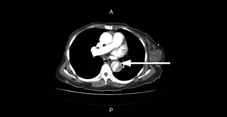
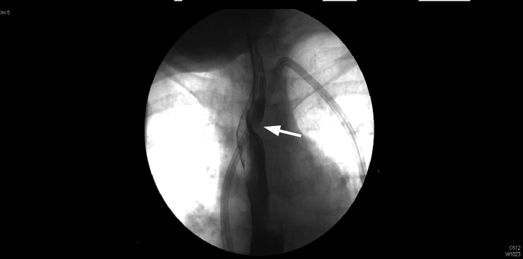
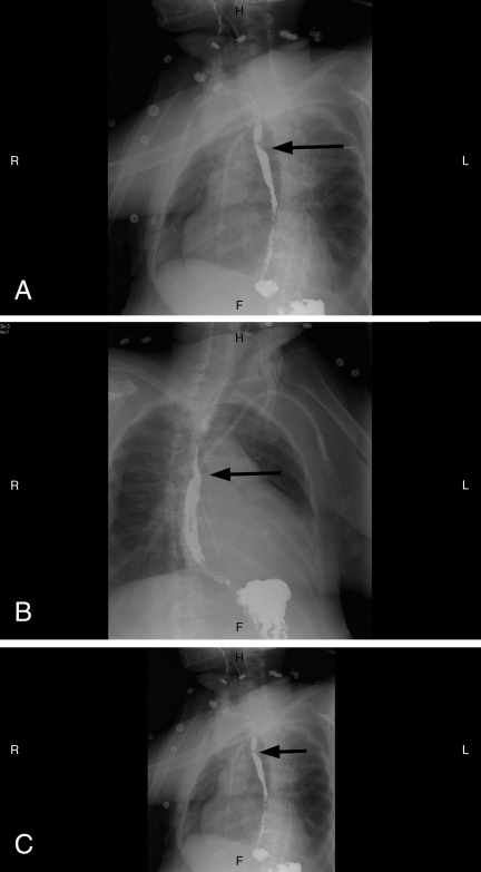
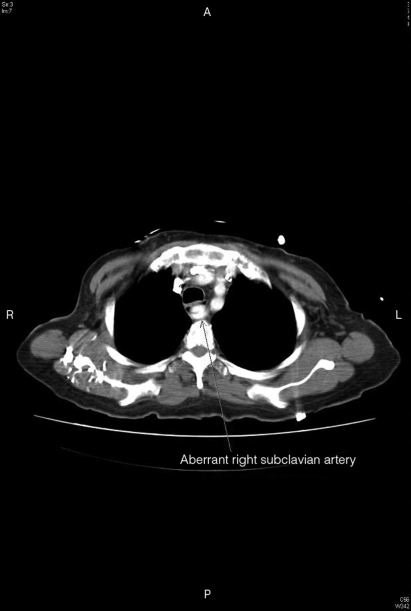
A 78-year-old African American woman with a past medical history of uncontrolled hypertension, type 2 diabetes mellitus, hyperlipidemia, ischemic stroke and end-stage renal disease presented to the emergency department of the North Chicago Veterans Administration Medical Center (Chicago, USA) with a chief complaint of severe excruciating backache. Initial physical examination revealed a blood pressure of 230/80 mmHg in the left arm and 216/72 mmHg in the right arm. The pulse was regular, at 88 beats/min. The patient was alert and oriented in time, place and person. All laboratory tests, including cardiac enzymes, were within normal limits, except for serum creatinine, which was 583 μmol/L. A 12-lead electrocardiogram performed at the time of admission revealed a first-degree atrioventricular block and an anterior infarct of indeterminate age, with diffuse T wave abnormalities. The patient underwent a computed tomography (CT) scan of the chest (with contrast), which revealed dissection of the aortic arch, descending thoracic aorta (Figure 1) and a dissecting aberrant right subclavian artery originating from the left aortic arch, distal to the origin of left subclavian artery. No stenotic lesions or aneurysm at origin of the aberrant vessel were visualized. The patient was transferred to the coronary care unit and was managed medically for type B aortic dissection with analgesics and an intravenous antihypertensive drug. On her fourth day of stay at the coronary care unit, the patient complained of new-onset dysphagia, primarily to solids. An esophagogram revealed notching on the esophagus, classic of an aberrant right subclavian artery (Figures 2, 3A, 3B and 3C). CT scan of the chest with contrast performed earlier at the time of admission correlated with the diagnosis of acute, dissecting aberrant right subclavian artery (Figure 4). Because the patient’s symptoms were mild and intermittent, and because she had multiple underlying comorbidities, surgical intervention was deferred. The patient was managed conservatively in the hospital and discharged after blood pressure stabilization with a scheduled follow-up appointment.
Figure 1.
Computed tomography of distal chest and abdomen with contrast demonstrating aortic dissection in the descending aorta. The arrow indicates separation within lumen indicative of dissection. A Anterior; P Posterior
Figure 2.
Anteroposterior fluoroscopic image of esophagus demonstrating an oblique extrinsic defect coursing from inferior (patient’s left) to superior (patient’s right), consistent with an aberrant right subclavian artery at the upper thoracic level, just above the aortic arch. Arrow indicates filling defect
Figure 3.
Oblique esophagogram images (A, B and C right [R], left [L] and R, respectively) demonstrating an oblique extrinsic defect coursing from the posteroinferior (patient’s L) to the anterosuperior (patient’s R), consistent with an aberrant R subclavian artery at the upper thoracic level, just above the aortic arch. Arrow indicates filling defect. F Foot; H Head
Figure 4.
Computed tomography of the proximal chest and abdomen demonstrating an aberrant right (R) subclavian artery arising from the aorta distal to the origin of the left (L) subclavian artery, compressing the proximal esophagus. A Anterior; P Posterior
Unfortunately, no follow-up information is available because the patient was lost to follow-up.
DISCUSSION
The most common embryological abnormality of the aortic arch is an aberrant right subclavian artery, which occurs in 0.5% to 1.8% of the population (1).
Current classification and developmental considerations
The abnormal origin of the right subclavian artery can be explained by the involution of the fourth vascular arch with the right dorsal aorta (2). The seventh intersegmental artery remains attached to the descending aorta, and this persistent intersegmental artery becomes the right subclavian artery. This leads to the aberrant artery, which often follows a retroesophageal course (Figure 2). David Bayford, a physician in London, England, originally described the syndrome as “dysphagia by freak of nature”; it is commonly referred to as ‘dysphagia lusoria’ and was first reported in 1794 (3).
CLINICAL SIGNIFICANCE
A review of the literature did not reveal any case of acute aortic dissection associated with new-onset dysphagia.
An aberrant right subclavian artery is a congenital anomaly and is usually asymptomatic, diagnosed by chance as a radiological finding, although it can cause compression of the trachea and the esophagus. Dyspnea is a common presentation in childhood, while dysphagia is the most frequent presentation in adulthood. In the past, cases have been reported of patients with Kommerell’s diverticulum and tracheal compression that simulated asthma (4). Surgical intervention in these patients can cause the symptoms to disappear.
The characteristic symptoms in adulthood are secondary to esophageal compression. In such patients, dysphagia to solids is the most common presenting complaint (5). An esophagogram may be the initial test of choice for these patients, and the diagnosis is confirmed by CT scan or magnetic resonance imaging.
Although arteriography is considered to be the gold standard, it is only recommended for patients undergoing surgery. Most cases reported in literature have occurred in younger age groups (younger than 35 years of age) who generally have symptoms related to tracheal or esophageal compression. Our patient developed new-onset dysphagia secondary to this rare anomaly at 78 years of age.
It is important to note that an aberrant right subclavian artery is associated with disorders such as Down’s, DiGeorge and Edwards’ syndromes. In the present case, our patient did not have a past medical history of the any of the above disorders.
MANAGEMENT
Asymptomatic patients are managed conservatively, while surgical intervention is recommended for patients presenting with respiratory distress or dysphagia.
In our patient, given her age, underlying comorbidities and mild intermittent dysphagia, conservative treatment was recommended.
REFERENCES
- 1.Richardson JV, Doty DB, Rossi NP, Ehrenhaft JL. Operation for aortic arch anomalies. Ann Thorac Surg. 1981;31:426–32. doi: 10.1016/s0003-4975(10)60994-0. [DOI] [PubMed] [Google Scholar]
- 2.Edwards JE. Congenital malformations of the heart and great vessels. Malformations of the thoracic aorta. In: Gould SE, editor. Pathology of the heart. 2nd edn. Springfield: Charles C Thomas; 1960. pp. 391–462. [Google Scholar]
- 3.Bayford D. An account of a singular case of obstructed deglutition. Memoirs Med Soc London. 1794;2:275–86. [Google Scholar]
- 4.Morel V, Corbineau H, Lecoz A, et al. Two cases of ‘asthma’ revealing a diverticulum of Kommerell. Respiration. 2002;69:456–60. doi: 10.1159/000064009. [DOI] [PubMed] [Google Scholar]
- 5.Guijarro JF, Moratinos P, de la Torres S. Disfagia lusoria causada por arteria subclavia izquierda aberrante y divertículo de Kommerell. Rev Esp Enferm Dig. 2002;94:290–7. [PubMed] [Google Scholar]


