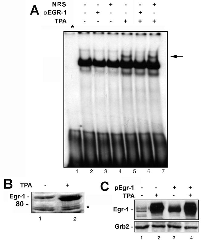
Figure 2. Egr-1 binds to the GRS element.
A. U-87 MG cells were treated with TPA as described in Materials and Methods and gel shifts were performed with nuclear extracts in the presence and absence of antibody to Egr-1 (α-Egr-1) or non-immune rabbit serum as indicated. Lane 1 - free probe. The arrow indicates the GBPi gel shift band. B. The nuclear extracts from Panel A were analyzed by Western blot for Egr-1 expression. The asterisk indicates a nonspecific band that indicates equivalent protein loading. C. In a separate experiment, U-87 MG cells were transfected with plasmid expressing Egr-1 and/or treated with TPA as described in Materials and Methods. Expression of Egr-1 was evaluated by Western blot with Grb2 as loading control.

