Abstract
Objectives
The aim of this study is to determine the prevalence of styloid process elongation (SPE) detected on panoramic radiographs (PRs) in Cappadocia region population in Turkey and to investigate the SPE incidences in relation to the age subgroups.
Methods
Between 2004 to 2007 years, a random sample of 750 PRs was collected from the data files and any questionable PR was excluded. Therefore, 698 PRs were included in the present study. The subjects were divided into six age subgroups: 10–19, 20–29, 30–39, 40–49, 50–59 and 60 years and older. Fifty-four (7.7%) patients demonstrated SPE at least one side.
Results
There were statistical differences between 10–19, 20–29 age subgroups and 30–39, 50–59 age subgroups in terms of the SPE prevalence, but not other subgroups.
Conclusions
According to our knowledge, this is the highest prevalence in comparison to the other Turkish reports and the first study in terms of the SPE prevalence in Cappadocia region population. Also, the subgroup analyse suggested that the age may not have a role in the elongation of the SP.
Keywords: Panoramic radiography, Styloid process elongation, Cappadocia region population
INTRODUCTION
The styloid process (SP) is a cylindrical, long cartilaginous bone located on the temporal bone. Many nerves and vessels such as carotid arteries are adjacent to the SP.1–3 The normal SP length is approximately 20–30 mm.4–9 The SP length which is longer than 30 mm was considered to be styloid process elongation (SPE).4,6,8,10 The SPE which is known as Eagle’s syndrome (ES) when it causes clinical symptoms as neck and cervicofacial pain.6,7,11 It is assumed that this symptoms and signs are due to the compression of the SP on some neural and vascular structures. More uncommonly, symptoms such as dysphagia, tinnitus, and otalgia may occur in patients with ES.1,12 It may also cause stroke due to the compression of carotid arteries.4,13 Although there are many suggested hypotheses, the exact etiology of calcified and ossified SPE is unknown.3,14
Eagle’s syndrome is diagnosed by both radiographical and physical examination. More commonly; a panoramic radiography (PR) is used to determine whether the SP is elongated. Computed tomography (CT) is useful for complementary information to that provided by PR.14,15
In the present study, our aim is to investigate the prevalence of SPE by PR and to analyze this prevalence in relation to gender and age subgroups in a Turkish population living in Cappadocia region. According to the literature, this is the first study investigating the SPE prevalence in this region.
MATERIALS AND METHODS
The study is based on 750 PRs consecutively retrieved from the archival records. All the PRs were taken between 2004 to 2007 at the Erciyes University Faculty of Dentistry, Department of Oral Diagnosis and Radiology. The PRs of the 750 Turkish patients with dental problems had originally been taken for routine examination and not for the investigation of the SPEs. It was not necessary to seek ethical approval as the PRs were essential for the routine clinical evaluation of the patients. Any PR who has questionable SP was excluded in the present study. The PRs were excluded whether radiograph quality is not good enough, stylohyoid complex was not clearly identified and superimposed on the temporal bone. The subjects were divided into six age subgroups: 10–19, 20–29, 30–39, 40–49, 50–59 and 60 years and older. The sample and regionality of the study, patient distributions according to age subgroups were the limitations of the current report.
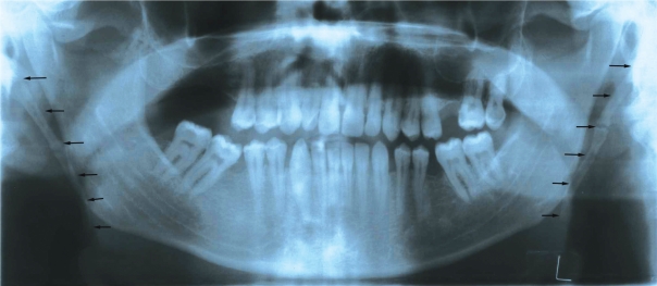
All the PRs were done and evaluated in the same fashion (Orthopantomography® OP100, Tuusula, Finland). The PRs were processed in relation to the manufacturer’s recommendations in an automatic film processor. For mineralized stylohyoid complexes and lengths of bilateral SPs were evaluated by using the measurement method of Jung et al’s study.8 In brief, the measurements were taken on the temporal bone’s frontal side. A thin transparent line is usually imagined between the SP shadows and the tympanic bone in this area on the PRs. This transparent line corresponds to the cleft between the SP and the temporal bone’s tympanic plate.8,16 The tip of the SP is its bony end including calcified parts of the ligament. All the PRs were viewed in subdued ambient light using transmitted light from a standard viewbox. The lengths of the SPE variants were measured using a true-toscale radiometric ruler (magnification factor: 1.4). The radiographs were investigated and the measurements were performed by the same author (Y.S). To check the intraobserver variations, measurements were repeated after one month on a subset of 300 PRs. Deviations of the mean length of the SP between first and second measurements were <2%. SPE can be assumed if either the SP or the adjacent stylohyoid ligament ossification shows an overall length in excess of 30 mm4,6,8,10 (Figure 1).
Figure 1.
Panoramic radiograph showing in a patient with bilateral elongated styloid styloid processes (arrows).
The observed results were analyzed with SPSS 15.0 (Statistical package for social science Inc., Chicago, Illinois, USA). t-test and Chi-Square tests were used for statistical analysis. P values less than 0.05 were accepted as statistically significant.
RESULTS
Seven hundred and fifty patients with dental problems were enrolled in the present study. The PRs of 52 patients who have questionable SPs were excluded. Therefore, on 698 (285 male; 413 female) of the 750 PRs, the length of the SP could be measured at least on one side. The mean age of these 698 (93.1%) patients was 34.9±14.1 years. Fifty four (7.7%) subjects demonstrated SPE at least one side (Table 1). The mean age was significantly higher in the patients with SPE than the patients without SPE (p=0.04). The mean age of these 54 (40 male; 14 female) subjects was 38.7±13.1 years. The mean ages for male and female patients with SPE were 39.1±14.0 and 37.5±10.7 years, respectively (Table 2). No significant difference in mean age between the two samples was detected (p=0.7). The mean SP length was not significantly different between the male (38.1±6.2) and the female (36.6±6.0) patients with SPE (p= 0.4).
Table 1.
SPE prevalence in relation to age and gender
| Age groups (years) | Female
|
Male
|
Total
|
|||
|---|---|---|---|---|---|---|
| n (%) | SPE | n (%) | SPE (%) | n (%) | SPE (%) | |
| 10–19 | 53 (12.8) | 0 (0.0) | 32 (11.2) | 3 (1.0) | 85 (12.2) | 3 (0.4) |
| 20–29 | 138 (33.4) | 4 (0.9) | 81 (28.4) | 7 (2.4) | 219 (31.4) | 11 (1.6) |
| 30–39 | 77 (18.6) | 6 (1.4) | 61 (21.4) | 10 (3.5) | 138 (19.7) | 16 (2.3) |
| 40–49 | 78 (18.9) | 1 (0.2) | 49 (17.2) | 10 (3.5) | 127 (18.2) | 11 (1.6) |
| 50–59 | 49 (11.9) | 3 (0.7) | 43 (15.1) | 8 (2.8) | 92 (13.2) | 11 (1.6) |
| 60+ | 18 (4.3) | 0 (0.0) | 19 (6.6) | 2 (0.7) | 37 (5.3) | 2 (0.3) |
| Total | 413 (59.2) | 14 (3.4) | 285 (40.8) | 40 (14.0) | 698 (100) | 54 (7.7) |
Table 2.
Age distribution of elongated SP according to gender
| Male
|
Female
|
Total
|
||
|---|---|---|---|---|
| n=40 | n=14 | n= 54 | P | |
| Mean | 39.1 | 37.5 | 38.7 | 0.7 |
| Std. Deviation | 14.0 | 10.7 | 13.1 | |
| Minimum | 16 | 26 | 16 | |
| Maximum | 68 | 57 | 68 |
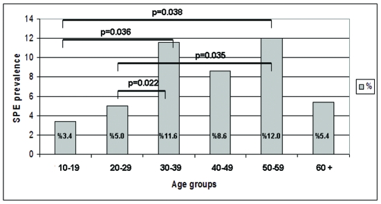
The SPE incidences of the patients in relation to the six age groups are summarized in Figure 2. There were statistical differences between 10–19, 20–29 age subgroups and 30–39, 50–59 age subgroups in terms of the SPE prevalence, but not other subgroups (p= 0.036, 0.038, 0.022, 0.035, respectively).
Figure 2.
Styloid process elongation prevalences in relation to age subgroups in patients with dental problems.
DISCUSSION
The elongation of the SP and structural changes in stylohyoid ligament with its clinical symptoms and signs were first described by Eagle. Therefore, it is also called as the Eagle’s syndrome.11,17 ES is diagnosed by both physical and radiographical examination. The SP palpation in the tonsillar fossa is indicative of SPE which are not normally palpable. Palpation of the tip of the SP should exacerbate the symptoms associated with this syndrome. If highly suspicious for ES, confirmation can be done by radiographical imaging.15 Generally, a PR rather than CT is used to detect if the SP is elongated.14 There are many vessels such as carotid arteries and nerves adjacent to the SP.1–3 The signs and symptoms with this syndrome are due to the anatomic relationship between the SP and its surrounding structures.1,12 The symptoms can be confused with some disorders including a wide variety of facial neuralgias, oral, dental and, temporomandibular diseases. Therefore, a detailed differential diagnosis for SPE should be done.18
The reported radiographic prevalence of the SPE varies from less than 2% to greater than 30% in the literature.6,9,19–22 There are several reports investigating the SPE prevalence in Turkish population. Ilguy et al. evaluated the PRs of 860 subjects in terms of SPE. Of these patients, 32 patients (3.7%) had SPEs.6 In another study, the SPE prevalence on the PRs was investigated for the 900 adult patients with dental problems and the prevalence was 1.1%.22 Also, Bozk r et al. investigated the SPE prevalence in 200 edentulous patients who were over 50 years old. It was found that the SPE prevalence was 4%.20 As in our study, the length of SP and/or stylohyoid ligament, which are longer than 30 mm were considered to be SPE in all these Turkish reports.6,22 According to our knowledge, the present study is the first report investigating the SPE prevalence and evaluating the subgroups in terms of this prevalence on PRs in Cappadocia region population. In the current study, the prevalence was 7.7%. In Turkey, there are many ethnic groups and regions. So, ethnic groups are changing according to the different regions. All the Turkish reports investigating SPE including our study were done in the different regions of Turkey. Therefore, regional factors including dietary factors and ethnicity may be important for the different SPE prevalances in these reports. There were statistical differences between 10–19, 20–29 age subgroups and 30–39, 50–59 age subgroups in terms of the SPE incidence, but not other subgroups. Therefore, no relationship could be established between the SPE and increasing patient age as in Correll et al’s study.23 However, the patient abnormal distributions according to age subgroups was the limitation of this interpretation in the current report.
As a result, SPEs found as incidental findings on PRs may be important clinically in not only patients with systemic diseases, but also normal population. Instead of many hypotheses and studies, the exact etiology of elongated SP is unknown. SPEs in the present study were detected by PRs in 7.7% of this population. According to our knowledge, this is the first study done in Cappadocia region population. We also investigated the SPE incidences of the patients in relation to the age groups. In conclusion, according to our knowledge, this is the highest prevalence in comparison to the other Turkish reports and the first study in terms of the SPE prevalence in Cappadocia region population. Also, the subgroup analyse suggested that the age may not have a role in the elongation of the SP.
REFERENCES
- 1.Gözil R, Yener N, Calgüner E, Araç M, Tunç E, Bahcelioḡlu M. Morphological characteristics of styloid process evaluated by computerized axial tomography. Ann Anat. 2001;183:527–535. doi: 10.1016/S0940-9602(01)80060-1. [DOI] [PubMed] [Google Scholar]
- 2.Krennmair G, Piehslinger E. Variants of ossification in the stylohyoid chain. Cranio. 2003;21:31–37. doi: 10.1080/08869634.2003.11746229. [DOI] [PubMed] [Google Scholar]
- 3.Camarda AJ, Deschamps C, Forest D. II. Stylohyoid chain ossification: a discussion of etiology. Oral Surg Oral Med Oral Pathol. 1989;67:515–520. doi: 10.1016/0030-4220(89)90265-x. [DOI] [PubMed] [Google Scholar]
- 4.Prasad KC, Kamath MP, Reddy KJ, Raju K, Agarwal S. Elongated styloid process (Eagle’s syndrome): a clinical study. J Oral Maxillofac Surg. 2002;60:171–175. doi: 10.1053/joms.2002.29814. [DOI] [PubMed] [Google Scholar]
- 5.Monsour PA, Young WG. Variability of the styloid process and stylohyoid ligament in panoramic radiographs. Oral Surg Oral Med Oral Pathol. 1986;61:522–526. doi: 10.1016/0030-4220(86)90399-3. [DOI] [PubMed] [Google Scholar]
- 6.Ilguy M, Ilguy D, Guler N, Bayirli G. Incidence of the type and calcification patterns in patients with elongated styloid process. J Int Med Res. 2005;33:96–102. doi: 10.1177/147323000503300110. [DOI] [PubMed] [Google Scholar]
- 7.Kursoglu P, Unalan F, Erdem T. Radiological evaluation of the styloid process in young adults resident in Turkey’s Yeditepe University faculty of dentistry. Oral Surg Oral Med Oral Pathol Oral Radiol Oral Endod. 2005;100:491–494. doi: 10.1016/j.tripleo.2005.05.061. [DOI] [PubMed] [Google Scholar]
- 8.Jung T, Tschernitschek H, Hippen H, Schneider B, Borchers L. Elongated styloid process: when is it really elongated? Dentomaxillofac Radiol. 2004;33:119–124. doi: 10.1259/dmfr/13491574. [DOI] [PubMed] [Google Scholar]
- 9.Zaki HS, Greco CM, Rudy TE, Kubinski JA. Elongated styloid process in a temporomandibular disorder sample: prevalence and treatment outcome. J Prosthet Dent. 1996;75:399–405. doi: 10.1016/s0022-3913(96)90032-3. [DOI] [PubMed] [Google Scholar]
- 10.Ramadan SU, Gokharman D, Tuncbilek I, Kacar M, Kosar P, Kosar U. Assessment of the stylohoid chain by 3D-CT. Surg Radiol Anat. 2007;29:583–588. doi: 10.1007/s00276-007-0239-8. [DOI] [PubMed] [Google Scholar]
- 11.Eagle WW. Elongated styloid process. Report of two cases. Arch Otolaryngol. 1937;25:548–587. doi: 10.1001/archotol.1949.03760110046003. [DOI] [PubMed] [Google Scholar]
- 12.Bafaqeeh SA. Eagle syndrome: classic and carotid artery types. J Otolaryngol. 2000;29:88–94. [PubMed] [Google Scholar]
- 13.Chuang WC, Short JH, McKinney AM, Anker L, Knoll B, McKinney ZJ. Reversible left hemispheric ischemia secondary to carotid compression in Eagle syndrome: surgical and CT angiographic correlation. Am J Neuroradiol. 2007;28:143–145. [PMC free article] [PubMed] [Google Scholar]
- 14.Murtagh RD, Caracciolo JT, Fernandez G. CT findings associated with Eagle syndrome. Am J Neuroradiol. 2001;22:1401–1402. [PMC free article] [PubMed] [Google Scholar]
- 15.Rechtweg JS, Wax MK. Eagle’s syndrome: a review. Am J Otolaryngol. 1998;19:316–321. doi: 10.1016/s0196-0709(98)90005-9. [DOI] [PubMed] [Google Scholar]
- 16.Okabe S, Morimoto Y, Ansai T, et al. Clinical significance and variation of the advanced calcified stylohyoid complex detected by panoramic radiographs among 80-year-old subjects. Dentomaxillofac Radiol. 2006;35:191–199. doi: 10.1259/dmfr/12056500. [DOI] [PubMed] [Google Scholar]
- 17.Eagle WW. Elongated styloid process, further observations and a new syndrome. Arch Otolaryngol. 1948;47:630–640. doi: 10.1001/archotol.1948.00690030654006. [DOI] [PubMed] [Google Scholar]
- 18.Aral IL, Karaca I, Güngör N. Eagle’s syndrome masquerading as pain of dental origin. Case report. Aust Dent J. 1997;42:18–19. doi: 10.1111/j.1834-7819.1997.tb00090.x. [DOI] [PubMed] [Google Scholar]
- 19.Keur JJ, Campbell JP, McCarthy JF. The clinical significance of the elongated styloid process. Oral Surg Oral Med Oral Pathol. 1986;61:399–404. doi: 10.1016/0030-4220(86)90426-3. [DOI] [PubMed] [Google Scholar]
- 20.Bozk r MG, Boḡa H, Dere F. The evaluation of elongated styloid process in panoramic radiographs in edentulous patients. Tr. J. of Medical Science. 1999;29:481–485. [Google Scholar]
- 21.Grossman JR, Tarsitano JJ. The styloid-stylohyoid syndrome. J Oral Surg. 1977;35:555–560. [PubMed] [Google Scholar]
- 22.Erol B. Radiological assessment of elongated styloid process and ossified stylohyoid ligament. Journal of Marmara University Dental Faculty. 1996;2:554–556. [PubMed] [Google Scholar]
- 23.Correlly RW, Jensen JL, Taylor JB, Rhyne RR. Mineralization of the stylohyoid-stylomandibular ligament complex. Oral Surg Oral Med Oral Pathol. 1979;48:286–291. doi: 10.1016/0030-4220(79)90025-2. [DOI] [PubMed] [Google Scholar]


