Abstract
Objectives
To examine the effects of dental fluorosis and total and self-etch bonding systems on microleakage of Class-V composite restorations in permanent molar teeth.
Methods
Teeth were classified as three main groups according to Thylstrup-Fejerskov Index (TFI) as TFI=0, TFI=1–3 and TFI=4. Total and self-etching/bonding procedures were determined for each main group. Total-etching procedures were acid-etching for 30s and acid-etching for 60s with Single Bond/total-etch bonding system. Self-etching procedure was applied with Prompt-L-Pop/self-etch bonding system. 63 box-shaped Class-V cavities (4 × 2 × 2 mm) were prepared on mid-buccal/palatinal/lingual surfaces of teeth for totalling nine test groups (n=7). Restorations with composite material (Charisma) polymerized with halogen unit for 40s. Teeth were thermocycled between +5°C – +55°C (×500), immersed in 0.5% basic-fuchsin solution (37°C, 24h) and separated longitudinally in bucco-lingual direction. Dye penetration was examined under stereomicroscope (3.2 × 10).
Results
Microleakage levels were higher in teeth of TFI=4 than TFI=0 occlusally or cervically (P<.05). In TFI=0; total-etched teeth for 30s have statistically shown more leakage than total-etched teeth for 60s occlusally or cervically (P<.05). In TFI=4; microleakage levels were significantly higher for 30s than 60s cervically (P<.05). For all TFI levels, microleakage was commonly increased with self-etch system than total-etch system (P<.05). Generally, higher leakage was present at cervical margins than occlusal margins (P<.05).
Conclusions
Microleakage has increased by severity of dental fluorosis. Generally, more leakage was observed in total-etched teeth for 30s than 60s. Microleakage was commonly higher in self-etched teeth than total-etched teeth. More leakage was present at cervical margins than occlusal margins.
Keywords: Class-V restorations, Composites, Dental fluorosis, Microleakage
INTRODUCTION
Anti-cariogenic and positive effects of fluorides on teeth and carious lesions were proved in dentistry.1–4 However, common using of fluoride-containing products such as foods, soft drinks, supplements and some dental materials have resulted in increased prevalence of dental fluorosis in many countries over the past few decades.5–8 Dental fluorosis is also endemic in several parts of the world.9–11
Fluorosed enamel is characterized by an outer hypermineralized surface and porosity of subsurface layer.9 Fluorosed teeth sometimes need to be restored for functional or aesthetic reasons.6 Composite resin applications may be practiced for the treatment of moderately or severely fluorosed teeth.5 Composite restorations often depend largely on micromechanical retention obtained from etched enamel with the acid-etching technique which was first described by Buonocore.12 Hypermineralized surface of fluorosed enamel is difficult to acid-etch.13 Bonding of restorations on fluorosed teeth involves etching the acid-resistant enamel and may necessitate prolonging the etching time.9
Microleakage is the marginal permeability to bacterial, chemical and molecular invasion at the interface between the teeth and restorations. It may cause discoloration, recurrent caries and pulpal pathology.14 It was decreased by use of acid-etching technique15,16 and bonding systems.15,17,18
Recently, bonding agents may be classified as total-etch (TE) and self-etch (SE) bonding systems according to their application steps. TE bonding systems are consisted of two or three application steps. First step includes an acid-etching procedure. Following steps are composed by separated or combined applications of primer and adhesive solutions. SE bonding systems have self-etching efficiency on tooth structures. 2 step SE adhesives rely on the use of a separate self-etching (which is combined to the priming step) before application of the resin. 19–21
Our null hypothesis is that, dental fluorosis and different etching/bonding procedures may affect microleakage levels of Class V composite restorations on permanent teeth. The purpose of this in-vitro study was to examine the microleakage of Class-V composite resin restorations using total and self-etch bonding systems on fluorosed permanent molar teeth.
MATERIALS AND METHODS
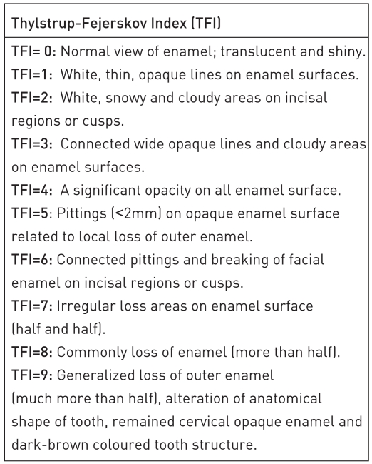
In study, freshly extracted, non-carious, fluorosed molar permanent teeth were used. All teeth were cleaned, polished and immersed in distilled water containing 0.1% thymol solution, and stored in distilled water in room temperature until used. Teeth were classified according to Thylstrup-Fejerskov Index (TFI)22,23 (Figure 1) and separated into 3 main groups as TFI=0, TFI=1–3 and TFI=4.
Figure 1.
Two different bonding systems with different etching procedures were used;
▪ Single Bond (SB) (3M, St Paul, MN, USA, Lot no: 55144)/TE system: SB was applied with different etching times of 30 seconds (s) and 60s with a 35% phosphoric acid etching-gel (Multipurpose Etching Gel; 3M, USA, Lot no: 20020613).
▪ Prompt-L-Pop (PLP) (ESPE, Germany, Lot no: 118642)/SE system: PLP was directly applied without etching step separately.
All etching and bonding procedures were shown in Table 1. Group names were coded as 0/1/4 and a/b/c where numbers represent the TFI scores. Small letters represent the bonding procedure including acid etching of enamel for 30s and application of SB total etch bonding system (a), acid etching of enamel for 60s and application of SB total etch bonding system (b) and application of PLP self-etching bonding system (c).
Table 1.
All test groups according to TFI levels/bonding/etching procedures with codes (n=7).
| TFI levels | Bonding/etching procedures
|
||
|---|---|---|---|
| Groups a | Groups b | Groups c | |
| Groups 0 | Group 0a | Group 0b | Group 0c |
| Groups 1 | Group 1a | Group 1b | Group 1c |
| Groups 4 | Group 4a | Group 4b | Group 4c |
Groups 0: Non-fluorosed teeth in TFI=0
Groups 1: Fluorosed-teeth in TFI=1–3
Groups 4: Fluorosed-teeth in TFI=4
Groups a: SB total-etch bonding system/30s acid-etching procedure
Groups b: SB total-etch bonding system/60s acid-etching procedure
Groups c: PLP self-etch bonding system/no acid-etching procedure
Totally nine test groups were determined (n=7). All test groups were shown in Table 1. A total of 63 standardized, box-shaped Class-V cavities were prepared (sizes: 4 mm length × 2 mm width × 2 mm depth) on the mid-buccal and mid-palatinal/lingual surfaces of the randomly selected teeth by the same researcher. A high speed dental torque (KaVo Super Torque, Lux 2, 640 B, P 294779, Germany) and fissure diamond burs (North Bel, FG Coarse, lot no. 200000011267, Italy) were used. Burs were renewed after each five cavity preparations. Cavity margins were placed in enamel. Enamel margins were beveled with a 45° angle. The depth of cavities was millimetrically standardized using a periodontal probe.
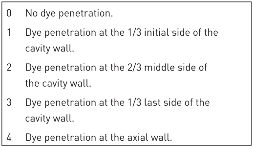
A composite resin restorative material (Charisma; Heraeus-Kulzer, lot no.110067, Shade A2, Germany) was placed into the cavities with bulk-technique and polymerized using a halogen light unit (Hilux 200, Benlioglu Dental, Turkey, 450 mW/cm2) for 40s. All specimens were immersed in water at 37°C for 24 hours after finishing and polishing procedures with a polishing set (Soft-lex polishing discs; 3M, St Paul, Minneapolis, USA). Teeth were then thermocycled between water baths held at +5°C and +55°C with a transfer time of 10s and a dwell time of 30s for 500 cycles. A double coated nail varnish was applied to the whole surface of the teeth except for the restorations and approximately 1 mm of tooth surfaces adjacent to the restorations. Then, all specimens were immersed in 0.5% basic-fuchsin solution for 24 hours at 37°C. After removal from the solution, the teeth were rinsed in tap water and embedded vertically in autopolymerizing acrylic resin blocks (Orthocryl EQ, Dentaurum, Germany) and separated in a bucco-lingual direction through the centre of each tooth with a water-cooled diamond saw (Micracut Precision Cutter, Metkon, Turkey). The specimens were evaluated under a stereomicroscope at 32X magnification (Leica, MZ12/AG, Switzerland, CH 9435) for dye penetration. Dye penetration scores at cavity walls were determined occlusally and cervically according to Alavi and Kianimanesh24 (Figure 2). Kruskal-Wallis H-Test, Mann-Whitney U-Test and Wilcoxon Signed Range Test were used for statistical evaluation of results (P<.05).
Figure 2.
The scoring method of dye penetration according to Alavi and Kianimanesh.24
RESULTS
Significant differences were present between groups named 0, 1 and 4 occlusally and cervically according to Kruskal-Wallis Test (Table 2) (P=.000). Comparisons between all test groups and the statistical differences were shown occlusally and cervically according to Mann Whitney U Test (Table 3) (P<.05).
Table 2.
Statistical differences between teeth in three levels of TFI for occlusal and cervical margins according to Kruskal-Wallis Test (P<.05 = *).
| Chi-Square | df | P | |
|---|---|---|---|
| Occlusal | 34.52 | 8 | .000* |
| Cervical | 47.21 | 8 | .000* |
Table 3.
Statistical differences between TFI/bonding/etching groups according to Mann-Whitney U Test for occlusal and cervical margins (P<.05 = *).
| Significant differences between test groups | Values of Mann-Whitney U Test | P<.05 = *
|
|||
|---|---|---|---|---|---|
| Oclusally | Cervically | ||||
| 0a | 0b | 10.5 | 7.5 | .037* | .015* |
| 0c | 10 | 10.5 | .03* | .037* | |
| 1a | 12.5 | 23.5 | .054 | .872 | |
| 1b | 24.5 | 15 | 1 | .091 | |
| 1c | 5 | 5 | .007* | .006* | |
| 4a | 5 | 3 | .006* | .004* | |
| 4b | 12.5 | 14 | .054 | .122 | |
| 4c | 5 | 0 | .007* | .001* | |
| 0b | 0c | 2 | 1.5 | .002* | .002* |
| 1a | 2.5 | 7.5 | .002* | .015* | |
| 1b | 10.5 | 14 | .037* | .107 | |
| 1c | 1 | 0 | .001* | .001* | |
| 4a | 1 | 0 | .001* | .001* | |
| 4b | 2.5 | 3 | .002* | .004* | |
| 4c | 1 | 0 | .002* | .001* | |
| 0c | 1a | 21 | 13.5 | .591 | .116 |
| 1b | 10 | 3 | .030* | .002* | |
| 1c | 16 | 15 | .225 | .091 | |
| 4a | 17.5 | 9 | .298 | .020* | |
| 4b | 21 | 21 | .591 | .53 | |
| 4c | 14.5 | 0 | .165 | .001* | |
| 1a | 1b | 12.5 | 15 | .054 | .091 |
| 1c | 13 | 8.5 | .1 | .028* | |
| 4a | 14 | 6 | .122 | .013* | |
| 4b | 24.5 | 16.5 | 1 | .253 | |
| 4c | 12 | 1 | .08 | .002* | |
| 1b | 1c | 5 | 0 | .007* | .001* |
| 4a | 5 | 0 | .006* | .001* | |
| 4b | 12.5 | 6 | .054 | .007* | |
| 4c | 5 | 0 | .007* | .001* | |
| 1c | 4a | 22 | 16.5 | .705 | .244 |
| 4b | 13 | 12.5 | 1 | .054 | |
| 4c | 21.5 | 2 | .678 | .020* | |
| 4a | 4b | 14 | 7.5 | .122 | .015* |
| 4c | 19.5 | 7.5 | .473 | .020* | |
| 4b | 4c | 12 | 0 | .08 | .001* |
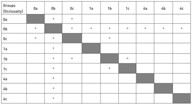
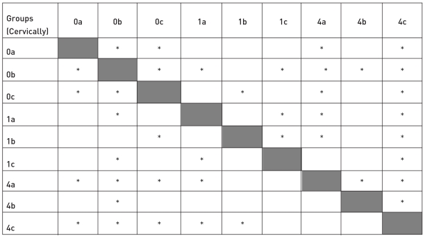
Microleakage was generally found less in groups named b than groups named a. There were significant differences between group named 0a and group named 0b for both occlusally (P=.037) and cervically (P=.015). Difference was not significant between group named 1a and group named 1b occlusally (P=.054) or cervically (P=.091). Statistically less microleakage was observed in group named 4b than group named 4a cervically (P=.015). Microleakage levels were commonly increased in groups named c than groups named a and than groups named b occlusally (P<.05). The significant differences were between group named 0c and group named 0a (P=.03), between group named 0c and group named 0b (P=.002) occlusally. There were not significant differences between group named 1c and group named 1a (P=.1) and between group named 4c and group named 4a (P=.473) and between group named 4c and group named 4b (P=.08) occlusally. There was significant differences between group named 1c and group named 1b (P=.007). The significant differences were present between group named 0c and group named 0a (P=.037) and between group named 0c and group named 0b (P=.002) and between group named 1c and group named 1b (P=.001) cervically. There were statistically significant differences between group named 0b and group named 1a (P=.002), between group named 0b and group named 1b (P=.037) and between group named 0b and group named 1c (P=.001) occlusally. Significant differences were found between group named 0b and group named 4a (P=.001) and between group named 0b and group named 4b (P=.002) and between group named 0b and group named 4c (P=.002) occlusally. Differences were not significant between group named 0a and group named 1a (P=.054) and between group named 0a and group named 1b (P=1) occlusally. The significant difference was present between group named 0a and group named 1c (P=.007) occlusally. Differences were not significant between group named 0a and group named 1a (P=.872) and between group named 0a and group named 1b (P=.091) cervically. The significant difference was present between group named 0a and group named 1c (P=.006) cervically. Statistically significant differences were present between group named 0a and group named 4a (P=.004) and between group named 0a and group named 4c (P=.001) cervically. There were significant differences between group named 0b and group named 1a (P=.015) and between group named 0b and group named 1c (P=.001) cervically. The differences were statistically significant between group named 0b and group named 4a (P=.001) and between group named 0b and group named 4b (P=.004) and between group named 0b and group named 4c (P=.001) cervically. Statistically significant differences were present between group named 4c and group named 0c (P=.001), and between group named 4c and group named 1c (P=.02) cervically. There were significant differences between group named 4c and group named 4a (P=.02) and between group named 4c and group named 4b (P=.001) cervically. Statistically significant difference was present between occlusal and cervical margins (P=.000). All results and statistical differences were shown in Table 2–6 and Figures 3 and 4.
Figure 3.
Statistical differences between all groups (occlusally) (* = P<.05 ).
Figure 4.
Statistical differences between all groups (cervically) (* = P<.05 ).
DISCUSSION
In this study, the hypothesis was accepted that, dental fluorosis and different etching/bonding procedures affect microleakage levels of Class-V composite restorations on permanent teeth. Microleakage is examined with several in-vitro studies such as dye-penetration tests, chemically agents, compressed air, neutron activation analysis, bacterial studies, radioisotope materials etc.25–28. The most frequent using methods are conventional and dependable dye-penetration tests.29,30 Basic fuchsin (0.5–2%),24,30–33 methylene blue (0.2–2%),34–40 silver nitrate (50%),41,42 crystal violet (0.05%), eritrosin (2%),43 and Rodhamine B (0.2%)44 are commonly using dye-penetration solutions.
Recently, more improved another techniques are also used for determining the leakage of dental materials. For example; sliding of dental tissues and examination of leakage with ammoniacal AgNO3 solutions under scanning electron microscopy (SEM) using the backscattered electron mode45 or contemporary field emission in-lens SEM (FEI-SEM)46,47 or transmission electron microscopy (TEM).45,46,48 Another new examination technique is confocal microscopy. It is for comparing with the current microscopic techniques for examining restorative dental procedures and dental materials and can be considered as being midway between optical and electron microscopy.49 This technique that can be used both in the clinic and the high resolution microscopy suite for fluorescent structures within semi transparent materials such as cells and dental hard tissues.50,51 The laser scanning type microscope (CLSM) and the real-time direct view of tandem scanning microscopes (TSM) are basically two types of confocal optical microscope.49 All confocal scanning optical microscopes are suitable for making high-resolution images of many structures in teeth under near normal conditions. The number of applications widens considerably if the microscope can operate at high speed. The high-frame speed of TSM enables real-time examination of teeth in vivo and experimental procedures examined microscopically on extracted teeth can include the observation of the fluid flow and application of adhesives.52 In addition, new imaging techniques such as multi-photon laser excitation of dyes give the potential of greater depth penetration and improved resolution.51 All these techniques are more sophisticated, relative and ultra-informative methods for leakage tests. However, these techniques require exhaustive equipments, detailed technical precisions and knowledge and rather expensive hardware. Therefore, using of these more advanced methods may not be easy by all researchers every time.
In this study, a conventional and dependable dye-penetration method with basic fuchsin solution was used for examination of microleakage. It was reported that, basic fuchsin may be prepared in 2% concentration,53 otherwise 0.5% concentration was generally preferred24,30–33,54 as in the present study.
One-slice cutting technique is common used in conventional dye-penetration/microleakage examinations under conventional microscopy.31–33,37–40,54–60 Also, more sensitive multi-slices cutting technique may be used for leakage studies, but this technique is usually preferred for more improved ultra-microscopical examinations e.g. SEM/TEM or confocal microscopy.45–47,49–52 In the present study, conventional one-slice cutting technique was used for conventional dye-penetration/microleakage test. Already, multi-slices cutting technique may not be suitable for fluorosed teeth owing to their hypermineralized and very easily breakable enamel structure because of multi-slices cutting technique requires a series of very thin and smooth slices of tooth structures.
For composite restorations, incremental placement technique has been suggested to reduce microleakage28,59 and to produce greater resistance to cuspal fractures.61 Otherwise, it was reported that, bulk placement technique is also suitable for composite restorations of 2-mm thickness and this technique is commonly used in microleakage studies.17,33,34,38,39,42,62 In the present study, bulk placement technique was used in standardized 2-mm depth of cavities of composite restorations.
Marginal microleakage is an important factor which causes to the clinical failure of restorations.14,16 Some determinants may affect the microleakage such as structure of enamel,13 acid-etching time,15 bonding systems,17,18 cavity designs and C-factor.63,64
In this study, more microleakage was found in fluorosed teeth than non-fluorosed teeth and microleakage levels increased depending on the severity of dental fluorosis. These higher microleakage levels may be explained by a weaker bonding of composite restorations on fluorosed teeth because of the pitted and detachable fluoroapatite structure of fluorosed enamel which has hypermineralized surface layer and quite extensive subsurface porosity.9
In dentistry, using of the acid-etching technique has reduced the microleakage.15,16 However, demineralization rates of enamel is affected from the type and concentration of etching agents.65,66 Otherwise, the depth of etch of fluorosed teeth is influenced not only by the type and concentration of acid etchants, but also by the etching time and the chemical composition of the enamel. Because the fluoroapatite in the hypermineralized surface layer of fluorosed teeth is more resistant to acid dissolution than the hydroxyapatite in non-fluorosed teeth, it has often been suggested the etching time of fluorosed enamel be doubled.6,13,67 Therefore, in the present study, two different acid-etching times were applied on the fluorosed teeth with total-etch bonding system. Etching time of 30s was applied as normal etching time, and etching time of 60s was applied as doubled etching time. In the study, less microleakage levels were observed in total-etch bonding groups with etching time of 60s than total-etch bonding groups with etching time of 30s. These results on etching times of 60s and 30s supported the recommendations of previous researchers about doubled etching time on fluorosed teeth.6,13,67
The results of the study indicated that, microleakage scores were generally increased in self-etch groups than total-etch groups. Likewise, more microleakage was found with self-etch bonding systems in some previous studies.36,60,62 Bedran de Castro et al58 reported that, a self-etching primer system obtained higher values of microleakage compared with an one-bottle total-etch system. Owens and Johnson59 observed in their study that, the use of a total-etch system significantly reduced microleakage than a self-etch system. In another study, Salim et al69 indicated that, the results suggested the application of a conventional two-bottle bonding system used with a total-etch technique is better than that of a self-etching adhesive system. In a different study, a multi-step total-etch adhesive system exhibited significantly less leakage at the enamel margin than the self-etch adhesive systems.57 Brandt et al56 reported in their study that, only two self-etch bonding systems between totaling six different self-etch bonding systems could be clinically acceptable alternatives to clinically proven a total-etch bonding system and the other self-etch products showed more microleakage. Deliperi et al39 declared that, significantly more dye-penetration was observed in one-step self-etch adhesive group than another self-etch adhesives and a total-etch adhesive. On the other hand, some other studies indicated that microleakage was not found significantly different between self and total-etch bonding systems.41,43,70 Deliperi et al55 reported in their study that, microleakage was not affected by the type of adhesive or its application technique. In another study performed by Santini et al,71 there was no significant difference in leakage between the self-etching groups and a total-etch group in enamel margins. In a study made by Owens et al,57 there were no significant difference among the adhesive groups when the dentin margins were evaluated.
The results of the current study have supported the findings of previous studies about more leakage with self-etch bonding systems than total-etch bonding systems.36,39,56–60,62,69 Higher leakage scores in the present study may be related with inadequate demineralization of enamel by using self-etching bonding system. Additionally, the inefficiency of self-etching bonding system on fluorosed teeth may be related on hypermineralized/acid-resistant enamel.
Finally, there are only two microleakage studies on fluorosed teeth in literature.68,33 Furthermore, there are not any study comparing total and self-etching/bonding systems or normal and prolonged etching times on microleakage of permanent fluorosed teeth. Therefore, the results of the current study are rather important for;
▪ Adding more knowledge on microleakage levels of composites on fluorosed teeth.
▪ Getting some information about microleakage levels of fluorosed teeth with different bonding systems.
▪ Supporting the suggestions of previous researchers about prolonged etching time on fluorosed teeth for better adhesion.6,13,67
On the other hand, it has been reported that, leakage could not been completely eliminated in composite restorations even though using of total-etch bonding systems yet.24,34 Also, in the present study, microleakage could not been absolutely eradicated in spite of using of a total-etch bonding system in fluorosed or non-fluorosed teeth.
Microleakage levels were found higher at cervical margins than occlusal margins of teeth in this study (P<.05). More leakage was also reported at cervical margins than occlusal margins in previous studies.24,72 The findings of the current study were parallel with their results. This condition may be explained with thinner and weaker structure of enamel at cervical areas.
C-factor is defined as the ratio of the bonded surface area to the unbonded or free surface area.63 It may also play a role in marginal sealing. A high C-factor is a risk in bonding procedure. Because increased polymerization stresses may occur by high rates of C-factor.63,64 The box-shaped cavity design was recommended by some previous researchers in their studies to resemble most closely the clinical situation resulting in a C-factor of about 3, where relatively high shrinkage stresses can be expected.30,73,74 Box-shaped cavity design was used to determine the C-factor relatively in this study.
This study was performed on examination of microleakage levels of composite restorations with different bonding systems on fluorosed teeth. It is hoped that, the findings of the current study may help to next restorative studies on fluorosed teeth. However, these findings must be supported with other future researches.
CONCLUSIONS
Microleakage was found significant in fluorosed teeth than non-fluorosed teeth. Leakage was affected by dental fluorosis and increased by severity of fluorosis.
Generally less microleakage was observed at total-etch bonding groups with 60s etching procedure than total-etch bonding groups with 30s etching procedure. 60s etching procedure has reduced microleakage in particularly TFI=4.
Commonly more microleakage was found in all self-etch bonding groups than total-etch bonding groups.
Generally more microleakage was observed in cervical margins than occlusal margins.
Table 4.
Descriptives for occlusal and cervical margins according to Wilcoxon Signed Rank Test.
| Margins | n | Mean | SD | Min | Max |
|---|---|---|---|---|---|
| Occlusal | 63 | 1.25 | 0.82 | 0 | 4 |
| Cervical | 63 | 1.81 | 1.08 | 0 | 4 |
Table 5.
Statistical differences between occlusal and cervical margins according to Wilcoxon Signed Rank Test (P<.05 = *).
| Significant differences between occlusal-cervical margins | |
|---|---|
| Z | − 4.19718 |
| P | .000* |
ACKNOWLEDGEMENTS
This study was made as second part of the PhD Thesis of correspondence author which was performed at Department of Paedodontics, Faculty of Dentistry, Ankara University, in April 2004.
REFERENCES
- 1.Bryant BS, Retief DH, Bradley EL, Denys FR. The effect of topical fluoride treatment on enamel fluoride uptake and the tensile bond strength of an orthodontic bonding resin. Am J Orthod. 1985;87:294–302. doi: 10.1016/0002-9416(85)90004-1. [DOI] [PubMed] [Google Scholar]
- 2.Whitford GM, Ekstrand J. Summary of Session I: Metabolism of fluoride. J Dent Res. 1990;69:513. [Google Scholar]
- 3.Hallgren A, Oliveby A, Twetman S. Salivary fluoride concentrations in children with glass ionomer cemented orthodontic appliances. Caries Res. 1990;24:239–241. doi: 10.1159/000261274. [DOI] [PubMed] [Google Scholar]
- 4.Croll TP. Light-hardened glass-ionomer-resin cement restoration adjacent to a bonded orthodontic bracket: A case report. Quintessence Int. 1994;25:65–67. [PubMed] [Google Scholar]
- 5.Pang DTV, Phillips CL, Bawden JW. Fluoride intake from beverage consumption in a sample of North Carolina children. J Dent Res. 1992;71:1382. doi: 10.1177/00220345920710070601. [DOI] [PubMed] [Google Scholar]
- 6.Al-Sugair MH, Akpata ES. Effect of fluorosis on etching of human enamel. J Oral Rehabil. 1999;26:521–528. doi: 10.1046/j.1365-2842.1999.00391.x. [DOI] [PubMed] [Google Scholar]
- 7.Leverett D. Prevalence of dental fluorosis in fluoridated and nonfluoridated communities-a preliminary investigation. J Public Health Dent. 1986;46:184–187. doi: 10.1111/j.1752-7325.1986.tb03140.x. [DOI] [PubMed] [Google Scholar]
- 8.Awliya WY, Akpata ES. Effect of fluorosis on shear bond strength of glass ionomer-based restorative materials to dentin. J Prosthet Dent. 1999;81:290–294. doi: 10.1016/s0022-3913(99)70271-4. [DOI] [PubMed] [Google Scholar]
- 9.Ateyah N, Akpata E. Factors affecting shear bond strength of composite resin to fluorosed human enamel. Oper Dent. 2000;25:216–222. [PubMed] [Google Scholar]
- 10.Dean HT, Arnold FA, Evolve E. Domestic water and dental caries. V. Additional studies of the relation of fluoride in domestic waters to dental caries in 4.425 white children, aged 12–14 years of 13 cities in 4 states. Public Health Report. 1942;57:1155–1179. [Google Scholar]
- 11.Akpata ES, Fakiha Z, Khan N. Dental fluorosis in 12–15-year-old rural children exposed to fluorides from well drinking water in the Hail region of Saudi Arabia. Community Dent Oral Epidemiol. 1997;25:324–327. doi: 10.1111/j.1600-0528.1997.tb00947.x. [DOI] [PubMed] [Google Scholar]
- 12.Buonocore MG. A simple method of increasing the adhesion of acrylic filling materials to enamel surfaces. J Dent Res. 1955;34:849–883. doi: 10.1177/00220345550340060801. [DOI] [PubMed] [Google Scholar]
- 13.Opinya GN, Pameijer CH. Tensile bond srtength of fluorosed Kenyan teeth using the acid etch technique. Int Dent J. 1986;36:225–229. [PubMed] [Google Scholar]
- 14.Wieczkowski G Jr, Yu XY, Davis EL, Joynt RB. Microleakage in various dentin bonding agent/composite resin systems. Oper Dent. 1992;5(Suppl):62–67. [PubMed] [Google Scholar]
- 15.Retief DH, Denys FR. Adhesion to enamel and dentin. Am J Dent. 1989;2:133–144. [PubMed] [Google Scholar]
- 16.Barkmeier WW, Cooley RL. Laboratory evaluation of adhesive systems. Oper Dent. 1992;5(Suppl):50–61. [PubMed] [Google Scholar]
- 17.Neme AL, Evans DB, Maxson BB. Evaluation of dental adhesive systems with amalgam and resin composite restorations: comparison of microleakage and bond strength results. Oper Dent. 2000;25:512–519. [PubMed] [Google Scholar]
- 18.Hembree JH, Andrews JT. In vitro microleakage of several acid-etch composite systems. J Dent Res. 1976;55(Special issue B) Abstract No:309. [PubMed] [Google Scholar]
- 19.Bouillaguet S, Gysi P, Wataha JC, Ciucchi B, Cattani M, Godin CH, Meyer JM. Bond strength of composite to dentin using conventional, one-step and self-etching adhesive systems. J Dent. 2001;29:55–61. doi: 10.1016/s0300-5712(00)00049-x. [DOI] [PubMed] [Google Scholar]
- 20.Zheng L, Pereira PN, Nakajima M, Sano H, Tagami J. Relationship between adhesive thickness and microtensile bond strength. Oper Dent. 2001;26:97–104. [PubMed] [Google Scholar]
- 21.Van Meerbeek B, Vargas M, Inoue S, Yoshida Y, Peumans M, Lambrechts P, Vanherle G. Adhesives and cements to promote preservation dentistry. Oper Dent. 2001;6(Suppl):119–144. [Google Scholar]
- 22.Thylstrup A, Fejerskov O. Clinical appearance of dental fluorosis in permanent teeth in relation to histologic changes. Commun Dent Oral Epidemiol. 1978;6:315–328. doi: 10.1111/j.1600-0528.1978.tb01173.x. [DOI] [PubMed] [Google Scholar]
- 23.Fejerskov O, Larsen MJ, Richards A, Baelum V. Dental tissue effects of fluoride. Adv Dent Res. 1994;8:15–31. doi: 10.1177/08959374940080010601. [DOI] [PubMed] [Google Scholar]
- 24.Alavi AA, Kianimanesh N. Microleakage of direct and indirect composite restorations with three dentin bonding agents. Oper Dent. 2002;27:19–24. [PubMed] [Google Scholar]
- 25.Taylor MJ, Lynch E. Microleakage. J Dent. 1992;20:3–10. doi: 10.1016/0300-5712(92)90002-t. [DOI] [PubMed] [Google Scholar]
- 26.Charlton DG, Moore BK. In vitro evaluation of two microleakage detection tests. J Dent. 1992;20:55–58. doi: 10.1016/0300-5712(92)90015-5. [DOI] [PubMed] [Google Scholar]
- 27.Bayne SC, Hermann HO, Edward J. Update on dental composite restorations. JADA. 1994;125:687–701. doi: 10.14219/jada.archive.1994.0113. [DOI] [PubMed] [Google Scholar]
- 28.Puckett A, Fitchie J, Hembree J, Jr, Smith J. The effect of incremental versus bulk fill techniques on the microleakage of composite resin using a glass-ionomer liner. Oper Dent. 1992;17:186–191. [PubMed] [Google Scholar]
- 29.Kemp-Scholte CM, Davidson CL. Overhang of class V composite resin restorations from hygroscopic expansion. Quintessence Int. 1989;20:551–553. [PubMed] [Google Scholar]
- 30.Friedl KH, Schmalz G, Hiller KA, Markl A. Marginal adaptation of Class V restorations with and without “soft-start polymerization”. Oper Dent. 2000;25:26–32. [PubMed] [Google Scholar]
- 31.Attar N, Korkmaz Y. Effect of two-light-emitting diode (LED) and one halogen curing light on the microleakage of Class V flowable composite restorations. J Contemp Dent Pract. 2007;8:80–88. [PubMed] [Google Scholar]
- 32.Nalcaci A, Ulusoy N. Effect of thermocycling on microleakage of resin composites polymerized with LED curing techniques. Ouintessence Int. 2007;38:433–439. [PubMed] [Google Scholar]
- 33.Küçükeşmen Ç, Sönmez H, Üşümez A, Küçükeşmen HC. Effects of dental fluorosis on microleakage from Class-V ormocer restorations in permanent molar teeth. Fluoride. 2007;40:134–139. [Google Scholar]
- 34.Manhart J, Chen HY, Mehl A, Weber K, Hickel R. Marginal quality and microleakage of adhesive class V restorations. J Dent. 2001;29:123–130. doi: 10.1016/s0300-5712(00)00066-x. [DOI] [PubMed] [Google Scholar]
- 35.Ferrari M, Vichi A, Mannocci F, Davidson CL. Sealing ability of two “compomers” applied with and without phosphoric acid treatment for Class V restorations in vivo. J Prosthet Dent. 1998;79:131–135. doi: 10.1016/s0022-3913(98)70205-7. [DOI] [PubMed] [Google Scholar]
- 36.Amaral CM, Hara AT, Pimenta LA, Rodrigues Microleakage of hydrophilic adhesive systems in Class V composite restorations. Am J Dent. 2001;14:31–33. [PubMed] [Google Scholar]
- 37.Owens BM, Johnson WW, Harris EF. Marginal permeability of self-etch and total-etch adhesive systems. Oper Dent. 2006;31:60–67. doi: 10.2341/04-185. [DOI] [PubMed] [Google Scholar]
- 38.Deliperi S, Bardwell DN, Wegley C, Congiu MD. In vitro evaluation of giomers microleakage after exposure to 33% hydrogen peroxide: self-etch vs total-etch adhesives. Oper Dent. 2006;31:227–232. doi: 10.2341/05-16. [DOI] [PubMed] [Google Scholar]
- 39.Deliperi S, Bardwell DN, Wegley C. Restoration interface microleakage using one total-etch and three self-etch adhesives. Oper Dent. 2007;32:179–184. doi: 10.2341/06-54. [DOI] [PubMed] [Google Scholar]
- 40.Aranha AC, Pimenta LA. Effect of two different restorative techniques using resin-based composites on microleakage. Am J Dent. 2004;17:99–103. [PubMed] [Google Scholar]
- 41.Pontes DG, Tavares De Melo A, Monnerat AF. Microleakage of new all-in-one adhesive systems on dentinal and enamel margins. Quintessence Int. 2002;33:136–139. [PubMed] [Google Scholar]
- 42.Heping LI, Burrow MF, Tyas MJ. The effect of load cycling on the nanoleakage of dentin bonding systems. Dent Mater. 2002;18:111–119. doi: 10.1016/s0109-5641(01)00029-x. [DOI] [PubMed] [Google Scholar]
- 43.Gordan VV, Vargas MA, Cobb DS, Denehy GE. Evaluation of asidic primers in microleakage of class V composite resin restorations. Oper Dent. 1998;23:244–249. [PubMed] [Google Scholar]
- 44.Corona S, Borsatto Mc, Palma Dibb RG, Ramos RP, Brugnera A, Pecora JD. Microleakage of class V resin composite restorations after bur, air-abrasion or Er:YAG laser preparation. Oper Dent. 2001;26:491–497. [PubMed] [Google Scholar]
- 45.Reis AF, Arrais CAG, Novaes PD, Carvalho RM, De Goes MF, Giannini M. Ultramorphological analysis of resin-dentin interfaces produced with water-based single-step and two-step adhesives: nanoleakage expression. J Biomed Mater Res Part B: Appl Biomater. 2004;71B:90–98. doi: 10.1002/jbm.b.30076. [DOI] [PubMed] [Google Scholar]
- 46.Suppa P, Breschi L, Ruggeri A, Mazzotti G, Prati C, Chersoni S, Di Lenarda R, Pashley DH, Tay FR. Nanoleakage within the hybrid layer: A correlative FEISEM/TEM investigation. J Biomed Mater Res Part B: Appl Biomater. 2005;73B:7–14. doi: 10.1002/jbm.b.30217. [DOI] [PubMed] [Google Scholar]
- 47.Yuan Y, Shimada Y, Ichinose S, Tagami J. Qualitative analysis of adhesive interface nanoleakage using FE-SEM/EDS. Dent Mater. 2007;23:561–569. doi: 10.1016/j.dental.2006.03.015. [DOI] [PubMed] [Google Scholar]
- 48.Reis AF, Giannini M, Pereira PN. Long-term TEM analysis of the nanoleakage patterns in resin-dentin interfaces produced by different bonding strategies. Dent Mater. 2007;23:1164–1172. doi: 10.1016/j.dental.2006.10.006. [DOI] [PubMed] [Google Scholar]
- 49.Watson TF. Applications of confocal scanning optical microscopy to dentistry. Br Dent J. 1991;9(171):287–291. doi: 10.1038/sj.bdj.4807695. [DOI] [PubMed] [Google Scholar]
- 50.Watson TF, Boyde A. Confocal light microscopic techniques for examining dental operative procedures and dental materials. A status report for the American Journal of Dentistry. Am J Dent. 1991;4:193–200. [PubMed] [Google Scholar]
- 51.Watson TF, Azzopardi A, Etman M, Cheng PC, Sidhu SK. Confocal and multi-photon microscopy of dental hard tissues and biomaterials. Am J Dent. 2000;13(Spec):19D–24D. [PubMed] [Google Scholar]
- 52.Watson TF. Applications of high-speed confocal imaging techniques in operative dentistry. Scanning. 1994;16:168–173. doi: 10.1002/sca.4950160302. [DOI] [PubMed] [Google Scholar]
- 53.Zyskind D, Frenkel A, Fuks A, Hirschfeld Z. Marginal leakage around V-shaped cavities restored with glass ionomer cements: an in vitro study. Quintessence Int. 1991;22:41–45. [PubMed] [Google Scholar]
- 54.Araujo Fde O, Vieira LC, Monteiro Junior S. Influence of resin composite shade and location of the gingival margin on the microleakage of posterior restorations. Oper Dent. 2006;31:556–561. doi: 10.2341/05-94. [DOI] [PubMed] [Google Scholar]
- 55.Deliperi S, Bardwell DN, Papathanasiou A, Kastali S, Garcia-Godoy F. Microleakage of a microhybrid composite resin using three different adhesive placement techniques. J Adhes Dent. 2004;6:135–139. [PubMed] [Google Scholar]
- 56.Brandt PD, de Wet FA, du Preez IC. Self-etching bonding systems: In-vitro micro-leakage evaluation. SADJ. 2006;61:248,250–251. [PubMed] [Google Scholar]
- 57.Owens BM, Johnson WW, Harris EF. Marginal permeability of self-etch and total-etch adhesive systems. Oper Dent. 2006;31:60–67. doi: 10.2341/04-185. [DOI] [PubMed] [Google Scholar]
- 58.Bedran de Castro AK, Pimenta LA, Amaral CM, Ambrosano GM. Evaluation of microleakage in cervical margins of various posterior restorative systems. J Esthet Restor Dent. 2002;14:107–114. doi: 10.1111/j.1708-8240.2002.tb00159.x. [DOI] [PubMed] [Google Scholar]
- 59.Owens BM, Johnson WW. Effect of insertion technique and adhesive system on microleakage of Class V resin composite restorations. J Adhes Dent. 2005;7:303–308. [PubMed] [Google Scholar]
- 60.Hannig M, Fu B. Effect of air-abrasion and resin composite on microleakage of Class V restorations bonded with self-etching primers. J Adhes Dent. 2001;3:265–272. [PubMed] [Google Scholar]
- 61.Wieczkowski G, Jr, Joynt RB, Klockowski R, Davis EL. Effects of incremental versus bulk fill technique on resistance to cuspal fracture of teeth restored with posterior composites. J Prosthet Dent. 1988;60:283–287. doi: 10.1016/0022-3913(88)90269-7. [DOI] [PubMed] [Google Scholar]
- 62.Luo Y, Lo ECM, Wei SHY, Tay FR. Comparison of pulse activation vs conventional light-curing on marginal adaptation of a compomer conditioned using a total-etch or a self-etch technique. Dent Mater. 2002;18:36–48. doi: 10.1016/s0109-5641(01)00018-5. [DOI] [PubMed] [Google Scholar]
- 63.Feilzer AJ, de Gee AJ, Davidson CL. Setting stress in composite resin in relation to configuration of the restoration. J Dent Res. 1987;66:1636–1639. doi: 10.1177/00220345870660110601. [DOI] [PubMed] [Google Scholar]
- 64.Kubo S, Yokota H, Yokota H, Hayashi Y. The effect of light-curing modes on the microleakage of cervical resin composite restorations. J Dent. 2004;32:247–254. doi: 10.1016/j.jdent.2003.11.005. [DOI] [PubMed] [Google Scholar]
- 65.Brännström M, Nordenvall KJ. The effect of acid etching on enamel, dentin and the inner surface of the resin restoration. A SEM investigation. J Dent Res. 1977;56:917–923. doi: 10.1177/00220345770560081501. [DOI] [PubMed] [Google Scholar]
- 66.Brännström M, Nordenvall KJ, Malmgren O. The effect of various pretreatment methods of the enamel in bonding procedures. Am J Orthod. 1978;74:522–530. doi: 10.1016/0002-9416(78)90027-1. [DOI] [PubMed] [Google Scholar]
- 67.Christensen GJ. Clinical factors affecting adhesion. Oper Dent. 1992;5(Suppl):24. [PubMed] [Google Scholar]
- 68.Kırzýoglu Z, Özay Ertürk M, Karay lmaz H, Orhan H. Effects of dental fluorosis and salivary contamination on microleakage of four different restorative materials in primary molars. Fluoride. 2006;39:220–227. [Google Scholar]
- 69.Salim S, Santini A, Husham A. A in-vitro study of microleakage around Class V cavities bonded with a self-etching material versus a conventional two-bottle system. Prim Dent Care. 2006;13:107–111. doi: 10.1308/135576106777795545. [DOI] [PubMed] [Google Scholar]
- 70.Gillet D, Nancy J, Dupuis V, Dorignac G. Microleakage and penetration depth of three types of materials in fissure sealant. Self-etching primer vs etching: an in vitro study. J Clin Pediatr Dent. 2002;26:175–178. doi: 10.17796/jcpd.26.2.31h2381422840n3n. [DOI] [PubMed] [Google Scholar]
- 71.Santini A, Ivanovic V, Ibbetson R, Milia E. Influence of marginal bevels on microleakage around Class V cavities bonded with seven self-etching agents. Am J Dent. 2004;17:257–261. [PubMed] [Google Scholar]
- 72.Ozturk AN, Usumez A, Ozturk B, Usumez S. Influence of different light sources on microleakage of class V composite resin restorations. J Oral Rehabil. 2004;31:500–504. doi: 10.1111/j.1365-2842.2004.01273.x. [DOI] [PubMed] [Google Scholar]
- 73.Bausch JR, De Lange K, Davidson CL, Peters A, De Gee AJ. Clinical significance of polymerization shrinkage of composite resins. J Prosthet Dent. 1982;48:59–67. doi: 10.1016/0022-3913(82)90048-8. [DOI] [PubMed] [Google Scholar]
- 74.Kinomoto Y, Torii M. Photoelastic analysis of polymerization contraction stresses in resin composite restorations. J Dent. 1998;26:165–171. doi: 10.1016/s0300-5712(96)00083-8. [DOI] [PubMed] [Google Scholar]


