Abstract
Plant defense is based on a complex response triggered by unfavorable external impacts. The redox state of the cells and its temporal alteration, the oxidative burst, is an important regulatory element of this defense response. Data collected during the last years have caused us to change the previous, strongly simplified theory on signaling which had been based on a speculative, rather sequential mechanism. In the framework of signal transduction, H2O2 signaling pathway(s) is/are only a special part of signal transduction but interacting with other pathways it/they influence the whole transducting system in several points. Our results show that in complexity and in basic regulatory mechanisms (transients, oscillation, tuning, signaling pattern) H2O2 signaling is comparable with other pathways, of which we have more detailed cognition, and our present knowledge makes developing a new theory on this aspect necessary.
Key Words: oxidative burst, elicitors, hydrogen peroxide, location, timing, long term monitoring, signal transduction
Introduction
Plants have developed advanced response to struggle with adverse biotic and abiotic impacts of their environment. One component of this complex defense response is the oxidative burst, production of “excess” reactive oxygen species (ROSs)1,2 leading to the expression of defense genes3 and characteristic changes of metabolism.4 Because of its relative long lifetime and medium reactivity, H2O2 plays a central role in the defense mechanism:4 as a second messenger, H2O2 acts directly in the signal transduction system while as a potent oxidative factor it has a particular effect on the redox state of the cell and modifies several outcomes of regulation and metabolism indirectly.2
In case of general developmental5,6 and physiological7 processes and defense responses called forth by different pathogens or stresses, there is a number of H2O2 sources: e.g., superoxide dismutase, cell wall bounded peroxidases, photosynthesis, disordered function of mitochondria,2,4,8 probably with different timing and regulation. In addition, H2O2 generation byplays occur because of the cross-talk of other signaling pathways.4,9 Our results also point to the fact that the previously described one or two H2O2 peaks1,10,11 after triggering the plant cells are only part of the whole oxidative response manifested through H2O2 production.12 A potential reason for the contradictory results is the different manner of measurements, namely, authors established the H2O2 generation activity from the supernatant (extracellular space)9,12,13 or the cells5,12,14 of the used model. Similarly, there are obscurities in the H2O2 quantification, inasmuch as, by implication, authors accept the measured values as general rates; however, there are some indications that at least in some cases strong polarization exists in H2O2 formation within the cell.5,6,12,15–17 The produced H2O2 acts in the framework of a multi-component redox system of the plant cell2 and the scavenging system effectively modulates the influence of oxidative factors.2,4 Glutathion,17 ascorbate19,20 and other enzymatic and nonenzymatic reductive systems2,4,9 together operate with H2O2 and other ROS species2,4,9,17–20 to maintain the redox state and redox sensing regulates metabolism on the basis of the cumulative effect of the individual redox components.2,4
In their paper, Trewavas and Malhó21 pointed out the complexity of Ca2+ signaling, its multiple-influential feature and its connections with other transduction pathways. They emphasized that spatial and timing characteristics of “unit” signals act in concert to create the actual Ca2+ fingerprint. In 2002, Trewavas22 went one step further when he summarized in his excellent work the characterization of regulation, thereby giving a quintessential description of it.
In this addendum, we discuss our previous results focusing on the regulatory characteristics of H2O2 measured and suggest the intrinsic regulatory characters of it.
Timing of H2O2 Formation
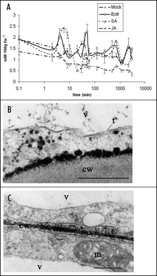
Similarly to other second messengers (e.g., cAMP,23,24 Ca2+ 21), H2O2 shows significant changes in its concentration after various stress situations. We have detected a number of H2O2 transients in fungal elicitor challenged Rubia tinctorum suspension cultures. Most of the authors observed alterations in H2O2 levels, one or two maxima, during the first hour of elicitor treatments or pathogen-host interactions.3,8,10,11 Only some papers implied three or more transients.12,13 Our results show that there is a rapid but not short H2O2 accumulation and an evident oscillation is detected not ending even at 48 hours after treatments. However, time-points, durations and amplitudes of the transients induced by the fungal cell wall hydrolysate, jasmonic acid (JA) and salicylic acid (SA) treatments were different during the prolonged challenge. Figure 1A shows the summarized, log scaled data of treatment effects on H2O2 level measured in the cells up to 48 hours after exposition to elicitors. Vitality of cells decreased during the experiments but up to 48 hours it was not significant. Changes of H2O2 level in the supernatant were obvious but the fine and quick alterations were not detectable. These facts point to the importance of data collecting methods and some uncertainty regarding the earlier published final conclusions.
Figure 1.
H2O2 accumulation in elicited Rubia tinctorum suspension culture cells. (A) Timing of H2O2 formation in untreated and elicited cultures. Mock (Mock), Botrytis cinerea fungal cell wall hydrolysate (Bot), jasmonic acid (JA), salicylic acid (SA). The 0.1 value of the log scale indicate 0 minuntes, other time-points are 2, 4, 6, 8, 10, 20, 30, 40, 50, 60, 120, 240, 480, 720, 1140, 2160, 2880 minutes. (B) Ce perhydroxids detected after 30 min fungal elicitor treatment. (C) Ce precipitates in the cell wall after 8 hours fungal elicitor treatment. v, vacuole; t, tonoplast; cw, cell wall; m, mitochondrion; Bar: 200 nm.
Tuning System
The observed oscillation of H2O2 level shows interesting minimal values between the maxima, which are significantly lower than the start level. These transient minima are general features of transduction systems and constitute the lifeblood of signal transduction regulation. 21,24,25 The accelerated production and “normal” metabolism of the signaling compound do not create such a profile except when there is only a temporary balance between the enhanced production and increased metabolic activity acting in concern but being out of mesh. Oscillation of H2O2 level in a wide range between maximal and minimal values is a result of the momentary rate of H2O2 generating processes and scavenging systems of the cell. In this way, apart from oxidative burst events there are also antioxidant dominancy periods in the redox state. The origin of these maxima and minima is rather poorly understood because of the lack of exact timing observations on the different H2O2 formation and scavenging elements. However, according to Trewavas' signaling system description, the observed features suggest a strictly fine-tuned mechanism in the case of H2O2 signaling also.
Location of H2O2 Generation
Fluorescence microscopic and cytochemical (Ce and DAB) investigations revealed topography of stress or pathogen attack caused H2O2 production in some model systems.3,13,15–17 H2O2 accumulation also occurred in spots during certain developmental processes (fiber secondary wall differentiation, lignification of xylem5,6). In cells after osmotic stress, the main H2O2 generation place was the tonoplast but to some extent H2O2 production was cytoplasmic endosomes.26 In our elicitor treated R. tinctorum cells, H2O2 formation appeared locally in the plasma membrane and cytoplasm at short-term (30 minute) sampling while later (one to eight hours) Ce perhydroxids were deposited mainly in the cell wall (Fig. 1B and C, respectively). The appearance of precipitate also differed. Cytoplasmic and plasma membrane related precipitates were rough and more or less vesicular or seemed to be of vesicular origin but cytochemical reaction resulted deposits were finely dispersed in the cell wall. The observed local feature of diaminobenzidine or Ce precipitates (or fluorescence signal) is reasonable and regular after pathogen attacks15–17,27 but in case of elicitor induced defense responses of suspension culture cells the triggering signals were direction independent. Interestingly, H2O2 generation seemed to be also a local event in these cells, affecting only a restricted cell area: apart from the visible electron density gradient, it was also proved by the sharp decrease of Ce content of the nearby cell wall regions detected by electron energy loss spectroscopy (EELS).
Conclusions
It is obvious from the above-mentioned facts that H2O2 utilizing metabolism and signaling pathways are multifunctional and complex systems. H2O2 formation and metabolism as well as their regulation comprise universal regulatory elements (feedback and/or feed-forward loops, amplification, transients, fine-tuning, oscillation, etc) and in several points there are cross-talk possibilities between the H2O2 pathway(s) and other signaling routes. Its/their coexistence and cooperation with various pathways does/do not exclude triggering or strong moderation of other transduction pathways (e.g., lipid, Ca2+, MAPK kinase signaling refs. 1, 2, 24 and 28–30). The coordinating effect can be manifested through influence on the redox state and may regulate processes in a cell, organ, organism or even between organisms (pathogenic or symbiotic partner and host interaction).13
The timing, tuning and localization features of H2O2 signaling elements point to the multi-source H2O2 total amount and the multi-element scavenging system interacting in a harmonized manner. An important principle is that signaling events are not only sequential even in the framework of one signaling pathway.2,4,22 The fine-tuning mechanism contains at least a few elements from the generation and scavenging side also. These elements and their regulation events are highly coordinated. H2O2 generation and scavenging system interaction and their regulation in cross-talk with other signal transduction pathways create a signal specific pattern resulting in a characteristic outcome after triggering. H2O2 signaling apart from other signaling pathways is also related to H2O2 utilizing metabolic processes (e.g., lignification, cell wall protein cross-link formation),28 which influence the signal amount. The relatively low average H2O2 level in the living systems queried its real antimicrobial effect15 but taking into consideration the strong spatial limit of H2O2 production, the local concentration of H2O2 might be high enough to injure microbes.
Footnotes
Previously published online as a Plant Signaling & Behavior E-publication: http://www.landesbioscience.com/journals/psb/article/4582
References
- 1.Wojtaszek P. Oxidative burst: An early plant response to pathogen infection. Biochem J. 1997;322:681–692. doi: 10.1042/bj3220681. [DOI] [PMC free article] [PubMed] [Google Scholar]
- 2.Foyer CH, Noctor G. Oxidant and antioxidant signaling in plants: A re-evaluation of the concept of oxidative stress in a physiological context. Plant Cell Environm. 2005;28:1056–1071. [Google Scholar]
- 3.Orozco-Cárdenas ML, Narváez-Vásquez J, Ryan CA. Hydrogen peroxide acts as a second messenger for the induction of defense genes in tomato plants in response to wounding, systemin, and methyl jasmonate. Plant Cell. 2001;13:179–191. [PMC free article] [PubMed] [Google Scholar]
- 4.Rojkind M, Domínguez-Rosales JA, Nieto N, Greenwel P. Role of hydrogen peroxide and oxidative stress in healing responses. Cell Mol Life Sci. 2002;59:1872–1891. doi: 10.1007/PL00012511. [DOI] [PMC free article] [PubMed] [Google Scholar]
- 5.Potikha TS, Collins CC, Johnson DI, Delmer DP, Levine A. The involvement of hydrogen peroxide in the differentiation of secondary walls in cotton fibers. Plant Physiol. 1999;119:849–858. doi: 10.1104/pp.119.3.849. [DOI] [PMC free article] [PubMed] [Google Scholar]
- 6.Ros Barceló A. Xylem parenchyma cells deliver the H2O2 necessary for lignification in differenciating xylem vessels. Planta. 2005;220:747–756. doi: 10.1007/s00425-004-1394-3. [DOI] [PubMed] [Google Scholar]
- 7.Kolla VA, Vavasseur A, Ranghavenra AS. Hydrogen peroxide production is an early event during bicarbonate induced stomatal closure in abaxial epidermis of Arabidopsis. Planta. 2007;225:1421–1429. doi: 10.1007/s00425-006-0450-6. [DOI] [PubMed] [Google Scholar]
- 8.Park J, Choi HJ, Lee S, Lee T, Yang Z, Lee Y. Rac-related GTP-binding protein in elicitor-induced reactive oxygen generation by suspension-cultured soybean cells. Plant Physiol. 2000;124:725–732. doi: 10.1104/pp.124.2.725. [DOI] [PMC free article] [PubMed] [Google Scholar]
- 9.Romero-Puertas MC, McCarthy I, Gómez M, Sandalio LM, Corpas FJ, Del Río LA, Palma JM. Reactive oxygen species-mediated enzymatic systems involved in the oxidative action of 2,4-dichlorophenoxyacetic acid. Plant Cell Environm. 2004;27:1135–1148. [Google Scholar]
- 10.Mur LAJ, Kenton P, Draper J. In Planta measurements of oxidative burst elicited by avirulent and virulent bacterial pathogens suggests that H2O2 is insufficient to elicit cell death in tobacco. Plant Cell Environm. 2005;28:548–561. [Google Scholar]
- 11.Miyata K, Miyashita M, Nose R, Otake R, Miyagawa H. Development of a colorimetric assay for determining the amount of H2O2 generated in tobacco cells in response to elicitors and its application to study of the structure-activity relationship of flagellin-derived peptides. Biosci Biotechnol Biochem. 2006;70:2138–2144. doi: 10.1271/bbb.60104. [DOI] [PubMed] [Google Scholar]
- 12.Bóka K, Orbán N, Kristóf Z. Dynamics and localization of H2O2 production in elicited cells. Protoplasma. 2007;230:89–97. doi: 10.1007/s00709-006-0225-8. [DOI] [PubMed] [Google Scholar]
- 13.Olmos E, Martiínez-Solano JR, Piqueras A, Hellín E. Early steps in the oxidative burst induced by cadmium in cultured tobacco cells (BY-2 line) J Exp Bot. 2003;54:291–301. doi: 10.1093/jxb/erg028. [DOI] [PubMed] [Google Scholar]
- 14.Baptista P, Martins A, Pais MS, Tavares RM, Lino-Neto T. Involvement of reactive oxygen species during early stages of ectomycirrhiza establishment between Castanea sativa and Pisolithus tinctorius. Mycorrhiza. 2007;17:185–193. doi: 10.1007/s00572-006-0091-4. [DOI] [PubMed] [Google Scholar]
- 15.Thordal-Christensen H, Zhang Z, Wei Y, Collinge DB. Subcellular localization of H2O2 signal in plants: H2O2 accumulation in papillae and hypersensiyive response during the barley-powdery mildew interaction. Plant J. 1997;11:1187–1194. [Google Scholar]
- 16.Bestwick CS, Brown IR, Bennett MHR, Mansfield JW. Localization of hydrogen peroxide accumulation diring the hypersensitive reaction of lettuce cells to Pseudomonas syringae pv phaseolicola. Plant Cell. 1997;9:209–221. doi: 10.1105/tpc.9.2.209. [DOI] [PMC free article] [PubMed] [Google Scholar]
- 17.Vanacker H, Carver TLW, Foyer CH. Early H2O2 accumulation in mesophyll cells leads to induction of glutathione during the hyper-sensitive response in the barley-powder mildew interaction. Plant Physiol. 2000;123:1289–1300. doi: 10.1104/pp.123.4.1289. [DOI] [PMC free article] [PubMed] [Google Scholar]
- 18.Tewari RK, Kumar P, Sharma PN, Bisht SS. Modulation of oxidative stress responsive enzymes by excess cobalt. Plant Sci. 2002;162:381–388. [Google Scholar]
- 19.Cano A, Hernández-Ruiz J, Arnao MB. Changes in hydrophilic antioxidant activity in Avena sativa and Triticum aestivum leaves of different age during de-etiolation and high light-treatment. J Plant Res. 2006;119:321–327. doi: 10.1007/s10265-006-0275-1. [DOI] [PubMed] [Google Scholar]
- 20.Teixeira FK, Menezes-Benavente L, Galvao VC, Margis R, Margis-Pinheiro M. Rice ascorbate peroxidase gene family encodes functionally diverse isoforms localized in different subcellular compartments. Planta. 2006;224:300–314. doi: 10.1007/s00425-005-0214-8. [DOI] [PubMed] [Google Scholar]
- 21.Trewavas A, Malhó R. Ca2+ signaling in plant cells: The big nerwork! Curr Opin Plant Biol. 1998;1:428–433. doi: 10.1016/s1369-5266(98)80268-9. [DOI] [PubMed] [Google Scholar]
- 22.Trewavas A. Plant cell signal transduction: The emerging phenotype. Plant Cell. 2002;14:S3–S4. doi: 10.1105/tpc.141360. [DOI] [PMC free article] [PubMed] [Google Scholar]
- 23.Cooke CJ, Smith CJ, Walton TJ, Newton RP. Evidence that cyclic AMP is involved in the hypersensitive response of Medicago sativa to a fungal elicitor. Phytochem. 1994;35:889–895. [Google Scholar]
- 24.Tengholm A. Cyclic AMP: Swing that message! Cell Mol Life Sci. 2007;64:382–385. doi: 10.1007/s00018-007-6440-4. [DOI] [PMC free article] [PubMed] [Google Scholar]
- 25.Fromm J, Lautner S. Electrical signals and their physiological significance in plants. Plant Cell Environm. 2007;30:249–257. doi: 10.1111/j.1365-3040.2006.01614.x. [DOI] [PubMed] [Google Scholar]
- 26.Leshem Y, Melamed-Book N, Cagnac O, Ronen G, Nishri Y, Solomon M, Cohen G, Levine A. Supresson of Arabidopsis vesicle-SNARE expression inhibited fusion of H2O2-containing vesicles with tonoplast and increased salt tolerance. PNAS. 2006;103:18008–18013. doi: 10.1073/pnas.0604421103. [DOI] [PMC free article] [PubMed] [Google Scholar]
- 27.Iwano M, Che FS, Goto K, Tanaka N, Takayama S, Isogai A. Electron microscopic analysis of the H2O2 accumulation preceding hypersensitive cell death induced by an incompatible strain of Pseudomonas avenae in cultured rice cells. Mol Plant Pathol. 2002;3:1–8. doi: 10.1046/j.1464-6722.2001.00087.x. [DOI] [PubMed] [Google Scholar]
- 28.Zhao J, Davis LC, Verpoorte R. Elicitor signal transduction leading to production of plant secondary metabolites. Biotechn Adv. 2005;23:283–333. doi: 10.1016/j.biotechadv.2005.01.003. [DOI] [PubMed] [Google Scholar]
- 29.Daxberger A, Nemak A, Mithöfer A, Fliegmann W, Hirt H, Ebel J. Activation of members of a MAPK module in β-glucan elicitor-mediated non-host resistance of soybean. Planta. 2007;225:1559–1571. doi: 10.1007/s00425-006-0442-6. [DOI] [PubMed] [Google Scholar]
- 30.Berken A. ROPs in the spotlight of plant signal transduction. Cell Mol Life Sci. 2006;63:2446–2459. doi: 10.1007/s00018-006-6197-1. [DOI] [PMC free article] [PubMed] [Google Scholar]


