Figure 2.
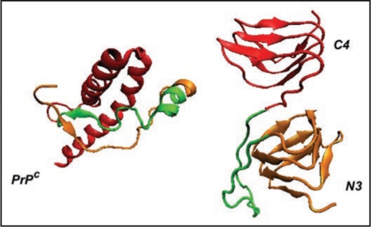
Comparison of PrP with LHBH structure. On the left we show human PrP (adopted from PDB structure 1QM0,31) with three color coded regions: residues 90–145 (orange), residues 146–167 (green), and residues 168–230. In the LHBH structure, which is the building block for tetramers (like that shown in Fig. 1), residues 90–145 go into a LHBH (N3, after ref. 7), 146–167 into a loop, and 168–230 into another LHBH (C4, present work).

