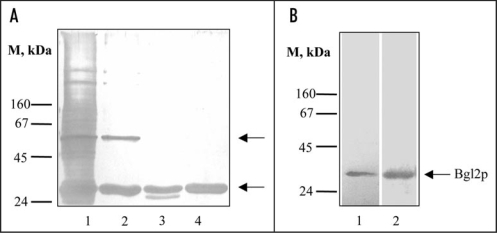
Figure 1.
Analysis of non-covalently bound proteins (SEPs) from the cell wall of S. cerevisiae. (A) SDS-PAGE of SEPs. Lane 1, SEPs extracted from wild type cell wall with hot Laemmli sample buffer (5 min, 100°C). Lane 2, SEPs extracted from cell walls after incubation (1 hour at 37°C) with 1% SDS; lane 3, with 1% SDS and proteinase K (10 mg/ml 1 hour at 37°C) step by step; lane 4, after incubation with 1% SDS and trypsin (2.5 mg/ml 30 min at 37°C) step by step. Upper arrow, presumptive Exg1p (61 kDa); lower arrow, Bgl2p (29 kDa). M stands for molecular weight markers, masses of marker proteins indicated on the figure (kDa). Proteins were separated on two-step 10/12% resolving gel and visualized by silver staining. (B) Silver stained SDS-PAGE (lane 1) and Western blot analysis (lane 2) of DMSO extracts of wild type cell walls. Proteins were extracted from cell walls, separated on 10% SDS gels and transferred to a nitrocellulose membrane. The transferred proteins were probed with an anti-Bgl2p antibody.

