Abstract
Gluteal muscle contracture is common after repeated intramuscular injections and sometimes is sufficiently debilitating to require open surgery. We asked whether arthroscopic release of gluteal muscle contracture using radiofrequency energy would decrease complications with clinically acceptable results. We retrospectively reviewed 108 patients with bilateral gluteal muscle contractures (57 males, 51 females; mean age, 23.7 years). We used inferior, anterosuperior, and posterosuperior portals. With the patient lying laterally, we developed and enlarged a potential space between the gluteal muscle group and the subcutaneous fat using blunt dissection. Under arthroscopic guidance through the inferior portal, we débrided and removed fatty tissue overlying the contractile band of the gluteal muscle group using a motorized shaver introduced through the superior portal. Radiofrequency then was introduced through the superior portal to gradually excise the contracted bands from superior to inferior. Finally, hemostasis was ensured using radiofrequency. Patients were followed a minimum of 7 months (mean, 17.4 months; range, 7–42 months). At last followup, the adduction and flexion ranges of the hip were 45.3° ± 8.7° and 110.2° ± 11.9°, compared with 10.4° ± 7.2° and 44.8° ± 14.1° before surgery. No hip abductor contracture recurred and no patient had residual hip pain or gluteal muscle wasting. We found gluteal muscle contracture could be released effectively with radiofrequency energy.
Level of Evidence: Level IV, therapeutic study. See the Guidelines for Authors for a complete description of levels of evidence.
Electronic supplementary material
The online version of this article (doi:10.1007/s11999-008-0595-7) contains supplementary material, which is available to authorized users.
Introduction
Gluteal muscle contractures (GMC) in adults are relatively common in many ethnicities, especially in Chinese. They usually are associated with repeated intramuscular injection into the gluteal region during childhood [1, 2, 7, 13]. An incidence of 1% to 2.5% in childhood has been reported [12, 14, 17]. Compared with Americans and Europeans, GMC is more widely reported in Chinese. This may be the result of more use of benzyl alcohol as a dissolvent for penicillin for intramuscular injections in Chinese in the 1970s and 1980s [1, 7], as several authors have suggested benzyl alcohol is the main cause of GMC [12, 13].
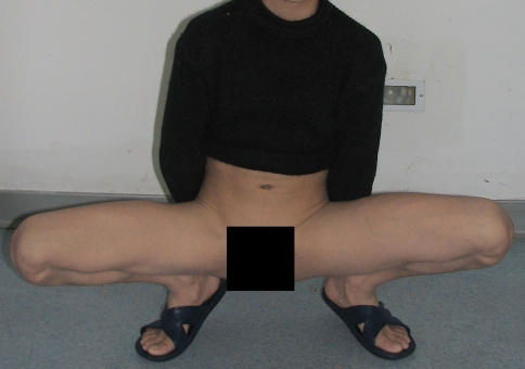
Patients with GMC typically present with hip abduction and external rotation when crouching. The knees cannot be brought together and are separated in a frog-leg position (Fig. 1). Stretching exercises usually are prescribed for nonoperative management. However, once the contractures are established, exercises and stretching of the shortened muscles do not produce improvement unless surgery is performed [2]. Reduction of the contractures and improvement of function with open release have been reported [2, 9, 18]. However, extensive surgical trauma [2, 18], hematoma formation [2, 18], wound complications [2, 18], and slow recovery [2, 18] are drawbacks of this traditional open surgery. Patients requiring Z-lengthening usually need 1 to 3 months to achieve full range of active hip flexion [18]. In a series of 187 patients with open excision, there were 62 cases of severe cicatrical band formation, six cases of hematoma formation, three cases of wound infection, and one case of wound dehiscence [5]. We reasoned using an arthroscopic technique might avoid extensive surgical trauma and a large skin wound and would allow earlier rehabilitation of patients after GMC release. Radiofrequency energy also could help coagulation of any bleeding point during cutting of the contractile band. Therefore, in June 2001, we began releasing these contractures using a minimally invasive surgical technique with radiofrequency energy under arthroscopic guidance.
Fig. 1.
The patient is crouched with the lower limbs separated in a frog-leg position.
We asked whether arthroscopic release of GMC using radiofrequency energy would (1) decrease the complications associated with open surgery and (2) provide adequate hip adduction and flexion ranges of motion (ROM).
Materials and Methods
We retrospectively reviewed all 150 patients who had GMC release with radiofrequency energy under arthroscopic guidance from June 2001 to June 2005. The average duration of symptoms was 5.3 ± 1.1 years (range, 3–7 years). All had failed to improve with stretching exercises. We excluded 42 pediatric patients younger than 16 years. No patient had any of the following presentations we considered contraindications for surgery: (1) clinical or radiographic evidence of a spinal problem or lower limb neurologic problem; (2) clinical or radiographic evidence of hip dysplasia or subluxation; and (3) medically unfit for surgery under general anesthesia or spinal anesthesia. With the exclusions, we had 108 patients (216 hips; all were bilateral). There were 57 men and 51 women with an average age of 23.7 ± 5.9 years (range, 18–40 years). Patients were followed for a minimum of 7 months (mean, 17.4 months; range, 7–42 months). No patients were lost to followup. All patients provided informed consent for the operation and followed our rehabilitation protocol. This study was approved by the Institutional Review Board of our institution.
Detailed histories were taken from the patients with particular attention to the history of injury to the buttock and hip regions, history of injection around the gluteal region, premorbid and current activity levels, the need for walking aids, and mode of nonoperative treatments received (including medications, herbs, physiotherapy, and acupuncture). We also recorded the following parameters before the operation: (1) gluteal muscle wasting; (2) loss of lumbar lordosis or hyperlordotic lumbosacral spine; (3) gait pattern; (4) dimpling of skin around the gluteal region; (5) excessive external rotation of the hip at rest; (6) hip pain; (7) presence of a clicking sound during rotation of the hip; (8) adduction and flexion ROM of the hip; (9) presence of hamstring tightness; (10) Ober’s sign; (11) neurologic deficit; (12) squatting and crouching ability with both hips in a neutral position; and (13) cross-legged sitting. Gluteal muscle wasting was assessed by observing the outline of the gluteal region and was classified as normal or slightly or severely cone-shaped. Gait pattern was evaluated by observing the out-toe walking pattern. Hip pain was assessed by a pain visual analog scale.
All patients had a history of injections around the gluteal region. Although none had limitations with activities of daily living, they believed their activity levels were less than those of healthy people. All patients had an abnormal gait with out-toe walking. None of the patients could crouch with both knees close to each other or sit with their legs crossed. All patients had a clicking sound during rotation of the hip and a positive Ober’s sign. None had previous surgery before the index operation.
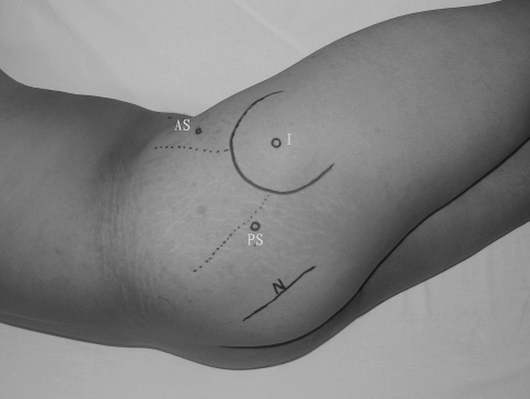
The patient was positioned in a lateral position while under general anesthesia or spinal anesthesia. Because all of our patients had both hips affected, we operated on one side first followed by the other side by turning and repositioning the patient. Under the guidance of an arthroscope, the border of the contracture was observed and all important anatomic landmarks were marked before surgery. We outlined the greater trochanter, anterior and posterior borders of the contracted gluteal muscle, and the sciatic nerve. We used a two- or three-portal technique to perform the operation, namely the inferior, anterosuperior (AS), and posterosuperior (PS) portals. With the hip in neutral position, we placed the inferior portal, which was used for introduction of the arthroscope, 3 cm directly inferior to the greater trochanter. The AS and PS portals, which were used as the drainage or the working portals for instruments, such as motorized shavers or the radiofrequency device, were placed superior to the greater trochanter. We placed the AS portal anterior to the anterior border of the contracted gluteal muscle, whereas the PS portal was placed posterior to the posterior border of the contracted gluteal muscle (Fig. 2). When the operator faced the patient’s back, we selected the PS portal for instrumentation and the AS portal was used for drainage; when the operator faced the patient’s abdomen, we selected the AS portal for instrumentation and the PS portal was used for drainage. The operator thus could complete the operations for both hips without changing his position or that of the monitor. During arthroscopic-assisted release, we enlarged the potential space between the gluteal muscle group and the subcutaneous fat using blunt dissection with a periosteal elevator. Normal saline (containing 1 mg adrenaline per 3000 mL normal saline) then was injected. This created a good working space and we presumed would reduce bleeding during the procedure. We then were able to clearly observe the anterior and posterior borders of the gluteal muscle group. The instrument was introduced through the AS and PS portals and the sciatic nerve was at least 20 mm distal to the operating site. Under arthroscopic guidance through the inferior portal, we used a motorized shaver introduced through the superior portal to débride and remove fatty tissue overlying the contractile band of the gluteal muscle group. A radiofrequency device (ArthroCare® Atlas® System with TriStar® 50 ArthroWand; ArthroCare Corporation, Sunnyvale, CA; shaft size: 3.0 mm, angle: 50°, bipolar, output power: 7/9 level, working temperature: approximately 53°C, mode: cut or coagulate) then was introduced through the superior portal and sectioned the contractile bands from superior to inferior (Fig. 3). Good hemostasis was maintained throughout surgery to allow clear observation of the surgical site during the entire procedure and to prevent hematoma formation after the operation. This was achieved by coagulation of any bleeding point using radiofrequency energy, increasing the pressure of saline inflow, and adding adrenaline to the saline solution. We monitored blood pressure, heart rate, and blood oxygen saturation of the patients regularly during surgery. Blood pressure was measured with a cuff and heart rate was counted once every 2 hours during the first 24 hours after surgery. During surgery, the operated hip was repeatedly flexed, externally rotated, abducted, and adducted to ensure good ROM. The average operating time for one side of GMC was 15.3 ± 9.8 minutes (range, 7–30 minutes).
Fig. 2.
The photograph shows the important anatomic landmarks on the patient during surgery. AS = anterosuperior portal; PS = posterosuperior portal; I = inferior portal; N = sciatic nerve.
Fig. 3.
The arthroscopic image shows the cutting of contractile bands using the radiofrequency device.
After surgery, the patients were asked to lie in a lateral position to compress one side with their body weight and a 2-kg ice bag was placed on the other side. The position was switched every 1 or 2 hours to ensure good hemostasis. We observed considerable exudation of fluid from the wound during the first 24 hours and thus wound dressings were changed at regular intervals.
Patient-controlled intravenous analgesia (sufentanil, 150 μg, and ondanestron hydrochloride, 16 mg per 100 mL saline) was given postoperatively. The analgesic was background infused at 2 mL/hour followed by a bolus dose at 0.5 mL/8 minutes. Most patients had analgesic stopped within 48 hours. Rehabilitation started 24 hours after surgery. Functional exercises, including (1) crouching with both knees close to each other, (2) abducting and adducting the upper limbs while lying in a lateral position, and (3) lying on the back, bringing the knees to the chest, clasping the hands to the front of the shin, and internally rotating the hips while keeping the pelvis as flat as possible, were performed to prevent scar formation and muscle wasting. These exercises were performed by the patients three to five times a day with 20 to 30 repetitions depending on their endurance. Patients were discharged from the hospital once they could move independently without any walking aids. We examined all patients to ensure there was no hematoma or early wound complications before discharging them. The average length of hospital stay was 3.2 ± 0.9 days (range, 2–5 days) after surgery.
All patients were followed at regular intervals after surgery. For the first 2 months, they were reassessed every 2 weeks to monitor wound complications and examine function of the hips. After that, they were seen for followup every 3 months. Most patients had stopped analgesic 48 hours postsurgery. For patients who still had pain, Fenbid® capsules (300 mg; GlaxoSmithKline, Philadelphia, PA) were given twice a day and for less than 1 week. We assessed adduction and flexion ROM, gait pattern, Ober’s sign, clicking sound during hip rotation, hip pain, and residual muscle wasting. All these parameters were assessed by a senior clinician blinded and not involved in the operations. Hip adduction and flexion ROM were used as the primary outcome of this study. They were measured three times and the mean values were taken. All patients returned for clinical followup for a minimum of 7 months (mean, 17.4 months; range, 7–42 months). We considered a followup greater than 7 months adequate because there was no change in hip adduction and flexion ROM and gait pattern 7 months after surgery.
Descriptive data are presented as mean ± standard deviation and range. Preoperative and postoperative adduction and flexion ROM of the hip were compared using a two-tailed paired t test. We analyzed the data with SPSS® (Version 11.0; SPSS Inc, Chicago, IL).
Results
No adverse effects on the hemodynamic status of the patients were observed during and after surgery. There were no complications related to the wound (infection, hematoma formation, breakdown of wound), and there was no evidence of sciatic nerve injury presenting as lower limb pain, numbness, or dysfunction or neurologic deficit in the study period.
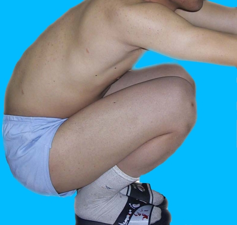
Adduction and flexion ROM were increased (both p < 0.001) at final followup: 10.4° ± 7.2° and 44.8° ± 14.1° before surgery versus 45.3° ± 8.7° and 110.2° ± 11.9° after surgery, respectively. Out-toe gaits were corrected with different degrees. All patients could crouch with both knees close to each other (Fig. 4) and sit with their legs crossed. No Ober’s sign or clicking sound during rotation of the hip was found. There was no recurrent contracture of the hip abductor, residual hip pain, or gluteal muscle wasting. A supplementary video shows the hip function of a patient before and 2 weeks after surgery (Supplemental materials are available with the online version of CORR). As shown in the video, the patient could cross her legs while sitting, could squat and crouch with both hips in a neutral position, showed improvement in adduction and flexion ROM of the hip, and had no clicking sound during rotation of the hip 2 weeks after surgery.
Fig. 4.
The patient is crouched with both knees close to each other after surgery.
Discussion
The superficial fibers and the upper deep part of the gluteal maximus end in a tendinous sheet, which passes lateral to the greater trochanter and is attached to the iliotibial band of the fascia lata, whereas the deep fibers of the lower part of the muscle are inserted into the greater trochanter. Muscle degeneration and fibrillation may occur by repeated intramuscular injection into the region, especially when benzyl alcohol is used as a dissolvent for penicillin for intramuscular injections [2, 3, 13] and thus flexibility of muscle fibers can be affected. Skin and subcutaneous fat tissue covering the outer upper quadrant of the buttocks also can be affected because of injection in this region, causing dimple-like depressions in the skin. Because of contractures of the gluteal muscle with skin depression, the buttocks look like a cone during squatting in bilaterally affected cases. Both hips are abducted and externally rotated. The knees are separated in a frog-leg position when the patient squats. When seated, the patient cannot cross his or her legs because of the contracture. The patient usually walks with an out-toe gait. GMC is common in patients in numerous countries, and particularly in Chinese patients [12, 17]. Its incidence rate in children is 1% to 2.5%, and adults also can be affected [12, 17]. Ma et al. [11] first reported intramuscular injection-associated GMC in children and introduced open excision to cut the contractile bands in 1978. Various techniques have been used for management of GMC since then. The purpose of our study was to review the results of arthroscopic release of GMC using radiofrequency energy with respect to (1) complications related to the surgery and (2) hip adduction and flexion ROM. Our hypothesis was that this method would reduce complications associated with open surgery and would have acceptable clinical outcomes. Our results showed GMC could decrease the complications associated with open surgery and provide adequate hip adduction and flexion ROM. Despite this, repeated intramuscular injections into the gluteal region should be avoided, especially when benzyl alcohol is used as a dissolvent.
Our study has several limitations. First, it was a retrospective review of a case series with no control group. We therefore cannot ensure the approach provided fewer complications and better outcomes than an open approach except as compared with published results. We did not collect factors that might influence the outcomes. There might be concern regarding the generalizability of the results to other patients with GMC because all the subjects were recruited from one hospital. Despite these limitations, our series was fairly large with 108 patients (216 hips, bilateral) and the followup was at least 7 months. A further prospective study should be done to confirm our results.
There were no complications related to the wound (infection, hematoma formation, breakdown of wound), and there was no evidence of sciatic nerve injury or neurologic deficit in the study period. There were improvements in clinical and functional outcomes at the last followup, with results comparable to those reported with traditional open surgery [2, 5, 18]. Open excision of the contracture fibers has been recognized as one of the most helpful procedures in the treatment of GMC, especially in regaining function of the lower limb and gait and improving appearance [2, 4]. However, the procedure requires a relatively large surgical wound and is associated with hematoma formation, low-grade infection, and delayed wound healing [2, 18]. In a series of 187 patients with open excision, there were 62 cases of severe cicatrical band formation, six cases of hematoma formation, three cases of wound infection, and one case of wound dehiscence [5]. Use of the radiofrequency vaporization technique in arthroscopy is regarded as a revolution in the history of arthroscopic surgery [8, 16]. Conventional electrosurgical devices remove target tissue by rapid heating (greater than 400°C), charring, and burning and therefore may cause inadvertent damage to the surrounding tissue. Uncontrolled bleeding may result, making arthroscopic observation difficult. However, radiofrequency devices use Coblation® (ArthroCare Corporation) technology to section the target tissue gently with minimal heating effect to the surrounding tissue [6]. With Coblation® (ArthroCare Corporation) technology, radiofrequency energy is used to excite the electrolytes in a conductive medium such as saline solution to create a precisely focused plasma whose energized particles have sufficient energy to break the molecular bond in tissue, causing tissue to dissolve at relatively low temperature (typically 40°C to 70°C). Numerous Coblation®-based devices are in fact designed to seal bleeding vessels [6, 10, 15]. In addition, the radiofrequency wand has the capability of cutting in all directions with low resistance and therefore allows fine manipulation in narrow spaces [8]. The use of radiofrequency energy in arthroscopic-assisted GMC release seemed to make the procedures easier and the data suggested the release was safe. Because of minimal surgical trauma, short operative time, and little obvious bleeding during the surgery, patients could start rehabilitation earlier. This might be an important factor for the success of our series. To minimize bleeding in the operative field, we added adrenaline (1 mg in 3000 mL) to the normal saline for irrigation. We observed no adverse effects on the hemodynamic status of the patients during and after surgery. Moreover, to avoid excessive bleeding during stripping and cutting of the muscle fibers, we advocate operating from the superficial layer to the deep layer. To accomplish complete excision, the hips should be mobilized to feel for any clicks or adhesion throughout the operation. Commonly missed areas include the contracture bands near the site where the gluteal muscle attaches to the posterior part of the greater trochanter, which may present as a clicking sound during hip mobilization.
Despite the advantages of the current surgical technique, one should not eliminate or minimize the choice of open surgery only because of the surgeon’s pursuit of a minimally invasive approach. In cases of uncontrolled bleeding or a deep contracture that are not reachable with an arthroscopic instrument, a small incision should be used.
We found the arthroscopic-assisted GMC release using radiofrequency energy was a good alternative when compared with traditional open surgery. It has the advantages of little surgical trauma, short operative time, little obvious bleeding during surgery, few surgical complications, earlier rehabilitation, and return of functional activities.
Electronic supplementary material
Acknowledgments
We thank the patients for participating in this study. We also are grateful to Dr. Mi Zhou, Dr. Zhi-Gand Wang, and Dr. Zhong-Li Li for help in data collection and for comments on the initial manuscript.
Footnotes
Each author certifies that he or she has no commercial associations (eg, consultancies, stock ownership, equity interest, patent/licensing arrangements, etc) that might pose a conflict of interest in connection with the submitted article.
Each author certifies that his or her institution has approved the human protocol for this investigation, that all investigations were conducted in conformity with ethical principles of research, and that informed consent for participation in the study was obtained.
Electronic supplementary material
The online version of this article (doi:10.1007/s11999-008-0595-7) contains supplementary material, which is available to authorized users.
References
- 1.Chung DC, Ko YC, Pai HH. [A study on the prevalence and risk factors of muscular fibrotic contracture in Jia-Dong Township, Pingtung County, Taiwan] [in Chinese]. Gaoxiong Yi Xue Ke Xue Za Zhi. 1989;5:91–95. [PubMed]
- 2.Fernandez de Valderrama JA, Esteve de Miguel R. Fibrosis of the gluteus maximus: a cause of limited flexion and adduction of the hip in children. Clin Orthop Relat Res. 1981;156:67–68. [PubMed]
- 3.Gao GX. Idiopathic contracture of the gluteus maximus in children. Arch Orthop Trauma Surg. 1988;107:277–279. [DOI] [PubMed]
- 4.Hang YS. Contracture of the hip secondary to fibrosis of the gluteus maximus muscle. J Bone Joint Surg Am. 1979;61:52–55. [PubMed]
- 5.He XJ, Li HP, Wang D, Wang B, Liu XG, Xu SY, Chen L. [Classification and management of the gluteal muscles contracture] [in Chinese]. Chin J Orthop. 2003;23:96–100.
- 6.High performance at low temperature. ArthroCare Sports Medicine US. Available at: www.arthrocaresportsmedicine.com/scnc_coblation_rf. Accessed June 28, 2008.
- 7.Huang Y, Li J, Lei W. [Gluteal muscle contracture: etiology, classification and treatment] [in Chinese]. Chin J Orthop. 1999;19:106–108.
- 8.Kramer J, Rosenthal A, Moraldo M, Muller KM. Electrosurgery in arthroscopy. Arthroscopy. 1992;8:125–129. [DOI] [PubMed]
- 9.Li K, Huang X, Tang J. Prospective efficacy of operative treatment of intramuscular injection caused gluteal muscle contracture [in Chinese]. Chin J Orthop. 1998;18:437–438.
- 10.Lopez M, Hayashi K, Fanton GS, Thabit G 3rd, Markel MD. The effect of radiofrequency energy on the ultrastructure of joint capsular collagen. Arthroscopy. 1998;14:495–501. [DOI] [PubMed]
- 11.Ma CX, Fang LG, Liu GL. [Injection caused gluteal muscle contracture] [in Chinese]. Chin J Orthop. 1978;16:345–346.
- 12.Peng M, Zhou Z, Zhou X. [Epidemiology of gluteal muscle contracture in Si Chuan Province] [in Chinese]. Chin J Pediatr Surg. 1989;10:356–358.
- 13.Sirinelli D, Oudjhane K, Khouri N. Case report 605: gluteal amyotrophy: a late sequela of intramuscular injection. Skeletal Radiol. 1990;19:221–223. [DOI] [PubMed]
- 14.Sun X. [An investigation on injectional gluteal muscle contracture in childhood in Mianyang City] [in Chinese]. Zhonghua Liu Xing Bing Xue Za Zhi. 1990;11:291–294. [PubMed]
- 15.Tasto JP, Ash SA. Current uses of radiofrequency in arthroscopic knee surgery. Am J Knee Surg. 1999;12:186–191. [PubMed]
- 16.Wang Y, Shi D, Gu Y. [Preliminary study on usage of ArthroCare radiofrequency in knee arthroscopic surgery] [in Chinese]. Chin J Orthop. 2001;21:172–175.
- 17.Zhang G, Zheng Z, Fu Z. [12459 cases of gluteal muscle contracture in children] [in Chinese]. Chin J Pediatr Surg. 1990;11:363–365.
- 18.Zhang KF, Li PF, Zhong-Ke LV, Ren HJ, Tang XF, Chun-Hui MA, Tang M. [Treatment of gluteus contracture with small incision: a report of 2518 cases] [in Chinese]. Chin J Orthop Trauma. 2007;20:851–852.
Associated Data
This section collects any data citations, data availability statements, or supplementary materials included in this article.


